. 2020 Mar;34(2):828-837.
doi: 10.1111/jvim.15673. Epub 2020 Jan 10.

Clinicopathological characteristics of histiocytic sarcoma affecting the central nervous system in dogs

Affiliations

Clinicopathological characteristics of histiocytic sarcoma affecting the central nervous system in dogs

Izumi Toyoda et al. J Vet Intern Med. 2020 Mar.

Abstract

Background: Histiocytic sarcoma affecting the central nervous system (CNS HS) in dogs may present as primary or disseminated disease, often characterized by inflammation. Prognosis is poor, and imaging differentiation from other CNS tumors can be problematic.

Objective: To characterize the clinicopathological inflammatory features, breed predisposition, and survival in dogs with CNS HS.

Animals: One hundred two dogs with HS, 62 dogs with meningioma.

Methods: Retrospective case series. Records were reviewed for results of cerebrospinal fluid (CSF) analysis, CBC, treatment, and outcome data.

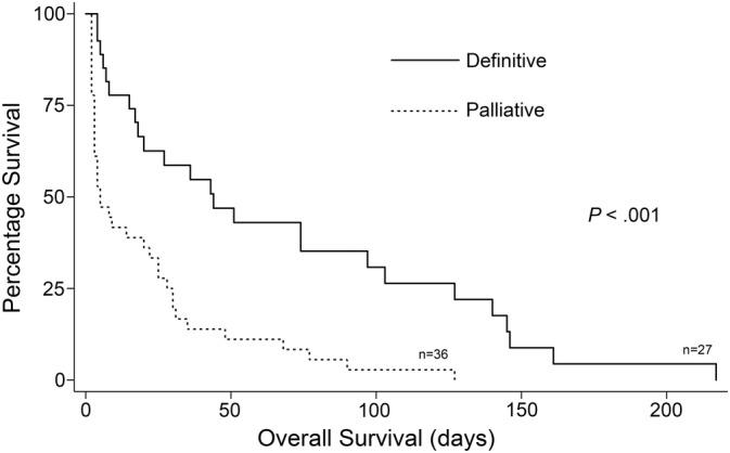
Results: Predisposition for CNS HS was seen in Bernese Mountain Dogs, Golden Retrievers, Rottweilers, Corgis, and Shetland Sheepdogs (P ≤ .001). Corgis and Shetland Sheepdogs had predominantly primary tumors; Rottweilers had exclusively disseminated tumors. Marked CSF inflammation was characteristic of primary rather than disseminated HS, and neoplastic cells were detected in CSF of 52% of affected dogs. Increased neutrophil to lymphocyte ratios were seen in all groups relative to controls (P <.008) but not among tumor subtypes. Definitive versus palliative treatment resulted in improved survival times (P < .001), but overall prognosis was poor.

Conclusions and clinical importance: Clinicopathological differences between primary and disseminated HS suggest that tumor biological behavior and origin may be different. Corgis and Shetland Sheepdogs are predisposed to primary CNS HS, characterized by inflammatory CSF. High total nucleated cell count and the presence of neoplastic cells support the use of CSF analysis as a valuable diagnostic test. Prognosis for CNS HS is poor, but further evaluation of inflammatory mechanisms may provide novel therapeutic opportunities.

Keywords: canine; central nervous system; cerebrospinal fluid; neoplasia.


Conflict of interest statement

Authors declare no conflict of interest.

Figures

Figure 1
Figure 1
A and B, Box plots of CSF total nucleated cell counts in primary CNS HS, disseminated CNS HS and meningiomas, regardless of corticosteroid administration. B, Expanded TNCC scale to allow assessment of low TNCC data points in disseminated CNS HS and meningioma groups. Boxes correspond to interquartile ranges (25th–75th percentiles); the horizontal line within the box is the median value (50th percentile). Whiskers represent lower and upper adjacent values (i.e. the lowest and highest observations that are still inside the region defined by the following limits: Lower limit: Q1−1.5 × IQR. Upper limit: Q3 + 1.5 × IQR). Significant differences between tumor groups are indicated by brackets. IQR, interquartile range
Figure 2
Figure 2
A and B, Box plots of CSF total protein in primary CNS HS, disseminated CNS HS and meningioma. A, All cases regardless of corticosteroid administration. B, Cases with no history of corticosteroid administration. Boxes correspond to interquartile ranges (25th–75th percentiles); the horizontal line within the box is the median value (50th percentile). Whiskers represent lower and upper adjacent values (i.e. the lowest and highest observations that are still inside the region defined by the following limits: Lower limit: Q1−1.5 × IQR. Upper limit: Q3 + 1.5 × IQR). Significant differences between tumor groups are indicated by brackets. Outlying data points are included with values in brackets. IQR, interquartile range
Figure 3
Figure 3
Cerebrospinal fluid cytospin preparations from dogs with primary CNS HS. A, Note the three extremely large, neoplastic, single round cells with large immature and atypical nuclei, admixed with numerous inflammatory cells comprising nondegenerate neutrophils and macrophages. B, There are two large neoplastic histiocytes, one of which is mitotic, admixed with numerous inflammatory cells. C, There is a markedly increased total nucleated cell count (TNCC) and numerous mitoses, which raises the index of suspicion for underlying primary CNS HS and should prompt a search for large, atypical round cells. Wright's‐Giemsa stain, scale bar = 10 μm
Figure 4
Figure 4
Boxplots of peripheral CBC neutrophil:lymphocyte ratios from cases of primary CNS HS, disseminated CNS HS, meningioma and control (clinically normal) dogs. Whiskers represent +/‐ 1.5 interquartile ranges. Significant differences between tumor groups are indicated by asterisks. Boxes correspond to interquartile ranges (25th–75th percentiles); the horizontal line within the box is the median value (50th percentile). Whiskers represent lower and upper adjacent values (i.e. the lowest and highest observations that are still inside the region defined by the following limits: Lower limit: Q1−1.5 × IQR. Upper limit: Q3 + 1.5 × IQR). Significant differences between tumor groups are indicated by brackets. Outlying data points are included with values in brackets. IQR, interquartile range
Figure 5
Figure 5
Kaplan‐Meier survival estimates for definitively and palliatively treated histiocytic sarcoma affecting the central nervous system (CNS HS) (primary and disseminated) cases that survived >1 day

References

    1. Affolter VK, Moore PF. Localized and disseminated histiocytic sarcoma of dendritic cell origin in dogs. Vet Pathol. 2002;39:74‐83. - PubMed
    1. Moore PF. A review of histiocytic diseases of dogs and cats. Vet Pathol. 2014;51:167‐184. - PubMed
    1. Abadie J, Hedan B, Cadieu E, et al. Epidemiology, pathology, and genetics of histiocytic sarcoma in the Bernese mountain dog breed. J Hered. 2009;100(Suppl 1):S19‐S27. - PMC - PubMed
    1. Moore PF, Rosin A. Malignant histiocytosis of Bernese Mountain dogs. Vet Pathol. 1986;23:1‐10. - PubMed
    1. Padgett GA, Madewell BR, Keller ET, Jodar L, Packard M. Inheritance of histiocytosis in Bernese Mountain dogs. J Small Anim Pract. 1995;36:93‐98. - PubMed