

The circadian clock protects against ionizing radiation-induced cardiotoxicity
- PMID: 31919902
- PMCID: PMC9677419
- DOI: 10.1096/fj.201901850RR
The circadian clock protects against ionizing radiation-induced cardiotoxicity
Abstract
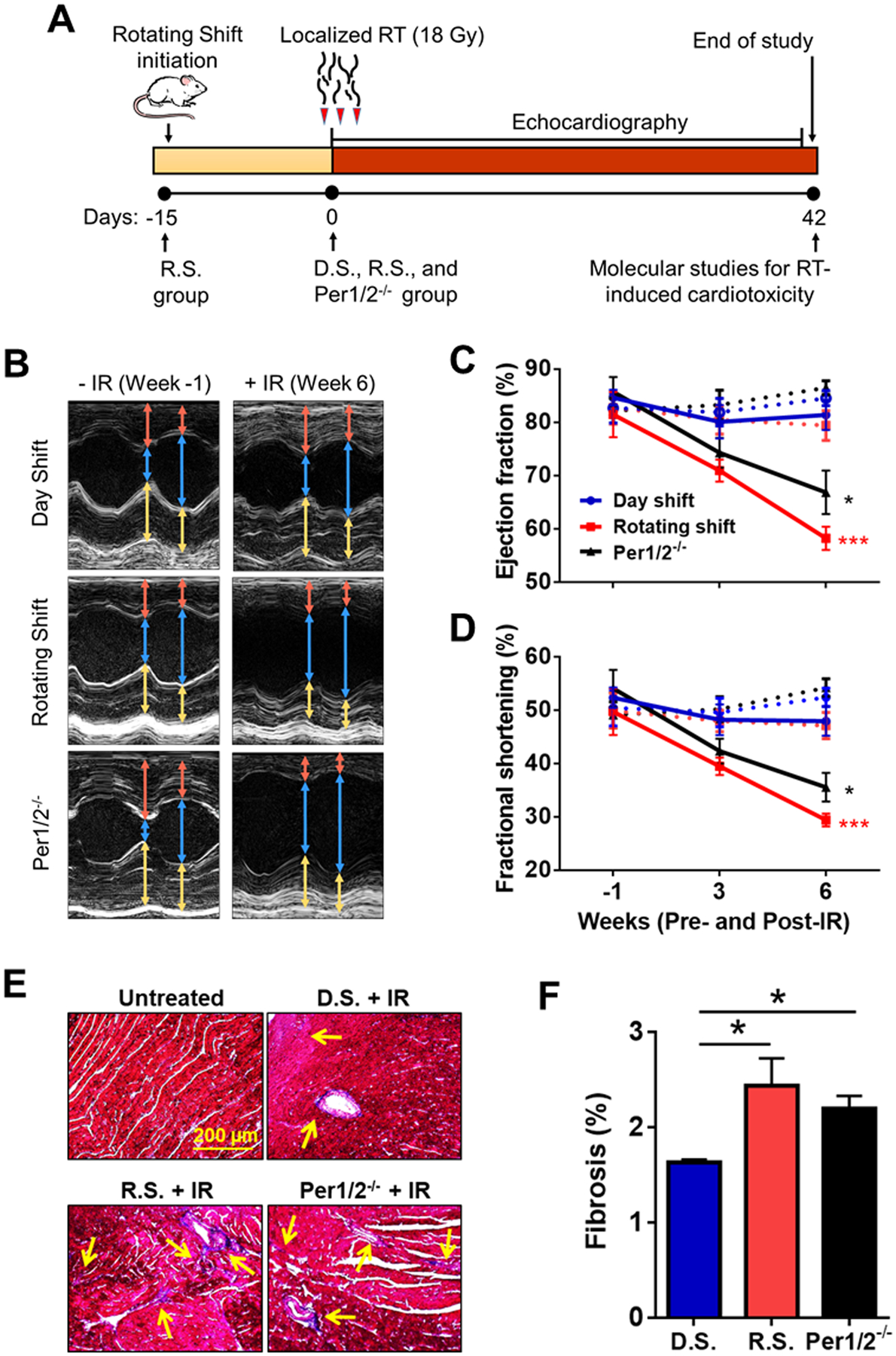
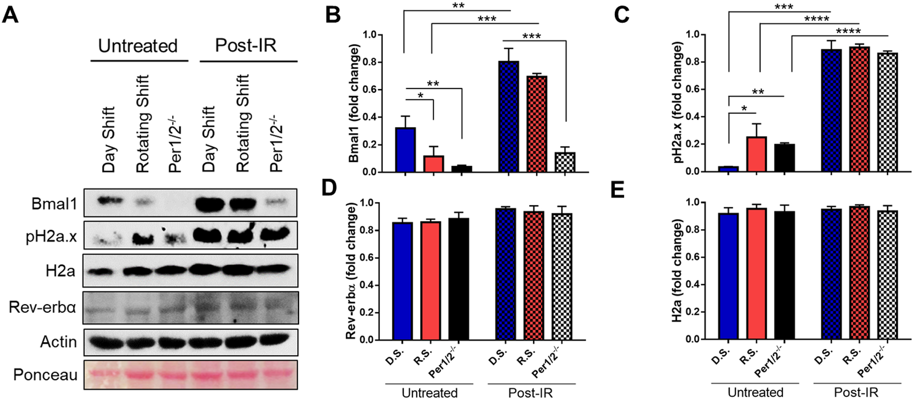
Radiation therapy (RT) is commonly used to treat solid tumors of the breast, lung, and esophagus; however, the heart is an unintentional target of ionizing radiation (IR). IR exposure to the heart results in chronic toxicities including heart failure. We hypothesize that the circadian system plays regulatory roles in minimizing the IR-induced cardiotoxicity. We treated mice in control (Day Shift), environmentally disrupted (Rotating Shift), and genetically disrupted (Per 1/2 mutant) circadian conditions with 18 Gy of IR to the heart. Compared to control mice, circadian clock disruption significantly exacerbated post-IR systolic dysfunction (by ultrasound echocardiography) and increased fibrosis in mice. At the cellular level, Bmal1 protein bound to Atm, Brca1, and Brca2 promoter regions and its expression level was inversely correlated with the DNA damage levels based on the state of the clock. Further studies with circadian synchronized cardiomyocytes revealed that Bmal1 depletion increased the IR-induced DNA damage and apoptosis. Collectively, these findings suggest that the circadian clock protects from IR-induced toxicity and potentially impacts RT treatment outcome in cancer patients through IR-induced DNA damage responses.
Keywords: Bmal1; circadian clock; heart; radiation; toxicity.
© 2020 Federation of American Societies for Experimental Biology.
Conflict of interest statement
Figures


Similar articles
-
The circadian clock protects against acute radiation-induced dermatitis.Toxicol Appl Pharmacol. 2020 Jul 15;399:115040. doi: 10.1016/j.taap.2020.115040. Epub 2020 May 15. Toxicol Appl Pharmacol. 2020. PMID: 32422325 Free PMC article.
-
Selenium is a modulator of circadian clock that protects mice from the toxicity of a chemotherapeutic drug via upregulation of the core clock protein, BMAL1.Oncotarget. 2011 Dec;2(12):1279-90. doi: 10.18632/oncotarget.411. Oncotarget. 2011. PMID: 22249125 Free PMC article.
-
Depletion of ATR selectively sensitizes ATM-deficient human mammary epithelial cells to ionizing radiation and DNA-damaging agents.Cell Cycle. 2014;13(22):3541-50. doi: 10.4161/15384101.2014.960729. Cell Cycle. 2014. PMID: 25483091 Free PMC article.
-
Effect of circadian clock mutations on DNA damage response in mammalian cells.Cell Cycle. 2012 Sep 15;11(18):3481-91. doi: 10.4161/cc.21771. Epub 2012 Aug 23. Cell Cycle. 2012. PMID: 22918252 Free PMC article.
-
Circadian Rhythm of NER and ATR Pathways.Biomolecules. 2021 May 11;11(5):715. doi: 10.3390/biom11050715. Biomolecules. 2021. PMID: 34064641 Free PMC article. Review.
Cited by
-
Basic helix-loop-helix ARNT like 1 regulates the function of immune cells and participates in the development of immune-related diseases.Burns Trauma. 2025 Jan 18;13:tkae075. doi: 10.1093/burnst/tkae075. eCollection 2025. Burns Trauma. 2025. PMID: 39830193 Free PMC article. Review.
-
Biological Adaptations of Tumor Cells to Radiation Therapy.Front Oncol. 2021 Nov 24;11:718636. doi: 10.3389/fonc.2021.718636. eCollection 2021. Front Oncol. 2021. PMID: 34900673 Free PMC article. Review.
-
The Impact of Genetic Variations on Radiotherapy Toxicity in Breast Cancer Patients: A Meta-Analysis of Acute and Late Skin Adverse Effects.Cancers (Basel). 2025 Jun 4;17(11):1880. doi: 10.3390/cancers17111880. Cancers (Basel). 2025. PMID: 40507362 Free PMC article. Review.
-
Long-lasting sex-specific alteration in left ventricular cardiac transcriptome following gamma and simGCRsim radiation.Sci Rep. 2025 Feb 18;15(1):5963. doi: 10.1038/s41598-025-89815-2. Sci Rep. 2025. PMID: 39966642 Free PMC article.
-
Chronoradiobiology of Breast Cancer: The Time Is Now to Link Circadian Rhythm and Radiation Biology.Int J Mol Sci. 2022 Jan 25;23(3):1331. doi: 10.3390/ijms23031331. Int J Mol Sci. 2022. PMID: 35163264 Free PMC article. Review.
References
-
- National Library of Medicine (U.S.). (2019) ClinicalTrials.gov linking patients to medical research. U.S. National Library of Medicine, National Institutes of Health, Dept. of Health & Human Services,, Bethesda, MD
-
- Sancar A, Lindsey-Boltz LA, Unsal-Kacmaz K, and Linn S (2004) Molecular mechanisms of mammalian DNA repair and the DNA damage checkpoints. Annu Rev Biochem 73, 39–85 - PubMed
-
- Darby SC, Ewertz M, McGale P, Bennet AM, Blom-Goldman U, Bronnum D, Correa C, Cutter D, Gagliardi G, Gigante B, Jensen MB, Nisbet A, Peto R, Rahimi K, Taylor C, and Hall P (2013) Risk of ischemic heart disease in women after radiotherapy for breast cancer. N Engl J Med 368, 987–998 - PubMed
-
- Boekel NB, Schaapveld M, Gietema JA, Russell NS, Poortmans P, Theuws JC, Schinagl DA, Rietveld DH, Versteegh MI, Visser O, Rutgers EJ, Aleman BM, and van Leeuwen FE (2016) Cardiovascular Disease Risk in a Large, Population-Based Cohort of Breast Cancer Survivors. Int J Radiat Oncol Biol Phys 94, 1061–1072 - PubMed
-
- Puukila S, Lemon JA, Lees SJ, Tai TC, Boreham DR, and Khaper N (2017) Impact of Ionizing Radiation on the Cardiovascular System: A Review. Radiat Res 188, 539–546 - PubMed
Publication types
MeSH terms
Substances
Grants and funding
LinkOut - more resources
Full Text Sources
Molecular Biology Databases
Research Materials
Miscellaneous
