

Differences in the relative counts of peripheral blood lymphocyte subsets in various age groups of pigs
- PMID: 31920218
- PMCID: PMC6921992
Differences in the relative counts of peripheral blood lymphocyte subsets in various age groups of pigs
Abstract
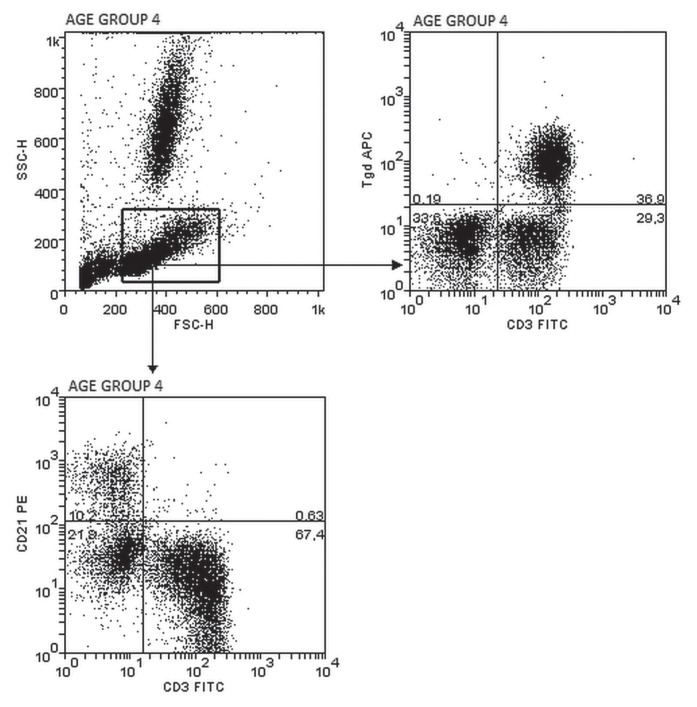
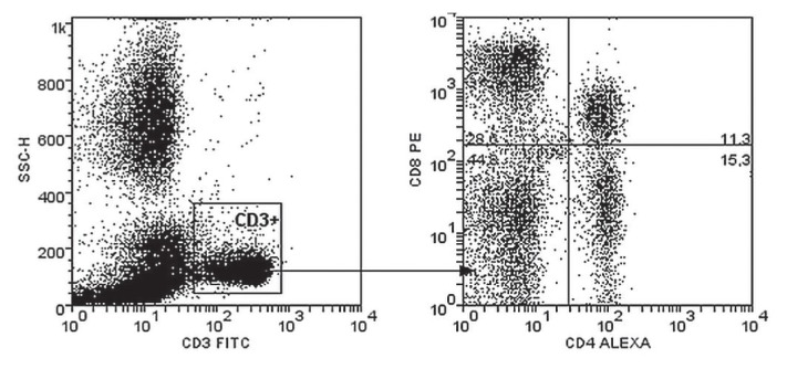
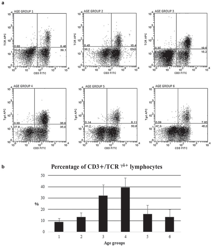
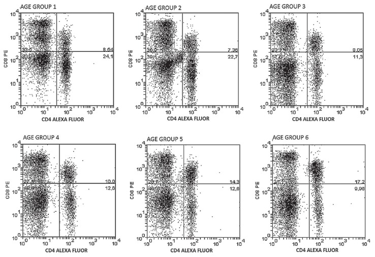
The aim of the study was to determine age-related changes in peripheral blood lymphocytes in pigs. Previous studies looking at age-related differences in lymphocyte subsets in porcine blood have not established reference ranges for these parameters. Moreover, most studies have concentrated on the dynamic changes in the first months of life, failing to continue observations in older animals. Therefore, in the present study, relative counts of various lymphocyte subpopulations (cytotoxic and helper T-cells, B-cells, and γδ T-cells) were evaluated to characterize the development of the cellular immune system at 28, 35, 135, and 200 days of age in growing pigs and adult sows (i.e., first and subsequent parity). In all examined groups, CD3+ cells constituted the largest percentage of cells. A statistically significant higher percentage of TCRγδ+CD3+ was noted in fatteners and gilts in comparison to other age groups. These results may be a reflection of antigenic pressure and show an immune response to viral or bacterial agents/environmental microbism.
L’objectif de la présente étude était de déterminer les changements associés à l’âge dans les lymphocytes du sang périphérique chez les porcs. Des études antérieures examinant les différences associées à l’âge dans les sous-populations de lymphocytes dans le sang porcin n’ont pas établi des écarts de référence pour ces paramètres. De plus, la plupart des études se sont concentrées sur les changements dynamiques dans les premiers mois de vie, omettant de continuer les observations chez les animaux plus âgés. Ainsi, dans la présente étude, les dénombrements relatifs des différentes sous-populations lymphocytaires (cellules-T cytotoxiques et helper, cellules-B, et cellules-T γδ) furent évalués afin de caractériser le développement du système immunitaire cellulaire à 28, 35, 135, et 200 jours d’âge chez des porcs en croissance et des truies adultes (i.e. première parité ainsi que les suivantes). Dans tous les groupes examinés, les cellules CD3+ constituaient le pourcentage le plus élevé de cellules. Un pourcentage significativement plus élevé de TCRγδ+CD3+ était noté chez les porcs en croissance et les cochettes comparativement aux autres groupes d’âge. Ces résultats pourraient être un reflet de la pression antigénique et montre une réponse immunitaire à des agents viraux ou bactériens du microbisme environnemental.(Traduit par Docteur Serge Messier).
Copyright and/or publishing rights held by the Canadian Veterinary Medical Association.
Figures

Similar articles
-
Reference ranges of lymphocyte subsets in healthy adults and adolescents with special mention of T cell maturation subsets in adults of South Florida.Immunobiology. 2014 Jul;219(7):487-96. doi: 10.1016/j.imbio.2014.02.010. Epub 2014 Mar 2. Immunobiology. 2014. PMID: 24661720
-
Effects of n-6:n-3 polyunsaturated fatty acid ratio on heterophil: lymphocyte ratio and T lymphocyte subsets in the peripheral blood of the Yangzhou gosling.Poult Sci. 2011 Apr;90(4):824-9. doi: 10.3382/ps.2010-01199. Poult Sci. 2011. PMID: 21406368
-
Blood lymphocyte T subsets reference values in blood donors by flow cytometry.Tunis Med. 2019 Feb;97(2):327-334. Tunis Med. 2019. PMID: 31539091
-
Immunophenotyping of blood lymphocytes in childhood. Reference values for lymphocyte subpopulations.J Pediatr. 1997 Mar;130(3):388-93. doi: 10.1016/s0022-3476(97)70200-2. J Pediatr. 1997. PMID: 9063413
-
Immunophenotyping of blood lymphocytes at birth, during childhood, and during adulthood in HIV-1-uninfected Ethiopians.Clin Immunol. 2003 Dec;109(3):338-46. doi: 10.1016/j.clim.2003.08.008. Clin Immunol. 2003. PMID: 14697749
Cited by
-
Genetic architecture of innate and adaptive immune cells in pigs.Front Immunol. 2023 Feb 6;14:1058346. doi: 10.3389/fimmu.2023.1058346. eCollection 2023. Front Immunol. 2023. PMID: 36814923 Free PMC article.
-
Effects of GHR Deficiency and Juvenile Hypoglycemia on Immune Cells of a Porcine Model for Laron Syndrome.Biomolecules. 2023 Mar 26;13(4):597. doi: 10.3390/biom13040597. Biomolecules. 2023. PMID: 37189345 Free PMC article.
-
Establishment of a piglet model for peritoneal metastasis of ovarian cancer.J Transl Med. 2022 Jul 21;20(1):329. doi: 10.1186/s12967-022-03533-1. J Transl Med. 2022. PMID: 35864492 Free PMC article.
References
-
- Becker BA, Misfeldt ML. Evaluation of the mitogen-induced proliferation and cell surface differentiation antigens of lymphocytes from pigs 1 to 30 days of age. J Anim Sci. 1993;71:2073–2078. - PubMed
-
- Rothwell L, Gramzinski RA, Rose ME, Kaiser P. Avian coccidiosis: Changes in intestinal lymphocyte populations associated with the development of immunity of Eimeria maxima. Parasite Immunol. 1995;17:525–533. - PubMed
-
- Comans-Bitter WM, de Groot R, van den Beemd R, et al. Immunophenotyping of blood lymphocytes in childhood. Reference values for lymphocyte subpopulations. J Pediatr. 1997;130:388–393. - PubMed
-
- Winnicka A, Kluciński W, Kawiak J, et al. Lymphocyte subpopulations, null cells and MHC II positive cells in peripheral blood of goats at different ages. Small Rumin Res. 1999;33:247–253.
-
- Faldyna M, Levá L, Knötigová P, Toman M. Lymphocyte subsets in peripheral blood of dogs — A flow cytometric study. Vet Immunol Immunopathol. 2001;82:23–37. - PubMed
MeSH terms
Substances
LinkOut - more resources
Full Text Sources
Medical