. 2019 Dec 18:10:887.
doi: 10.3389/fendo.2019.00887. eCollection 2019.

Curcumin Analogs Reduce Stress and Inflammation Indices in Experimental Models of Diabetes

Affiliations

Curcumin Analogs Reduce Stress and Inflammation Indices in Experimental Models of Diabetes

Saumik Biswas et al. Front Endocrinol (Lausanne). .

Abstract

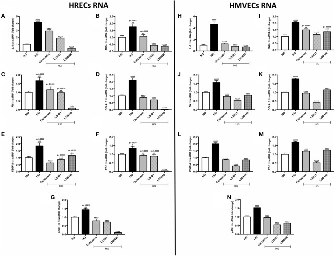
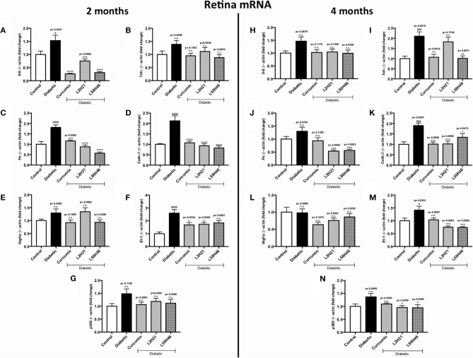
Chronic inflammation and oxidative stress lead to a multitude of adverse cellular responses in target organs of chronic diabetic complications. Curcumin, a highly investigated phytochemical, has been shown to exhibit both anti-inflammatory and antioxidant activities. However, the clinical application of curcumin has been greatly limited due to a poor pharmacokinetic profile. To overcome these limitations, we have generated analogs of curcumin to enhance bioavailability and offer a preferable pharmacokinetic profile. Here, we explored the effects of two mono-carbonyl curcumin analogs, L2H21 and L50H46, in alleviating indices of inflammation and oxidative stress in cell culture and mouse model of diabetic complications. Our results show that L2H21 and L50H46 normalize inflammatory mediators (IL-6 and TNF-α), extracellular matrix proteins (FN and COL4α1), vasoactive factors (VEGF and ET-1) and a key transcriptional coactivator (p300) in cultured human retinal microvascular endothelial cells (HRECs) and dermal-derived microvascular endothelial cells (HMVECs) challenged with high levels of glucose. These curcumin analogs also reduced glucose-induced oxidative DNA damage as evidenced by 8-OHdG labeling. We further show that treatment of streptozotocin-induced diabetic mice with curcumin analogs prevents cardiac and renal dysfunction. The preservation of target tissue function was associated with normalization of pro-inflammatory cytokines and matrix proteins. Collectively, our results show that L2H21 and L50H46 offer the anti-inflammatory and antioxidant activities as has been reported for curcumin, and may provide a clinically applicable therapeutic option for the treatment of diabetic complications.

Keywords: angiogenesis; curcumin analogs; diabetic complications; fibrosis; inflammation.


Figures

Figure 1
Figure 1
Curcumin analogs significantly reduced gene expressions associated with glucose-induced vascular damage in vitro. Left panel: RT-qPCR analyses showing increased mRNA levels of (A) IL-6, (B) TNF-α, (C) FN, (D) COL4α1, (E) VEGF-A, (F) ET-1, and (G) p300 in HRECs exposed to HG for 48 h. The administration of curcumin or its analogs (1-h pre-treatment) was able to dampen the glucose-induced levels of these transcripts. Right panel: RT-qPCR analyses showing elevated mRNA levels of (H) IL-6, (I) TNF-α, (J) FN, (K) COL4α1, (L) VEGF-A, (M) ET-1, and (N) p300 in HMVECs exposed to HG for 48 h. Similar to HRECs, HMVECs treated with curcumin or its derivatives also had significantly lower levels of high glucose-associated molecules [data expressed as a ratio to 18S ribosomal RNA (mean ± SEM); normalized to NG; #significance between NG vs. HG; *significance between HG vs. Treatment group; *, #P < 0.05, **,##P < 0.01, ***, ###P < 0.001, and ****, ####P < 0.0001; n = 6 from three independent experiments and performed in triplicates; NG (normal glucose) = 5 mM D-glucose; HG (high glucose) = 25 mM D-glucose; data analyzed by ANOVA followed by Tukey's post-hoc].
Figure 2
Figure 2
Curcumin and its analogs alter proteins associated with glucose-induced vascular damage in vitro. Left panel: Protein levels of (A) FN, (B) COL4α1, (C) VEGF-A, (D) ET-1, and (E) p300 in HRECs pre-treated with L2H21 or L50H46 before exposure to HG. Protein levels were measured by ELISA. HG increased the selected proteins in HRECs, while the curcumin derivatives significantly reduced these glucose-induced upregulations. Right panel: Protein levels of (F) FN, (G) COL4α1, (H) VEGF-A, (I) ET-1, and (J) p300 in cultured HMVECs as described for HRECs. Following the treatment of curcumin or its analogs in HMVECs incubated with HG, the protein markers exhibited downregulation [data expressed as a fold-change (mean ± SEM); normalized to NG; #significance between NG vs. HG; *significance between HG vs. Treatment group; *, #P < 0.05, **, ##P < 0.01, ***, ###P < 0.001, and ****, ####P < 0.0001; n = 6 from three independent experiments and performed in triplicates; n.s., not significant; NG (normal glucose) = 5 mM D-glucose; HG (high glucose) = 25 mM D-glucose; data analyzed by ANOVA followed by Tukey's post-hoc].
Figure 3
Figure 3
Effect of curcumin and its analogs on high glucose-induced oxidative DNA damage in HRECs and HMVECs. 8-OHdG staining was used to assess high glucose-induced oxidative DNA damage. Cells were exposed to HG for 48 h, with or without pre-treatment with curcumin or its analogs. The fluorescent images indicate that curcumin and its analogs decrease the level of glucose-induced oxidative DNA damage in both HRECs and HMVECs [original magnification at 20x; scale bar = 10 μm, same magnification for all images; NG (normal glucose) = 5 mM D-glucose; HG (high glucose) = 25 mM D-glucose; green stain = 8-OHdG and blue (DAPI) stain = nuclear counterstain; images were merged, and each image is representative of at least 3 separate experiments].
Figure 4
Figure 4
Effects of curcumin and its analogs on retinal transcripts at 2- and 4-months following induction of diabetes. Left panel: RT-qPCR analyses showing the upregulation of (A) Il-6, (B) Tnf-α, (C) Fn, (D) Col4α1, (E) Vegf-a, (F) Et-1, and (G) p300 mRNAs in the retinal tissues of diabetic mice at 2 months. Administration of curcumin or its analogs produced differential RNA expressions in the retinal tissues of diabetic mice. Right panel: RT-qPCR analyses demonstrating relative increases of (H) Il-6, (I) Tnf-α, (J) Fn, (K) Col4α1, (L) Vegf-a, (M) Et-1, and (N) p300 mRNAs in the retinal tissues of diabetic mice at 4 months. Treatment with curcumin or its analogs produced differential transcript expressions [data expressed as a ratio to β-actin (mean ± SEM); normalized to non-diabetic control; #significance between Control vs. Diabetic; *significance between Diabetic vs. Treatment group; *, #P < 0.05, **, ##P < 0.01, ***, ###P < 0.001, and ****, ####P < 0.0001; n = 6 animals/group; n.s., not significant; Control = non-diabetic controls; data analyzed by ANOVA followed by Tukey's post-hoc].
Figure 5
Figure 5
Effects of curcumin and its analogs on cardiac transcripts at 2 and 4 months. Left panel: RT-qPCR analyses showing increased mRNA levels of (A) Il-6, (B) Tnf-α, (C) Fn, (D) Col4α1, (E) Vegf-a, (F) Et-1, and (G) p300 in the cardiac tissues of diabetic mice at 2 months. Administration of curcumin, L2H21, or L50H46 produced differential expressions of these transcripts in the cardiac tissues of diabetic mice. Right panel: RT-qPCR analyses of (H) Il-6, (I) Tnf-α, (J) Fn, (K) Col4α1, (L) Vegf-a, (M) Et-1, and (N) p300 in cardiac tissues at 4 months. Treatment with curcumin or its analogs at this time point produced differential RNA expressions [data expressed as a ratio to β-actin (mean ± SEM); normalized to Control; #significance between Control vs. Diabetic; *significance between Diabetic vs. Treatment group; *, #P < 0.05, **, ##P < 0.01, ***, ###P < 0.001, and ****, ####P < 0.0001; n = 6 animals/group; n.s., not significant; Control = non-diabetic controls; data analyzed by ANOVA followed by Tukey's post-hoc].
Figure 6
Figure 6
Effects of curcumin and its analogs on renal transcripts at 2 and 4 months. Left panel: RT-qPCR analyses demonstrating significant upregulations of (A) Il-6, (B) Tnf-α, (C) Fn, (D) Col4α1, (E) Vegf-a, (F) Et-1, and (G) p300 mRNAs in the kidney tissues of diabetic mice at 2 months. Administration of curcumin or its analogs produced differential RNA expressions. Right panel: RT-qPCR analyses exhibiting relative upregulations of (H) Il-6, (I) Tnf-α, (J) Fn, (K) Col4α1, (L) Vegf-a, (M) Et-1, and (N) p300 transcripts in kidney tissues at 4 months. Treatment with curcumin or its derivatives also produced differential RNA expressions in the diabetic mice kidneys [data expressed as a ratio to β-actin (mean ± SEM); normalized to Control; #significance between Control vs. Diabetic; *significance between Diabetic vs. Treatment group; *, #P < 0.05, **, ##P < 0.01, ***, ###P < 0.001, and ****, ####P < 0.0001; n = 6 animals/group; n.s., not significant; Control = non-diabetic controls; data analyzed by ANOVA followed by Tukey's post-hoc].

References

    1. Khan Z, Chakrabarti S. Therapeutic targeting of endothelial dysfunction in chronic diabetic complications. Recent Pat Cardiovasc Drug Discov. (2008) 1:167–75. 10.2174/157489006777442531 - DOI - PubMed
    1. Biswas S, Chakrabarti S. Increased extracellular matrix protein production in chronic diabetic complications: implications of non-coding RNAs. Noncoding RNA. (2019) 5:30. 10.3390/ncrna5010030 - DOI - PMC - PubMed
    1. Khan Z, Farhangkhoee H, Chakrabarti S. Towards newer molecular targets for chronic diabetic complications. Curr Vasc Pharmacol. (2005) 4:45–57. 10.2174/157016106775203081 - DOI - PubMed
    1. Giacco F, Brownlee M. Oxidative stress and diabetic complications. Circ Res. (2010) 107:1058–70. 10.1161/CIRCRESAHA.110.223545 - DOI - PMC - PubMed
    1. Harrison DG. Endothelial function and oxidant stress. Clin Cardiol. (1997) 20(11 Suppl. 2):11–7. - PubMed