. 2019 Dec 20:10:2900.
doi: 10.3389/fmicb.2019.02900. eCollection 2019.

Diverse Expression of Antimicrobial Activities Against Bacterial Vaginosis and Urinary Tract Infection Pathogens by Cervicovaginal Microbiota Strains of Lactobacillus gasseri and Lactobacillus crispatus

Affiliations

Diverse Expression of Antimicrobial Activities Against Bacterial Vaginosis and Urinary Tract Infection Pathogens by Cervicovaginal Microbiota Strains of Lactobacillus gasseri and Lactobacillus crispatus

Fabrice Atassi et al. Front Microbiol. .

Abstract

We aimed to analyze the strain-by-strain expression of a large panel of antimicrobial activities counteracting the virulence mechanisms of bacterial vaginosis-associated Prevotella bivia CI-1 and Gardnerella vaginalis 594, pyelonephritis-associated Escherichia coli CFT073, and recurrent cystitis- and preterm labor-associated IH11128 E. coli by Lactobacillus gasseri and Lactobacillus crispatus clinical strains, and L. gasseri ATCC 9857 and KS 120.1, and L. crispatus CTV-05 strains isolated from the cervicovaginal microbiota of healthy women. All L. gasseri and L. crispatus strains exerted antimicrobial activity by secreted lactic acid, which killed the microbial pathogens by direct contact. Potent bactericidal activity was exerted by a very limited number of resident L. gasseri and L. crispatus strains showing the specific ability to a strain to produce and release antibiotic-like compounds. These compounds eradicated the microbial pathogens pre-associated with the surface of cervix epithelial cells, providing efficient protection of the cells against the deleterious effects triggered by toxin-producing G. vaginalis and uropathogenic E. coli. Furthermore, these compounds crossed the cell membrane to kill the pre-internalized microbial pathogens. In addition, all L. gasseri and L. crispatus cells exhibited another non-strain specific activity which inhibited the association of microbial pathogens with cervix epithelial cells with varying efficiency, partially protecting the cells against lysis and detachment triggered by toxin-producing G. vaginalis and uropathogenic E. coli. Our results provide evidence of strain-level specificity for certain antimicrobial properties among cervicovaginal L. gasseri and L. crispatus strains, indicating that the presence of a particular species in the vaginal microbiota is not sufficient to determine its benefit to the host. A full repertory of antimicrobial properties should be evaluated in choosing vaginal microbiota-associated Lactobacillus isolates for the development of live biotherapeutic strategies.

Keywords: Lactobacillus crispatus; Lactobacillus gasseri; antimicrobial; bacterial vaginosis; cervicovaginal microbiota; urinary tract infections.


Figures

FIGURE 1
FIGURE 1
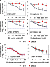
In vitro lactic acid-dependent and -independent killing activity by direct contact against BV-associated P. bivia CI-1 and Gardnerella. vaginalis 594 strains, pyelonephritis-associated E. coli strain CFT073, and recurrent cystitis- and preterm labor-associated E. coli strain IH11128. (A) Concentration-dependent killing activity of DL-lactic acid in BHI or DMEM. (B) Concentration-dependent killing activity of hydrogen peroxide in the presence of BHI or DMEM. Each value shown is the mean ±SD from three experiments. Student t-test, p < 0.01 compared to BHI. The dotted line shows the MBE99.99% value, defined as a reduction in the viable cell count of 4 log10.
FIGURE 2
FIGURE 2
Characteristics of the killing activity by direct contact of L. gasseri ATCC 9857, L. gasseri KS 120.1, and L. crispatus CTV-05 strains against BV-associated P. bivia CI-1 and G. vaginalis 594 strains, pyelonephritis-associated E. coli strain CFT073, and recurrent cystitis- and preterm labor- associated E. coli strain IH11128. (A) Lactic acid-dependent (BHI) and -independent (DMEM) killing activities exerted by each Lactobacillus strain in co-culture conditions. (B) Time-course of lactic acid-independent killing activity of L. gasseri ATCC 9857, L. gasseri KS 120.1, and L. crispatus CTV-05 CFCSs. (C) Concentration-dependent killing activity of L. gasseri KS 120.1 and L. crispatus CTV-05 CFCSs. (D) Effect of catalase treatment on the killing activity of L. crispatus CTV-05 CFCS. In (A), killing activity was determined after 4 h of direct contact with Lactobacillus cultures (18 h of culture adjusted to 108 CFU/ml Lactobacillus bacteria). In (A), the dotted line shows the MBE99.99% value, defined as a reduction in the viable cell count of 4 log10 CFU/ml. In (C,D), killing activity was determined in the presence of DMEM after 4 h of direct contact. Each value shown is the mean ±SD from three experiments. In (A), Student t-test, p < 0.01 compared to control. In (B), Student t-test, p < 0.01 at 3 and 4 h. In (C,D), Student t-test, p < 0.01 compared to control.
FIGURE 3
FIGURE 3
Inhibition of growth of BV-associated G. vaginalis 594, and recurrent cystitis- and preterm labor-associated E. coli strain IH11128. (A) Inhibition of G. vaginalis 594 growth after 24 h of co-culture with L. gasseri ATCC 9857, L. gasseri KS 120.1, or L. crispatus CTV-05 strains. (B) Inhibition of G. vaginalis 594 and UPEC IH11128 growth during a time-course of co-culture with L. gasseri ATCC 9857 CFCS. (C) Inhibition of UPEC IH11128 growth after 24 h of co-culture in the presence of 8-fold diluted L. gasseri KS 120.1, and L. crispatus CTV-05 CFCSs. Each value shown is the mean ±SD from three experiments. Student t-test, p < 0.01 compared to control.
FIGURE 4
FIGURE 4
Antagonistic activities of Lactobacillus cells and CFCSs against BV-associated G. vaginalis 594 and recurrent cystitis- and preterm labor-associated E. coli IH11128 adhering to HeLa cells. (A) Inhibition under competition conditions. (B) Inhibition under exclusion conditions. (C) Lack of inhibition under displacement conditions. (D) Killing of pre-adhering UPEC IH11128. (E) Killing of pre-internalized UPEC IH11128. In (A–C), 100% adhesion corresponds to 5.1 ± 0.5 CFU/ml for G. vaginalis 594 and 7.6 ± 0.4 CFU/ml for E. coli IH11128. Competition, exclusion and displacement experimental condition are described in section “Materials and Methods”. Each value shown is the mean ±SD from three experiments. Student t-test, p < 0.01 compared to control.
FIGURE 5
FIGURE 5
Protective effect of cells and CFCSs of L. gasseri ATCC 9857, L. gasseri KS 120.1, or L. crispatus CTV-05 strains against the cytotoxic activities of BV-associated G. vaginalis 594 and preterm labor and recurrent cystitis-associated E. coli IH11128. (A) Quantification of time-dependent G. vaginalis 594-induced cell-detachment in the presence, or not, of pre-colonizing Lactobacillus cells. (B) Quantification of time-dependent UPEC IH11128-induced cell detachment, in the presence, or not, of pre-colonizing Lactobacillus cells. (C) Micrographs illustrating the partial inhibition of UPEC IH11128-induced cell detachment in the presence of pre-colonizing Lactobacillus cells. (D) Quantification of cell detachment in cell monolayers infected with UPEC IH11128, showing the total inhibition of cell detachment in the presence L. gasseri KS 120.1 or L. crispatus CTV-05 CFCSs and the lack of inhibition in the presence of L. gasseri ATCC 9857 CFCS. (E) Micrographs illustrating the entire inhibition of UPEC IH11128-induced cell detachment in the presence L. gasseri KS 120.1 or L. crispatus CTV-05 CFCSs. Phase-contrast micrographs are representative of two separate experiments. The number of attached cells was monitored by phase-contrast light microscopy. Each value shown is the mean ±SD from three experiments. Student t-test, p < 0.01 compared to G. vaginalis 594 or UPEC IH11128.
FIGURE 6
FIGURE 6
Distribution of the killing activities and adhesion inhibition activity among L. gasseri and L. crispatus strains isolated from cervicovaginal samples of healthy women. (A) Killing activity against BV-associated G. vaginalis 594 was determined in the presence of BHI or DMEM. (B) Killing activity against recurrent cystitis- and preterm labor- associated UPEC IH11128 was determined in the presence of BHI or DMEM. (C) Competitive inhibition activity exerted by L. gasseri and L. crispatus strains against adhesion of UPEC IH11128 onto cervicovaginal epithelial HeLa cells. (D) Strain-by-strain expression of total killing activity (in the presence of BHI) versus inhibition of pathogen adhesion by Lactobacillus cells. (E) Strain-by-strain expression of killing activity independent of secreted lactic acid (in the presence of DMEM) versus inhibition of pathogen adhesion by Lactobacillus cells. In (A,B), the killing activity was measured after 4 h of direct contact with the pathogens (Control: 5 × 10⁸ CFU/ml). The dotted line shows the MBE99.99% value, defined as a reduction in the viable cell count of 4 log10 CFU/ml. In C, the inhibition of adhesion was measured after 3 h of incubation. 100% UPEC IH11128 adhesion corresponds to 7.1 ± 0.7 CFU/ml. Each value shown is the mean ±SD from three experiments. Graphs in (D,E) were constructed by plotting the killing activities (measured in the presence of BHI or DMEM) shown in Figure 1B versus the exclusion inhibition activities shown in Figure 1C.

References

    1. Acedo J. Z., Chiorean S., Vederas J. C., Van Belkum M. J. (2018). The expanding structural variety among bacteriocins from Gram-positive bacteria. FEMS Microbiol. Rev. 42 805–828. 10.1093/femsre/fuy033 - DOI - PubMed
    1. Alakomi H. L., Skytta E., Saarela M., Mattila-Sandholm T., Latva-Kala K., Helander I. M. (2000). Lactic acid permeabilizes gram-negative bacteria by disrupting the outer membrane. Appl. Environ. Microbiol. 66 2001–2005. 10.1128/aem.66.5.2001-2005.2000 - DOI - PMC - PubMed
    1. Amabebe E., Anumba D. O. C. (2018). The Vaginal microenvironment: the physiologic role of Lactobacilli. Front. Med. 5:181. 10.3389/fmed.2018.00181 - DOI - PMC - PubMed
    1. Amegashie C. P., Gilbert N. M., Peipert J. F., Allsworth J. E., Lewis W. G., Lewis A. L. (2017). Relationship between nugent score and vaginal epithelial exfoliation. PLoS One 12:e0177797. 10.1371/journal.pone.0177797 - DOI - PMC - PubMed
    1. Antonio M. A., Hawes S. E., Hillier S. L. (1999). The identification of vaginal Lactobacillus species and the demographic and microbiologic characteristics of women colonized by these species. J. Infect. Dis. 180 1950–1956. 10.1086/315109 - DOI - PubMed

LinkOut - more resources