. 2019 Dec 20:10:2932.
doi: 10.3389/fmicb.2019.02932. eCollection 2019.

Evidence of a Cellulosic Layer in Pandoravirus massiliensis Tegument and the Mystery of the Genetic Support of Its Biosynthesis

Affiliations

Evidence of a Cellulosic Layer in Pandoravirus massiliensis Tegument and the Mystery of the Genetic Support of Its Biosynthesis

Djamal Brahim Belhaouari et al. Front Microbiol. .

Abstract

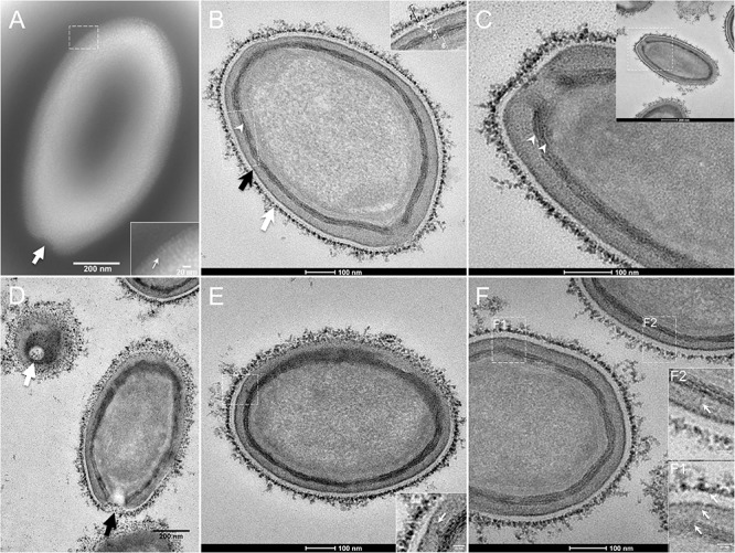
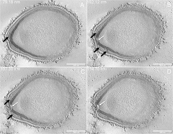
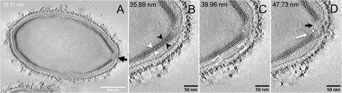
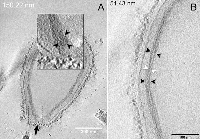
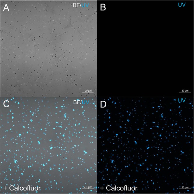
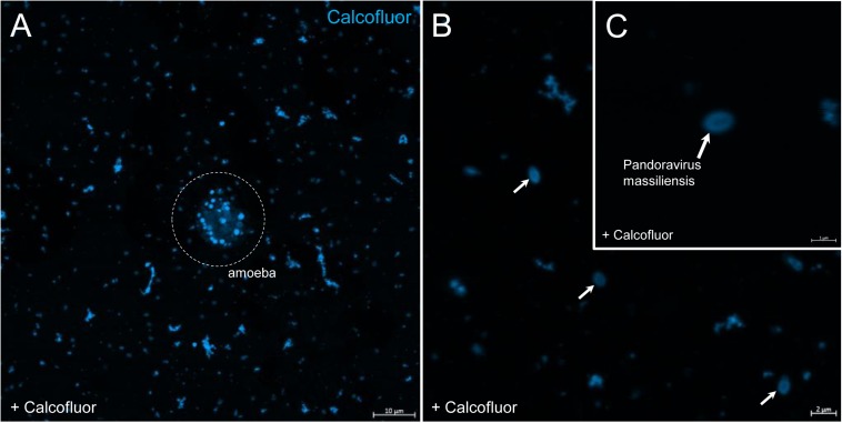
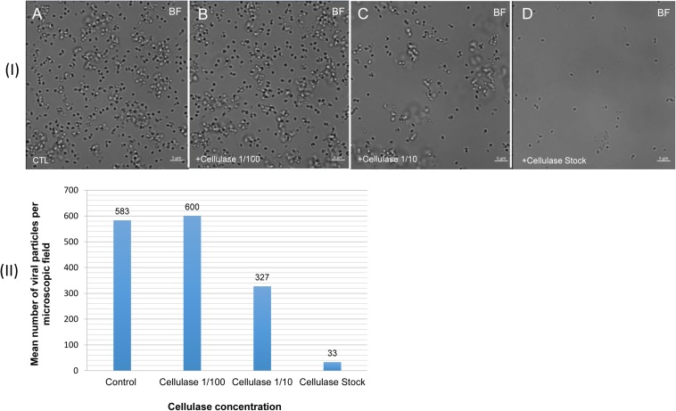
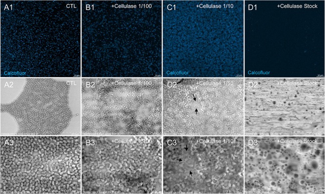
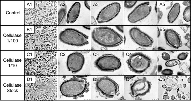
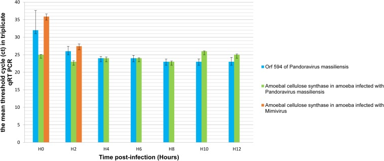
Pandoraviruses are giant viruses of ameba with 1 μm-long virions. They have an ovoid morphology and are surrounded by a tegument-like structure lacking any capsid protein nor any gene encoding a capsid protein. In this work, we studied the ultrastructure of the tegument surrounding Pandoravirus massiliensis virions and noticed that this tegument is composed of a peripheral sugar layer, an electron-dense membrane, and a thick electron-dense layer consisting in several tubules arranged in a helicoidal structure resembling that of cellulose. Pandoravirus massiliensis particles were stained by Calcofluor white, a fluorescent dye of cellulose, and the enzymatic treatment of particles by cellulase showed the degradation of the viral tegument. We first hypothesized that the cellulose tegument could be synthesized by enzymes encoded by the virus. Bioinformatic analyses revealed in P. massiliensis, a candidate gene encoding a putative cellulose synthase, with a homology with the BcsA domain, one of the catalytic subunits of the bacterial cellulose synthase, but with a low level of homology. This gene was transcribed during the replicative cycle of P. massiliensis, but several arguments run counter to this hypothesis. Indeed, even if this gene is present in other pandoraviruses, the one of the strain studied is the only one to have this BcsA domain and no other enzymes involved in the synthesis of cellulose could be detected, although we cannot rule out that such genes could have been undetected among the large proportion of Orfans of pandoraviruses. As an alternative, we investigated whether P. massiliensis could divert the cellulose synthesis machinery of the ameba to its own account. Indeed, contrary to what is observed in the case of infections with other giant viruses such as mimiviruses, it appears that the transcription of the ameba, at least for the cellulose synthase gene, continues throughout the growth phase of particles of P. massiliensis. Finally, we believe that this scenario is more plausible. If confirmed, it could be a unique mechanism in the virosphere.

Keywords: Pandoravirus; capsid; cellulose; cellulose synthase; giant virus.


Figures

FIGURE 1
FIGURE 1
Transmission electron microscopy (TEM) of Pandoravirus massiliensis. (A) Negative staining of a P. massiliensis particle: the ostiole (arrow) is located at the apex of the particle. Peripheral thin fibers can be observed enwrapping the particle (thin arrow in boxed zoomed region). (B) Ultrathin section showing (i) the most-peripheral sugars depicted by ruthenium red aggregates (thick white arrow and level 1 in boxed zoomed region); (ii) a thin electron-dense membrane (thick black arrow and level 3 in boxed zoomed region) and more centrally (iii) a thick bundle of tubules (thin white arrowhead and level 5 in boxed zoomed region). (C) Two thick tubules compose the inner-most thick layer (arrowheads). (D) Two particles with ostioles cut transversally or perpendicularly (white and black arrows). (E) The inner-most thick tubules with thick protrusions (white arrow in boxed zoomed region) toward the outer thin electron-dense membrane. (F) Thin fibers (white arrows in F1 and F2 boxed zoomed regions) projecting from the inner-most thick tubules, crossing the outer electron-dense membrane and reaching the peripheral ruthenium-red stained sugars.
FIGURE 2
FIGURE 2
Electron tomography of P. massiliensis particle from Movie 3. (A–D) Single planes in the tomogram from Movie 3 showing thick tubules protruding toward the periphery and the outer electron-dense membrane (black arrows); a thin tubule/membrane (white arrow) connects the thick tubules layers located on each side of the ostiole.
FIGURE 3
FIGURE 3
Electron tomography of P. massiliensis particle from Movie 4. (A) Single plane in the tomogram from Movie 4 showing a whole Pandoravirus particle and its ostiole located at one apex (arrow). (B). The inner-most layer is composed of two thick tubules well separated (black arrowheads) or contacting each other (white arrowheads). (C,D) Thin fibers (arrows) originating from the inner-most tubular thick layer projecting toward the peripheral sugars (white arrows, C), toward the inner core of the particle (white arrow, D) or at the level of the ostiole (black arrow, D).
FIGURE 4
FIGURE 4
Electron tomography of P. massiliensis particles from Movies 5 and 6. (A) Single plane in the tomogram from Movie 5 showing a Pandoravirus particle with its ostiole (black arrow). The magnified boxed region depicts a U-shaped thick tubule (black arrowheads) from the inner-most layer. (B) Single plane in Movie 6 from the zoomed-in tomogram from Movie 5 showing the helical structural arrangement of the two thick tubules (arrowheads) composing the inner-most layer of Pandoravirus particles with distant tubules (black arrowheads) or crossed tubules (white arrowheads).
FIGURE 5
FIGURE 5
Confocal imaging of Calcofluor staining of P. massiliensis. (A,B) Control Pandoravirus particles. (C,D) Calcofluor-stained Pandoravirus particles. (BF: brightfield; UV: ultraviolet).
FIGURE 6
FIGURE 6
Confocal imaging of Calcofluor staining of P. massiliensis. (A) Pandoravirus-infected ameba and single Pandoravirus particles stained with Calcofluor white. (B,C) Calcofluor-stained Pandoravirus particles showing an intense peripheral calcofluor signal and a less-stained central region.
FIGURE 7
FIGURE 7
Confocal imaging of cellulase-treated P. massiliensis (I) and estimation of the mean number of particles of P. massiliensis per microscopic field of observation after cellulase treatment (II). (IA) Control condition with untreated P. massiliensis particles. (IB–ID): cellulase-treated P. massiliensis particles. (II) The mean number of particles of P. massiliensis per microscopic field of observation after cellulase treatment was assessed by the ImageJ software.
FIGURE 8
FIGURE 8
Confocal imaging of Calcofluor-stained cellulase-treated P. massiliensis virions and scanning microscopy of cellulose treated P. massiliensis. (A1) Control condition with untreated P. massiliensis particles stained with Calcofluor-white. (B1,C1,D1) Cellulase-treated P. massiliensis particles stained with Calcofluor-white. (A2,A3) Control condition with untreated P. massiliensis. (B2,B3,C2,C3,D2,D3) Cellulase-treated P. massiliensis particles imaged with scanning microscopy on two magnification.
FIGURE 9
FIGURE 9
Transmission electron microscopy of cellulase-treated P. massiliensis particles. (A1–A5) Control condition with untreated P. massiliensis particles. (B1–B5) P. massiliensis particles treated with cellulase solution diluted at 1:100, (B3–B5) particles showed a defect of the envelope. (C1–C5) P. massiliensis particles treated with cellulase solution diluted at 1:10, (C3–C5) particles exhibited detachments of the different layers of the envelope. (D1–D5) P. massiliensis particles treated with cellulase solution stock. (D3–D5) Particles imaged in different stages of digestion from least to the most digested. (D5) “Ghost-like” particles presenting totally empty internal spaces and only a thin surrounding tegument (black arrow).
FIGURE 10
FIGURE 10
Representation of the mean threshold cycle (Ct) of triplicate qRT PCR on the RNA of P. massiliensis by targeting the predicted gene of the cellulose synthase (ORF594) and on RNA of A. castellanii infected once with P. massiliensis and a second time with Mimivirus by targeting the amoebal gene of the cellulose synthase, according to the time post-infection from 0 to 12 h post-infection.

References

    1. Aherfi S., Andreani J., Baptiste E., Oumessoum A., Dornas F. P., Andrade A. C. D. S. P., et al. (2018). A large open pangenome and a small core genome for giant pandoraviruses. Front. Microbiol. 9:1486. 10.3389/fmicb.2018.01486 - DOI - PMC - PubMed
    1. Andreani J., Aherfi S., Bou Khalil J. Y., Di Pinto F., Bitam I., Raoult D., et al. (2016). Cedratvirus, a double-cork structured giant virus, is a distant relative of pithoviruses. Viruses 8:300. 10.3390/v8110300 - DOI - PMC - PubMed
    1. Andreani J., Khalil J. Y. B., Baptiste E., Hasni I., Michelle C., Raoult D., et al. (2018). Orpheovirus IHUMI-LCC2: a new virus among the Giant Viruses. Front. Microbiol. 8:2643. 10.3389/fmicb.2017.02643 - DOI - PMC - PubMed
    1. Chen Y. W., Lee H. V., Juan J. C., Phang S.-M. (2016). Production of new cellulose nanomaterial from red algae marine biomass Gelidium elegans. Carbohydr. Polym. 151 1210–1219. 10.1016/j.carbpol.2016.06.083 - DOI - PubMed
    1. Clarke M., Lohan A. J., Liu B., Lagkouvardos I., Roy S., Zafar N., et al. (2013). Genome of Acanthamoeba castellanii highlights extensive lateral gene transfer and early evolution of tyrosine kinase signaling. Genome Biol. 14:R11. 10.1186/gb-2013-14-2-r11 - DOI - PMC - PubMed