. 2019 Dec 17:10:2763.
doi: 10.3389/fimmu.2019.02763. eCollection 2019.

RPS27, a sORF-Encoded Polypeptide, Functions Antivirally by Activating the NF-κB Pathway and Interacting With Viral Envelope Proteins in Shrimp

Affiliations

RPS27, a sORF-Encoded Polypeptide, Functions Antivirally by Activating the NF-κB Pathway and Interacting With Viral Envelope Proteins in Shrimp

Meng-Qi Diao et al. Front Immunol. .

Abstract

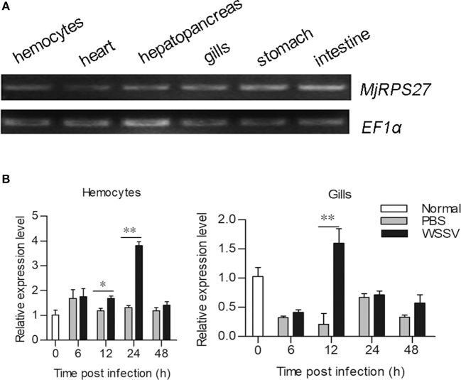
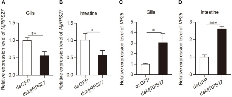
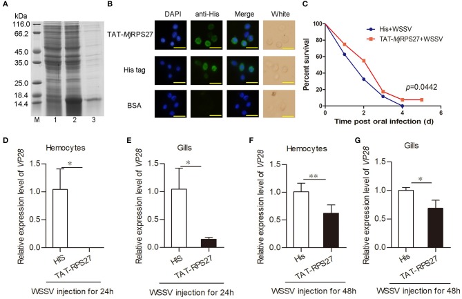
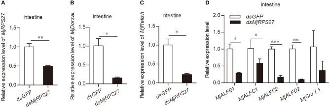
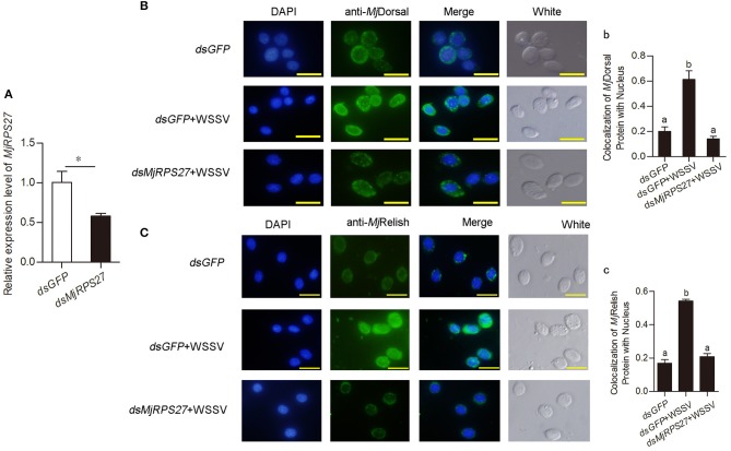
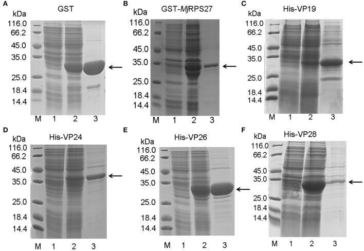
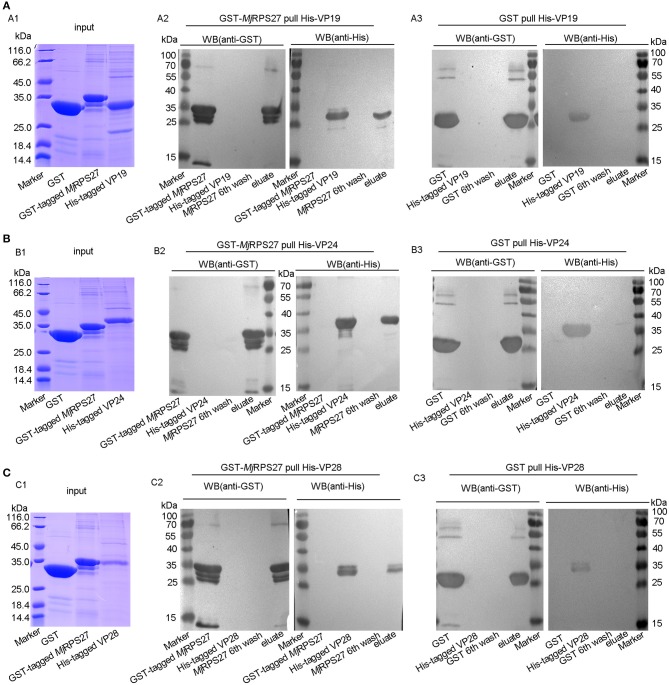
A small open reading frame (smORF) or short open reading frame (sORF) encodes a polypeptide of <100 amino acids in eukaryotes (50 amino acids in prokaryotes). Studies have shown that several sORF-encoded peptides (SEPs) have important physiological functions in different organisms. Many ribosomal proteins belonging to SEPs play important roles in several cellular processes, such as DNA damage repair and apoptosis. Several studies have implicated SEPs in response to infection and innate immunity, but the mechanisms have been unclear for most of them. In this study, we identified a sORF-encoded ribosomal protein S27 (RPS27) in Marsupenaeus japonicus. The expression of MjRPS27 was significantly upregulated in shrimp infected with white spot syndrome virus (WSSV). After knockdown of MjRPS27 by RNA interference, WSSV replication increased significantly. Conversely, after MjRPS27 overexpression, WSSV replication decreased in shrimp and the survival rate of the shrimp increased significantly. These results suggested that MjRPS27 inhibited viral replication. Further study showed that, after MjRPS27 knockdown, the mRNA expression level of MjDorsal, MjRelish, and antimicrobial peptides (AMPs) decreased, and the nuclear translocation of MjDorsal and MjRelish into the nucleus also decreased. These findings indicated that MjRPS27 might activate the NF-κB pathway and regulate the expression of AMPs in shrimp after WSSV challenge, thereby inhibiting viral replication. We also found that MjRPS27 interacted with WSSV's envelope proteins, including VP19, VP24, and VP28, suggesting that MjRPS27 may inhibit WSSV proliferation by preventing virion assembly in shrimp. This study was the first to elucidate the function of the ribosomal protein MjRPS27 in the antiviral immunity of shrimp.

Keywords: Relish; antimicrobial peptides; dorsal; kuruma shrimp; sORF encoded polypeptides; short open reading frame (sORF); white spot syndrome virus.


Figures

Figure 1
Figure 1
Tissue distribution and expression pattern of MjRPS27. (A) Tissue distribution of MjRPS27 mRNA. (B) Expression pattern of MjRPS27 in hemocytes and gills of shrimp challenged by WSSV as analyzed by qPCR. PBS injection shrimp served as a control. At least three shrimp were used for hemocytes and tissue collection at different time points. Significance was compared between the infected group and the same time point by t-test analysis, and significant difference was accepted at *p < 0.05; **p < 0.01.
Figure 2
Figure 2
VP28 expression was increased after knockdown of MjRPS27. (A,B) The RNAi efficiency of MjRPS27 was analyzed by qPCR 48 h post-dsRNA injection. The same amount of dsGFP injection served as a control. (C,D) After knockdown of MjRPS27, the shrimp were challenged with WSSV (5 × 10⁷ copies per shrimp), and the expression levels of VP28 were analyzed by qPCR 24 h post-WSSV challenge. Compared with the controls (dsGFP injection), the mRNA expression level of VP28 significantly increased. *p < 0.05; **p < 0.01; ***p < 0.001.
Figure 3
Figure 3
TAT–MjRPS27 overexpression inhibited WSSV proliferation. (A) Expression and purification of recombinant of TAT-MjRPS27. Lane M, protein marker; lane 1, total proteins from E. coli; lane 2, total proteins from E. coli after induced by IPTG; and lane 3, purified recombinant protein. (B) Immunocytochemistry to detect recombinant protein in hemocytes. Top panel, recombinant protein (TAT-MjRPS27) injection (10 μg/shrimp); middle, His tag protein injection; bottom, BSA injection. Bars = 20 μm. (C) Survival rate of shrimp injected with TAT-MjRPS27 (50 μg/shrimp); His tag protein injection served as a control. (D) VP28 expression in hemocytes of TAT-MjRPS27-WSSV-injected shrimp analyzed by qPCR at 24 h post-injection, His-WSSV-injected shrimp served as a control. (E) VP28 expression in gills of TAT-MjRPS27-WSSV-injected shrimp analyzed by qPCR at 24 h post injection, His-WSSV-injected shrimp served as a control. (F,G) VP28 expression in hemocytes (F) and Gills (G) of TAT-MjRPS27 and WSSV-injected shrimp was analyzed by qPCR 48 h post injection. His-WSSV-injected shrimp served as a control. PCR data were calculated using the 2−ΔΔCT method and expressed as the mean ± SD. Student's t-test was used to analyze significant differences among PCR data, and significant difference was accepted at *p < 0.05; **p < 0.01.
Figure 4
Figure 4
MjRPS27 was involved in the activation of the Toll and IMD pathways. (A) The RNA interference efficiency of MjRPS27 was analyzed by qPCR 48 h after dsMjRPS27 injection. The same amount of dsGFP was injected as a control. (B) After knockdown of MjRPS27, the expression level of MjDorsal was analyzed by qPCR. The mRNA expression level of MjDorsal was significantly downregulated compared with the control (dsGFP injection). (C) After knockdown of MjRPS27, the expression level of MjRelish was analyzed by qPCR. The mRNA expression level of MjRelish was significantly downregulated compared with the control (dsGFP injection). (D) After knockdown of MjRPS27, the expression levels of AMPs (MjALFB1, MjALFC1, MjALFC2, MjALFD2, and MjCruI 1) were analyzed by qPCR. The expression levels of AMPs, except MjCruI 1, were significantly downregulated compared with the control (injected dsGFP). *p < 0.05; **p < 0.01; ***p < 0.001.
Figure 5
Figure 5
The nuclear translocation of MjDorsal and MjRelish decreased after knockdown of MjRPS27 in shrimp. (A) Efficiency of MjRPS27 interference in gills; dsGFP injection served as a control. (B) Immunocytochemical analysis to detect the nucleus translocation of MjDorsal in hemocytes of shrimp after RNAi of MjRPS27; dsGFP-injected shrimp served as a control. Bars = 20 μm. (b) Statistics analysis of MjDorsal and nucleus co-localization in hemocytes. (C) Immunocytochemical analysis used to detect the nucleus translocation of MjRelish in hemocytes of shrimp after RNAi of MjRPS27; dsGFP-injected shrimp served as a control. Bars = 20 μm. (c) Statistics analysis of the colocalization of MjRelish with nucleus in hemocytes (details described in Materials and Methods). *p < 0.05.
Figure 6
Figure 6
Recombinant expression and purification of MjRPS27 and WSSV envelope proteins. (A) Recombinant expression and purification of GST tag protein. Lane M, protein marker; lane 1, total proteins from E. coli; lane 2, total proteins from E. coli after induction by IPTG; and lane 3, purified GST protein. (B) Recombinant expression and purification of MjRPS27. Lane M, protein marker; lane 1, total proteins from E. coli with pGEX4T-2/MjRPS27; lane 2, total proteins from E. coli after induction by IPTG; and lane 3, purified MjRPS27. (C) Recombinant expression and purification of WSSV VP19. Lane M, protein marker; lane 1, total proteins from E. coli with pET-32a(+)/VP19; lane 2, total proteins from E. coli after induction by IPTG; and lane 3, purified recombinant protein. (D) Recombinant expression and purification of WSSV VP24. Lane M, protein marker; lane 1, total proteins from E. coli with pET-32a(+)/VP24; lane 2, total proteins from E. coli after induction by IPTG; and lane 3, purified VP24. (E) Recombinant expression and purification of VP26. Lane M, protein marker; lane 1, total proteins from E. coli with pET-32a(+)/VP26; lane 2, total proteins from E. coli after induction by IPTG; and lane 3, purified VP26. (F) Recombinant expression and purification of WSSV VP28. Lane M, protein marker; lane 1, total proteins from E. coli with pET-30a(+)/VP28; lane 2, total proteins from E. coli after induction by IPTG; and lane 3, purified VP28. The arrows indicated the purified proteins.
Figure 7
Figure 7
MjRPS27 interacted with the WSSV envelope proteins, VP19, VP24, and VP28. (A) The interaction of MjRPS27 with VP19 was analyzed by GST pulldown. (A1) Input proteins (GST protein, GST-tagged MjRPS27, and His-tagged VP19) were analyzed by SDS-PAGE. (A2) GST-MjRPS27 pulldown His-VP19 was analyzed by Western blot: left panel, proteins used in the Pulldown assay were initially separated by SDS-PAGE, electrotransferred onto a nitrocellulose membrane, and analyzed by Western blot using anti-GST as primary antibody; right panel, SDS-PAGE-separated proteins were transferred onto the nitrocellulose membrane and analyzed by Western blot using anti-His as primary antibody. (A3) GST pulldown His-VP19 analyzed by Western blot: left panel, Western blot with anti-GST; right panel, Western blot using anti-His as primary antibody (control). (B) The interaction of MjRPS27 with VP24 was analyzed by GST pulldown. (B1) Input proteins (GST protein, GST-tagged MjRPS27 and His-tagged VP24) were analyzed by SDS-PAGE. (B2) GST-MjRPS27 pulldown His-VP24 was analyzed by Western blot: left panel, Western blot with anti-GST; right panel, Western blot analysis using anti-His as primary antibody. (B3) GST pulldown His-VP24 was analyzed by Western blot: left panel, Western blot with anti-GST; right panel, Western blot using anti-His as primary antibody (control). (C) The interaction of MjRPS27 with VP28 was analyzed by GST pulldown. (C1) Input proteins (GST protein, GST-tagged MjRPS27, and His-tagged VP28) were analyzed by SDS-PAGE. (C2), GST-MjRPS27 pulldown His-VP28 analysis: left panel, Western blot analysis with anti-GST; right panel, Western blot analysis using anti-His as primary antibody. (C3) GST pulldown His-VP28 analysis: left panel, Western blot analysis with anti-GST; right panel, Western blot analysis using anti-His as primary antibody (control).

Similar articles

Cited by

References

    1. Basrai MA, Hieter P, Boeke JD. Small open reading frames: beautiful needles in the haystack. Genome Res. (1997) 7:768–71. 10.1101/gr.7.8.768 - DOI - PubMed
    1. Khitun A, Ness TJ, Slavoff SA. Small open reading frames and cellular stress responses. Mol Omics. (2019) 15:108–16. 10.1039/C8MO00283E - DOI - PMC - PubMed
    1. Aspden JL, Eyre-Walker YC, Phillips RJ, Amin U, Mumtaz MA, Brocard M, et al. . Extensive translation of small Open reading frames revealed by Poly-Ribo-Seq. Elife. (2014) 3:e3528. 10.7554/eLife.03528 - DOI - PMC - PubMed
    1. Saghatelian A, Couso JP. Discovery and characterization of smORF-encoded bioactive polypeptides. Nat Chem Biol. (2015) 11:909–16. 10.1038/nchembio.1964 - DOI - PMC - PubMed
    1. White JW, Saunders GF. Structure of the human glucagon gene. Nucleic Acids Res. (1986) 14:4719–30. 10.1093/nar/14.12.4719 - DOI - PMC - PubMed

Publication types

LinkOut - more resources