. 2019 Dec 11:10:2766.
doi: 10.3389/fimmu.2019.02766. eCollection 2019.

Cancer Neoepitopes for Immunotherapy: Discordance Between Tumor-Infiltrating T Cell Reactivity and Tumor MHC Peptidome Display

Affiliations

Cancer Neoepitopes for Immunotherapy: Discordance Between Tumor-Infiltrating T Cell Reactivity and Tumor MHC Peptidome Display

Stina L Wickström et al. Front Immunol. .

Abstract

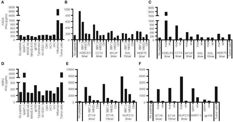
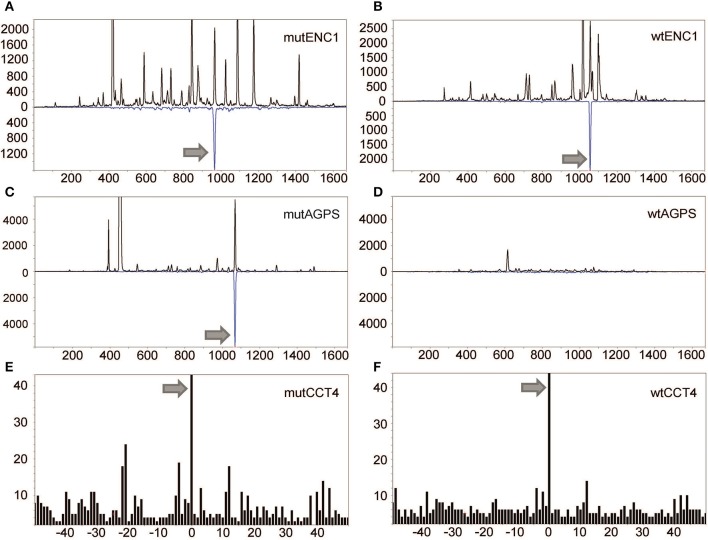
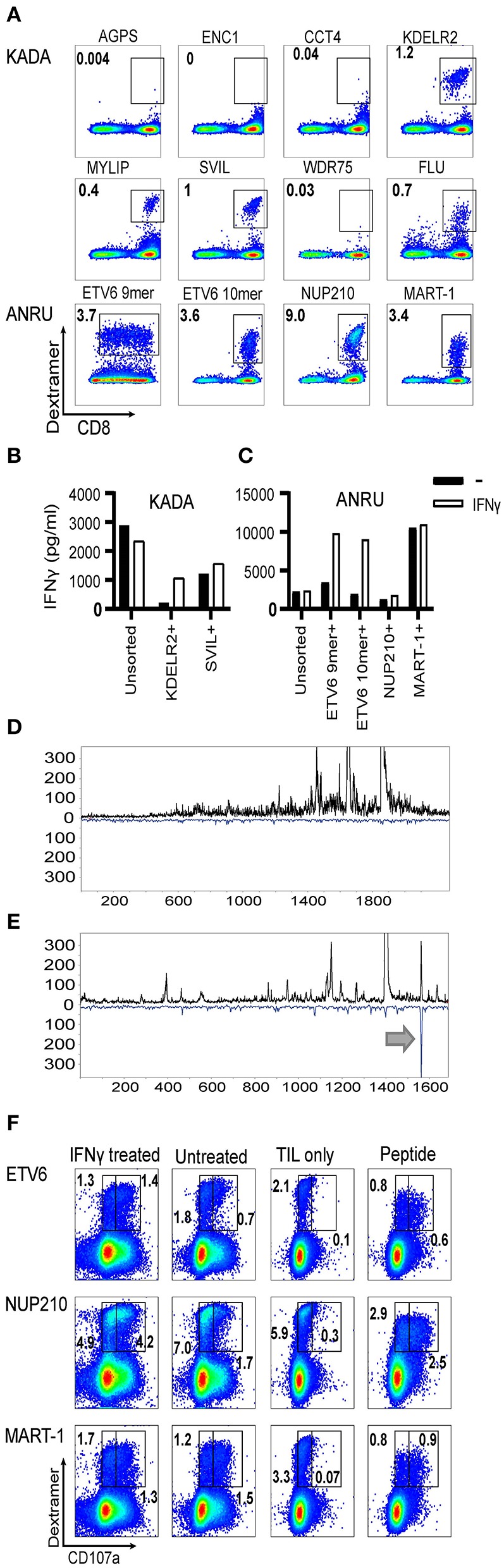
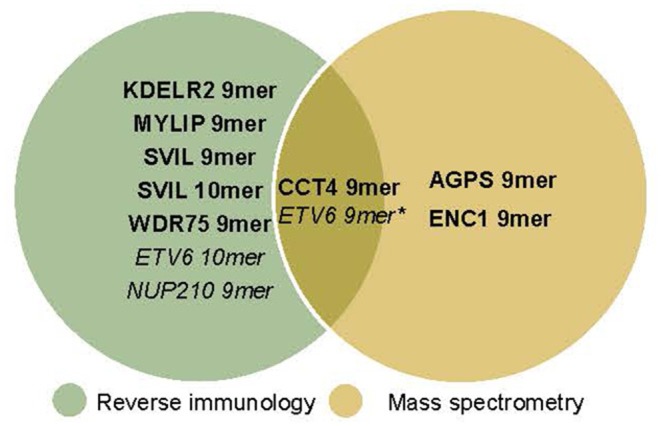
Tumor-infiltrating lymphocytes (TIL) are considered enriched for T cells recognizing shared tumor antigens or mutation-derived neoepitopes. We performed exome sequencing and HLA-A*02:01 epitope prediction from tumor cell lines from two HLA-A2-positive melanoma patients whose TIL displayed strong tumor reactivity. The potential neoepitopes were screened for recognition using autologous TIL by immunological assays and presentation on tumor major histocompatibility complex class I (MHC-I) molecules by Poisson detection mass spectrometry (MS). TIL from the patients recognized 5/181 and 3/49 of the predicted neoepitopes, respectively. MS screening detected 3/181 neoepitopes on tumor MHC-I from the first patient but only one was also among those recognized by TIL. Consequently, TIL enriched for neoepitope specificity failed to recognize tumor cells, despite being activated by peptides. For the second patient, only after IFN-γ treatment of the tumor cells was one of 49 predicted neoepitopes detected by MS, and this coincided with recognition by TIL sorted for the same specificity. Importantly, specific T cells could be expanded from patient and donor peripheral blood mononuclear cells (PBMC) for all neoepitopes recognized by TIL and/or detected on tumor MHC-I. In summary, stimulating the appropriate inflammatory environment within tumors may promote neoepitope MHC presentation while expanding T cells in blood may circumvent lack of specific TIL. The discordance in detection between physical and functional methods revealed here can be rationalized and used to improve neoantigen-targeted T cell immunotherapy.

Keywords: Immune peptidome; TIL; immunotherapy; mass spectrometry; neoepitopes; tumor.


Figures

Figure 1
Figure 1
Recognition of autologous tumor cells or peptides from shared tumor-associated antigens, from mutated genes, or from viruses by tumor-infiltrating lymphocytes. Tumor-infiltrating lymphocytes (TIL) and primary tumor cell lines were expanded from tumors of patients KADA and ANRU. The ability of TIL to recognize corresponding tumor cells or HLA-A2-transfected SK-OV-3 target cells pulsed with peptides from common tumor-associated antigens or viruses (A, KADA; D, ANRU), or HLA-A2-transfected SK-OV-3 cells pulsed with neoepitope peptides derived from mutated genes in tumor cells, in the absence or presence of anti-HLA-A2 (BB7.4) or -MHC-I (W6/32) blocking monoclonal antibodies (B, KADA; E, ANRU; only recognized peptides shown), or HLA-A2-transfected SK-OV-3 pulsed with neoepitope peptides compared to corresponding wild-type peptides (C, KADA; F, ANRU), and respond with IFN-γ secretion was assessed after 24 h by ELISA. Unpulsed HLA-A2-transfected SK-OV-3 target cells were used as a no peptide control and pulsed original SK-OV-3 cells (Ctrl) were used as a no HLA-A2 control as indicated.
Figure 2
Figure 2
Detection of mutated peptides presented on the tumor cell surface. LC-DIAMS Poisson detection plots for neoepitopes from mutated (A) and wild-type (B) ENC1 and mutated (C) and wild-type (D) AGPS from 500,000 cells. Top black traces are extracted ion chromatograms for m/z of the doubly charged precursor ion in units of counts per second. The inverted traces (blue) are Poisson chromatograms showing the number of events, scaled 10-fold (as a convenience in plotting), that can be embedded at fixed cutoff probability in the MS/MS spectrum of the DIA window containing the precursor m/z (17). Nanospray MS3 Poisson detection of cysteine-containing neoantigen peptide CCT4 (E) or the corresponding wild-type peptide (F) was performed from 1.5 million cells, as marked with a 0-offset Poisson peak (18).
Figure 3
Figure 3
Frequencies of neoantigen-specific T cells in TIL and assessment of their ability to recognize autologous tumor cells. HLA-A2/mutated peptide dextramers were produced for peptides found to activate TIL and/or found to be presented on tumor cell MHC-I. The dextramers were used to stain tumor-infiltrating lymphocytes (A), and anti-PE beads were used to enrich for stained cells followed by a rapid-expansion protocol. Thereafter, recognition of KADA (B) and ANRU (C) tumor cells by the sorted TIL was assessed by IFN-γ ELISA with or without IFN-γ pretreatment of the tumor. Only the specificities that could be significantly enriched by the sorting are shown. Unsorted cells were used as a control. The tumor cells were analyzed for neoepitope expression by MS and presented as LC-DIAMS Poisson detection plots for mutant ETV6 in untreated (D) and IFN-γ-treated ANRU tumor cells (E) with the arrow in the latter indicating detection. The total TIL population (F. ANRU) was co-cultured with untreated or IFN-γ pretreated autologous tumor cells, or with the different neoepitope peptides, analyzed by dextramer staining for the same epitope and for cell surface CD107a. As negative control, TIL alone were used and stained as described above. Dot plots are gated on lymphocytes/singlets/live cells and frequency indicates % dextramer+ out of CD8+ cells (A) and gated in the same way plus on CD8+ and frequency indicates % dextramer+CD07a or dextramer+CD107a+ of CD8+ cells (F).
Figure 4
Figure 4
Expansion of neoepitope peptide-specific T cells from PBMC. Dendritic cells from KADA (A) or ANRU (E) were loaded with their respective TIL-activating and/or tumor-presented peptides and used to stimulate autologous CD8 T cells. The same experiment was performed using three healthy donors for each peptide set (B–D, KADA neoepitopes; F–H, ANRU neoepitopes and MART-1). After 10 days of expansion, the cells were stained with corresponding HLA-A2/peptide dextramers and analyzed by flow cytometry. Dot plots are gated on lymphocytes/singlets/live cells and frequency indicates %dextramer+ out of CD8+ cells. For ANRU epitopes, the function of the neoepitope specific T cells was assessed by re-stimulation with ANRU tumor cells and evaluated by CD107a staining (I, ANRU; J–L, healthy donors). Only positive stainings are shown. Dot plots are gated on lymphocytes/singlets/live cells/CD8+ cells and frequency indicates % dextramer+CD07a or dextramer+CD107a+ of CD8+ cells.
Figure 5
Figure 5
Neoepitopes detected by reverse immunology vs. mass spectrometry. With reverse immunology TIL reactivity against 5/181 (KADA) and 3/49 (ANRU) of the predicted neoepitopes were observed. With mass spectrometry 3/136 (KADA, not all peptides could be analyzed) and 1*/38 (ANRU, not all peptides could be analyzed) of the predicted neoepitopes were detected on the cell surface. KADA epitopes are in Bold, ANRU epitopes are in Italic. *Detected after IFNγ treatment of the tumor.

References

    1. Feldman SA, Assadipour Y, Kriley I, Goff SL, Rosenberg SA. Adoptive cell therapy–tumor-infiltrating lymphocytes, T-cell receptors, and chimeric antigen receptors. Semin Oncol. (2015) 42:626–39. 10.1053/j.seminoncol.2015.05.005 - DOI - PMC - PubMed
    1. Morgan RA, Dudley ME, Wunderlich JR, Hughes MS, Yang JC, Sherry RM, et al. . Cancer regression in patients after transfer of genetically engineered lymphocytes. Science. (2006) 314:126–9. 10.1126/science.1129003 - DOI - PMC - PubMed
    1. Morgan RA, Chinnasamy N, Abate-Daga D, Gros A, Robbins PF, Zheng Z, et al. . Cancer regression and neurological toxicity following anti-MAGE-A3 TCR gene therapy. J Immunother. (2013) 36:133–51. 10.1097/CJI.0b013e3182829903 - DOI - PMC - PubMed
    1. Le DT, Uram JN, Wang H, Bartlett BR, Kemberling H, Eyring AD, et al. . PD-1 blockade in tumors with mismatch-repair deficiency. N Engl J Med. (2015) 372:2509–20. 10.1056/NEJMoa1500596 - DOI - PMC - PubMed
    1. Snyder A, Makarov V, Merghoub T, Yuan J, Zaretsky JM, Desrichard A, et al. . Genetic basis for clinical response to CTLA-4 blockade in melanoma. N Engl J Med. (2014) 371:2189–99. 10.1056/NEJMoa1406498 - DOI - PMC - PubMed

Publication types

MeSH terms