Clinical Trial
. 2019 Dec 11:10:2917.
doi: 10.3389/fimmu.2019.02917. eCollection 2019.

Cytotoxic T Cell-Derived Granzyme B Is Increased in Severe Plasmodium Falciparum Malaria

Affiliations
Clinical Trial

Cytotoxic T Cell-Derived Granzyme B Is Increased in Severe Plasmodium Falciparum Malaria

Lea-Christina Kaminski et al. Front Immunol. .

Abstract

In Plasmodium falciparum malaria, CD8+ T cells play a double-edged role. Liver-stage specific CD8+ T cells can confer protection, as has been shown in several vaccine studies. Blood-stage specific CD8+ T cells, on the other hand, contribute to the development of cerebral malaria in murine models of malaria. The role of CD8+ T cells in humans during the blood-stage of P. falciparum remains unclear. As part of a cross-sectional malaria study in Ghana, granzyme B levels and CD8+ T cells phenotypes were compared in the peripheral blood of children with complicated malaria, uncomplicated malaria, afebrile but asymptomatically infected children and non-infected children. Granzyme B levels in the plasma were significantly higher in children with febrile malaria than in afebrile children. CD8+ T cells were the main T cell subset expressing granzyme B. The proportion of granzyme B+ CD8+ T cells was significantly higher in children with complicated malaria than in uncomplicated malaria, whereas the activation marker CD38 on CD8+ T cells showed similar expression levels. This suggests a pathogenic role of cytotoxic CD8+ T cells in the development of malaria complications in humans.

Keywords: CD8+ T cells; Plasmodium falciparum; granzyme B; malaria; severe malaria.


Figures

Figure 1
Figure 1
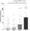
Granzyme B level in the plasma of P. falciparum infected children is associated with disease severity. Blood samples were taken from healthy, non-infected children (healthy donors, HD; n = 41) and asymptomatically infected children (AS, asymptomatically infected; n = 41) as well as from children with uncomplicated malaria (outpatients, OP; n = 35) and severe malaria (inpatients, IP; n = 32). Granzyme B levels in the plasma were measured by ELISA. Statistical significance was determined using Kruskal–Wallis Test with post-hoc Dunn's test: ****p < 0.0001; ***p < 0.001; **p < 0.01; *p < 0.05.
Figure 2
Figure 2
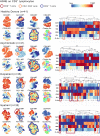
CD3+CD8+ T cells express high amounts of GrzB in P. falciparum infected children. CD3+ lymphocytes of all study participants were concatenated group wise (HD n = 41; AS n = 41; OP n = 36; IP n = 32). The cell number of all concatenated groups were downsampled to 106 cells/group. Hierarchical stochastic neighbor embedding (HSNE) analysis of all groups based on the expression of CD3, CD8, CD4, Granzyme B, CD38, and PD-1 were computed. Landmark development and clustering were complete data-driven. Landmark-based clusters were used for heat map generation. Color code represents mean fluorescence intensity (red = high; blue = low). Blue boxes indicate GrzB-producing CD4+ T cell subsets, red boxes indicate GrzB-producing CD8+ T cells. Orange boxes highlight CD3+CD4CD8 T cell subsets, producing GrzB.
Figure 3
Figure 3
Granzyme B expression in CD8+ T cells correlates with disease progression. Blood samples from children infected with P. falciparum but lacking symptoms (AS, n = 41), children treated as outpatients for uncomplicated malaria (OP, n = 35), children treated as inpatients for severe malaria (IP, n = 32), as well healthy, non-infected children (HD, n = 41) were stained with fluorescent labeled αCD3, αCD8, αCD4, αGranzyme B, αCD38, and αPD-1 antibodies for flow cytometric analyses. (A) Frequencies of CD8+ T cells among CD3+ cells were assessed in all 4 groups. (B) The CD8+CD3+ T cells were further analyzed for the expression of the effector molecule granzyme B, (D) CD38 and (E) PD-1 in all four groups. Statistical significance was determined using Kruskal–Wallis Test with post-hoc Dunn's test: ****p < 0.0001; ***p < 0.001; **p < 0.01; *p < 0.05. (C) The correlation between the frequency of GrzB expression by CD3+CD8+ T cells and parasitemia was analyzed in the blood of all infected children (n = 89), using Spearman rank correlation.

References

    1. Weiss WR, Sedegah M, Beaudoin RL, Miller LH, Good MF. CD8+ T cells (cytotoxic/suppressors) are required for protection in mice immunized with malaria sporozoites. Proc Natl Acad Sci USA. (1988) 85:573–6. 10.1073/pnas.85.2.573 - DOI - PMC - PubMed
    1. Chakravarty S, Cockburn IA, Kuk S, Overstreet MG, Sacci JB, Zavala F. CD8+ T lymphocytes protective against malaria liver stages are primed in skin-draining lymph nodes. Nat Med. (2007) 13:1035–41. 10.1038/nm1628 - DOI - PubMed
    1. Cockburn IA, Amino R, Kelemen RK, Kuo SC, Tse SW, Radtke A, et al. . In vivo imaging of CD8 T cell-mediated elimination of malaria liver stages. Proc Natl Acad Sci USA. (2013) 110:9090–5. 10.1073/pnas.1303858110. - DOI - PMC - PubMed
    1. Fernandez-Ruiz D, Ng WY, Holz LE, Ma JZ, Zaid A, Wong YC, et al. . Liver-resident memory CD8+ T cells form a front-line defense against malaria liver-stage infection. Immunity. (2016) 45:889–902. 10.1016/j.immuni.2016.08.011 - DOI - PubMed
    1. Epstein JE, Tewari K, Lyke KE, Sim BKL, Billingsley PF, Laurens MB, et al. . Live attenuated malaria vaccine designed to protect through hepatic CD8+ T cell immunity. Science. (2011) 334:475–80. 10.1126/science.1211548 - DOI - PubMed

Publication types