. 2019 Dec 18:10:2970.
doi: 10.3389/fimmu.2019.02970. eCollection 2019.

Coexpression of CCR7 and CXCR4 During B Cell Development Controls CXCR4 Responsiveness and Bone Marrow Homing

Affiliations

Coexpression of CCR7 and CXCR4 During B Cell Development Controls CXCR4 Responsiveness and Bone Marrow Homing

Saria Mcheik et al. Front Immunol. .

Abstract

The CXCL12-CXCR4 axis plays a key role in the retention of stem cells and progenitors in dedicated bone marrow niches. It is well-known that CXCR4 responsiveness in B lymphocytes decreases dramatically during the final stages of their development in the bone marrow. However, the molecular mechanism underlying this regulation and whether it plays a role in B-cell homeostasis remain unknown. In the present study, we show that the differentiation of pre-B cells into immature and mature B cells is accompanied by modifications to the relative expression of chemokine receptors, with a two-fold downregulation of CXCR4 and upregulation of CCR7. We demonstrate that expression of CCR7 in B cells is involved in the selective inactivation of CXCR4, and that mature B cells from CCR7-/- mice display higher responsiveness to CXCL12 and improved retention in the bone marrow. We also provide molecular evidence supporting a model in which upregulation of CCR7 favors the formation of CXCR4-CCR7 heteromers, wherein CXCR4 is selectively impaired in its ability to activate certain G-protein complexes. Collectively, our results demonstrate that CCR7 behaves as a novel selective endogenous allosteric modulator of CXCR4.

Keywords: B cells; CCR7; CXCR4; homing; lymphopoiesis.


Figures

Figure 1
Figure 1
Properties of B cell populations prepared from CCR7+/+ or CCR7−/− mice. (A) Expression of chemokine receptors in B cell subpopulations. BM B cell subpopulations were discriminated and sorted by flow cytometry and the expression of CXCR4, CCR7, CXCR5, and CCR6 was quantified by RT-qPCR using GAPDH and β-actin as references. (B) Cell surface expression of chemokine receptors. The cell surface expression of chemokine receptors in B cell subpopulations was estimated by flow cytometry using PE-conjugated anti-CXCR4 and APC-conjugated anti-CCR7, anti-CXCR5, and anti-CCR6 antibodies. Bars represent mean values ± SEM (n = 5) of the mean fluorescence index, detected in Pre-B, immature (i-B) or mature B cells (m-B) from CCR7+/+ (black bars) or CCR7−/− mice (white bars). The first black and white bar represents the mean fluorescence of control isotype. (C) Chemotaxis of BM B cells toward CXCL12. Transwell migration of BM B cells from CCR7+/+ (black dots) or CCR7−/− (white dots) mice in response to increasing concentrations of CXCL12. Migration index after a 2 h incubation were plotted for each subpopulation. All conditions were run in triplicates and the data, representative of two independent experiments, are presented as mean values ± SEM, *P < 0.05). (D) Enhanced chemotactic response to CXCL12 in mature B cells from CCR7−/− mice. Transwell migration of BM B cells from CCR7+/+ (black dots) and CCR7−/− (white dots) mice in response to 100 nM CXCL12. Migration indexes were plotted for each subpopulation. Data are represented as mean values ± SEM and dots correspond to individual mice (n = 6–8; **P < 0.005). (E) Blockade of CXCR4 inhibits CXCL12-elicited migration of CCR7−/− mature B cells. Transwell migration of BM mature B cells from CCR7−/− mice in response to 100 nM CXCL12 in the presence or not of 1 μM AMD3100 or 5 μg/ml MAB2165 (blocking anti-CXCR4 monoclonal). Data are presented as mean values ± SEM and dots correspond to individual mice (n = 6; ***P < 0.0005). (F) Blockade of CCR7 does not affect CXCR4 responsiveness in CCR7+/+ mature B cells. Transwell migration of BM mature B cells from CCR7+/+ mice in response to 100 nM CCL19 or CXCL12, and in the presence or not of 5 μg/ml MAB3477 (blocking anti-CCR7 monoclonal). Data are presented as mean values ± SEM, and dots correspond to individual mice (G) CCR7-deficiency abrogates the CCL19-dependent migration of mature B cells but does not affect the migration in response to CXCL13 or CCL20. Transwell migration of BM mature B cells from CCR7+/+ (black dots) and CCR7−/− (white dots) mice in response to 100 nM CCL19, CXCL13, or CCL20. Data are represented as mean values ± SEM, and dots correspond to individual mice (n = 5 to 8; *P < 0.05).
Figure 2
Figure 2
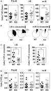
(A) Increased number of mature B cells in the BM of CCR7−/− mice. The number of B cell subpopulations was determined in the BM of CCR7+/+ (black dots) and CCR7−/− (white dots) mice. Data are represented as mean values ± SEM and dots correspond to individual mice (n = 10 to 13 mice; *P < 0.05; **P < 0.005). (B,C) Decreased egress of immature and mature B cells in the BM sinusoids of CCR7−/− mice. In vivo labeling of BM B cell subsets by injection of PE-conjugated anti-CD19 antibodies 4 min before sacrifice and tissue collection. The proportions of immature and mature B cells in the parenchyma (CD19; white) and sinusoids (CD19+; black) are displayed in (B) and the number of immature and mature B cells in sinusoids displayed in (C). Data for CCR7+/+ (black dots) and CCR7−/− (white dots) mice are represented as mean values ± SEM and dots correspond to individual mice (n = 8–9 mice; *P < 0.05). (D) B cell counts are comparable in the blood of CCR7+/+ and CCR7−/− mice. The number of B cell subpopulations was determined in the blood of CCR7+/+ (black dots) and CCR7−/− mice (white dots). Data are represented as mean values ± SEM and dots correspond to individual mice (n = 10–13 mice; *P < 0.05; **P < 0.005).
Figure 3
Figure 3
CCR7 deficiency results in increased homing of immature B cells to BM. B220+ cells purified from BM of CCR7+/+ (black dots) or CCR7−/− mice (white dots) were labeled with CFDA-SE and transferred into wild type recipients by injection into the retro-orbital venous plexus in combination or not with AMD3100. Two hours later, BM mononuclear cells were isolated and B cell subpopulations were analyzed by flow cytometry. Data are represented as mean values ± SEM and dots correspond to individual mice (n = 4–8 mice; **P < 0.005; ***P < 0.0005).
Figure 4
Figure 4
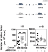
Increased number of B cells in the BM of CCR7−/− mice is not due to an alteration of BM environment. (A) Distribution and quantification of donor CD45.2+ (WT or CCR7−/−) B cells recovered from the BM of chimeric CD45.1+ recipients. (B) Distribution and quantification of donor CD45.1+ WT B cells recovered from the BM of chimeric CD45.2+ (WT or CCR7−/−) recipients. Data are represented as mean values ± SEM and dots correspond to individual mice (n = 8–12 mice; ***P < 0.005).
Figure 5
Figure 5
(A) Expression of CCR7 in Nalm-6 does not inhibit CXCR4 expression. Nalm-6 cells were stably transfected with a CCR7-encoding plasmid and cell surface expression of CXCR4 and CCR7 was monitored by flow cytometry. The data represent mean values of the mean fluorescent index ± SEM (n = 3). (B) Expression of CCR7 in Nalm-6 cells inhibits CXCL12-induced adhesion to VCAM-1. A suspension of Nalm-6 cells expressing CCR7 or not were stimulated with 1 μM CXCL12 for 1 min (black bars) or incubated in buffer (white bars) and then allowed to settle in VCAM-1-coated wells for 1 min. Uncoated wells were used as controls (dashed bars). Non-adherent cells were subsequently washed away and adherent cells were counted relative to the number of input cells. The data represent mean values ± SEM (n = 3; **P < 0.05; ***P < 0.005). (C) Expression of CCR7 in Nalm-6 cells inhibits CXCL12-induced chemotaxis. Migration of Nalm-6 cells expressing CCR7 (white dots) or not (black dots) was recorded in Transwells in response to increasing concentrations of CXCL12. The data represent mean values ± SEM (n = 3). (D) Expression of non-functional CCR7 mutants also inhibits CXCR4 responsiveness. Transwell migration of Nalm-6 cells stably expressing wild-type or non-functional CCR7 mutants was assayed in response to 10 nM CXCL12. Cells stably expressing CCR5 were used as control. The data represent mean values ± SEM (n = 3).
Figure 6
Figure 6
(A) CCR7 inhibits the CXCL12-induced activation of Gαi1 and Gαi2 proteins. Real-time measurement of BRET signal in HEK293T cells coexpressing Gαi1, Gαi2, or Gαi3 biosensors with CXCR4 only (black dots) or in a combination with CCR7 (white dots). Cells were stimulated for 1 min with increasing concentrations of CXCL12 after addition of coelenterazine 400. Results are expressed as ΔBRET, corresponding to the difference in BRET signal between Gαi-hRLuc8 and Gβ1γ2-GFP10, measured in the presence and absence of CXCL12. The data represent mean values ± SEM (n = 6). (B) CCR7 changes the basal BRET signal between CXCR4 and G proteins. HEK293T cells were transfected with hRLuc-Gαiβγ and CXCR4-Venus in combination or not with CCR7, and interaction between Gαiβγ and CXCR4 was investigated by measuring the energy transfer (BRET1) between the partners. The net BRET corresponds to the BRET measured between the two partners minus the BRET measured in cells expressing hRLuc-Gαiβγ only. The data represent mean values ± SEM (n = 5). (C) CCR7 inhibits the CXCL12-induced β-arrestin recruitment to the plasma membrane. HEK293T cells were transfected with the plasma membrane marker K-RasVenus, β-arrestin 2 Luc, and CXCR4 in combination or not with CCR7. The translocation of the β-arrestin 2 to the plasma membrane was recorded after stimulation with increasing concentration of CXCL12 by measuring the energy transfer (BRET) between the BRET energy donor β-arrestin 2 Luc and the BRET energy acceptor K-RasVenus. The data represent mean values ± SEM (n = 3).
Figure 7
Figure 7
(A) CCR7 interacts with CXCR4 in a BRET assay. HEK293T cells were transfected with a constant amount of the CCR7-hRLuc fusion and increasing amounts of the CXCR4-Venus fusion (black dots), or a constant amount of the CXCR4-hRLuc fusion and increasing amounts of the CCR7-Venus fusion (white dots), and heteromerization of CCR7 and CXCR4 was investigated by measuring the energy transfer (BRET1) between the two partners. As a control, an increasing amount of TSHR-Venus was used as BRET acceptor with CXCR4-hRLuc (formula image) or CCR7-hRLuc (♢) as donor. The Net BRET corresponds to the BRET measured between the two partners minus the BRET measured in cells expressing CXCR4-hRLuc or CCR7-hRLuc only. Data represent mean values ± SEM (n = 3). (B) CCR7 interacts with CXCR4 in a fluorescence complementation assay. HEK293T cells were transfected with CXCR4-V1, CXCR4-V2, CCR7-V1, and CCR7-V2 constructs, alone or as two by two combinations, and the fluorescence emission was recorded. As controls, TSHR-V1 and TSHR-V2 were cotransfected with the various CXCR4 and CCR7 constructs. Data represent mean values ± SEM (n = 3). (C) CCR7 interacts with CXCR4 at the plasma membrane. HEK293T cells were cotransfected with CXCR4-V1 and CCR7-V2 or CXCR4-V2 and CCR7-V1, and fluorescence was monitored by using fluorescent microscopy.
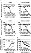
Figure 8
Figure 8
(A,B) Competition binding assays in CHO-K1 cells co-expressing CCR7 and CXCR4. Competition binding assays were performed on cells expressing CXCR4 or CCR7 only and on cells co-expressing CXCR4 and CCR7. Cells were incubated with 0.1 nM 125I-CXCL12 (A) or 0,1nM 125I-CCL19 (B) as tracers and increasing concentrations of unlabeled CXCL12 (black squares) or CCL19 (white squares) as competitors. The data were normalized for non-specific binding (0%) in the presence of 300 nM of competitor, and specific binding in the absence of competitor (100%). All points were run in triplicates and the data are presented as mean values ± SEM (n = 3). (C) CCR7 inhibits CXCR4 responsiveness in a calcium mobilization assay. Calcium mobilization assay was performed on CHO-K1 cells stably expressing the calcium-sensitive photoprotein aequorin and CXCR4 only or on cells co-expressing CXCR4 in combination with CCR7. Cells were loaded with coelenterazine H, stimulated with increasing concentrations of CXCL12 and the luminescence was recorded. The results were normalized for baseline activity (0%) and the maximal response obtained with 25 μM ATP (100%). All points were run in triplicates and the data are presented as mean values ± SEM (n = 3). (D) CCR7 inhibits the downmodulation of CXCR4 induced by CXCL12. CHO-K1 cells expressing CXCR4 only or in combination with CCR7 were either left untreated or stimulated for 90 min with increasing concentrations of CXCL12. Surface-bound CXCL12 was removed by an acid wash step and cell surface expression CXCR4 was estimated by FACS. The data were normalized for the expression of receptor in absence of stimulation (100%). All points were run in duplicates and the data represent mean values ± SEM (n = 3).

Similar articles

Cited by

References

    1. Lapidot T, Petit I. Current understanding of stem cell mobilization: the roles of chemokines, proteolytic enzymes, adhesion molecules, cytokines, and stromal cells. Exp Hematol. (2002). 30:973–81. 10.1016/S0301-472X(02)00883-4 - DOI - PubMed
    1. Juarez J, Bendall L, Bradstock K. Chemokines and their receptors as therapeutic targets: the role of the SDF-1/CXCR4 axis. Curr Pharm Des. (2004). 10:1245–59. 10.2174/1381612043452640 - DOI - PubMed
    1. Ponomaryov T, Peled A, Petit I, Taichman RS, Habler L, Sandbank J, et al. . Induction of the chemokine stromal-derived factor-1 following DNA damage improves human stem cell function. J Clin Invest. (2000). 106:1331–9. 10.1172/JCI10329 - DOI - PMC - PubMed
    1. Ceradini DJ, Kulkarni AR, Callaghan MJ, Tepper OM, Bastidas N, Kleinman ME, et al. . Progenitor cell trafficking is regulated by hypoxic gradients through HIF-1 induction of SDF-1. Nat Med. (2004). 10:858–64. 10.1038/nm1075 - DOI - PubMed
    1. Sugiyama T, Kohara H, Noda M, Nagasawa T. Maintenance of the hematopoietic stem cell pool by CXCL12-CXCR4 chemokine signaling in bone marrow stromal cell niches. Immunity. (2006). 25:977–88. 10.1016/j.immuni.2006.10.016 - DOI - PubMed

Publication types

MeSH terms