. 2019 Dec 31;10(1):e2020003.
doi: 10.5826/dpc.1001a03. eCollection 2020.

Update on Dermoscopy and Infectious Skin Diseases

Affiliations

Update on Dermoscopy and Infectious Skin Diseases

Vincenzo Piccolo. Dermatol Pract Concept. .

Abstract

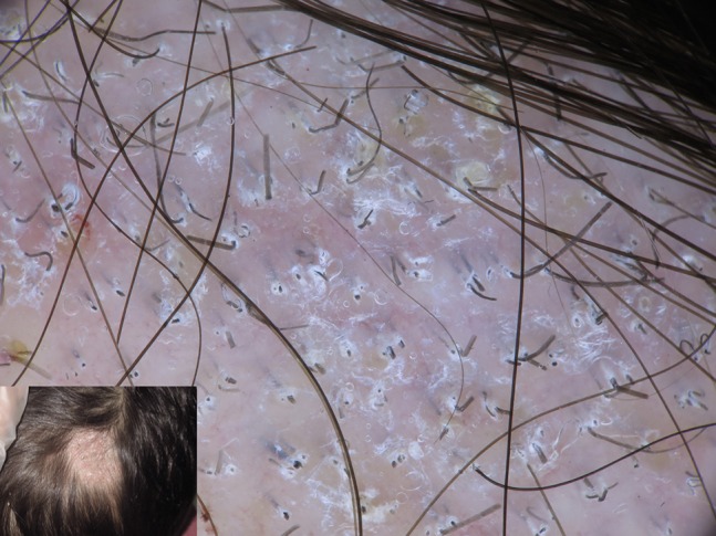
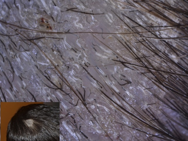
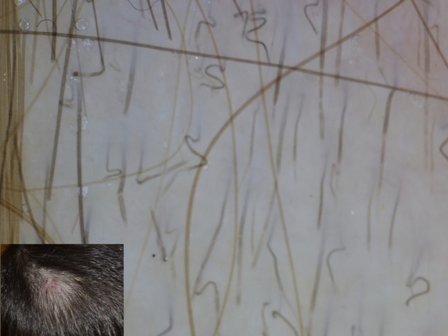
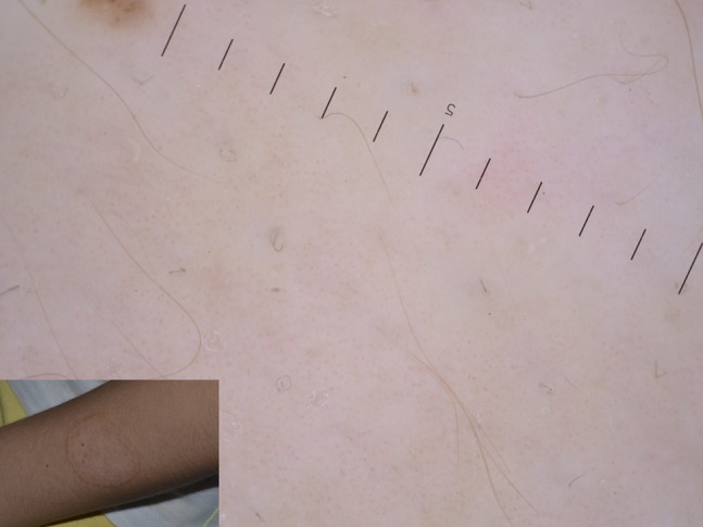
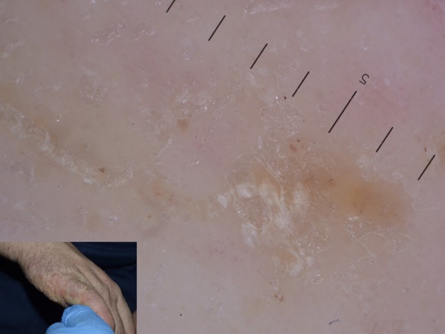
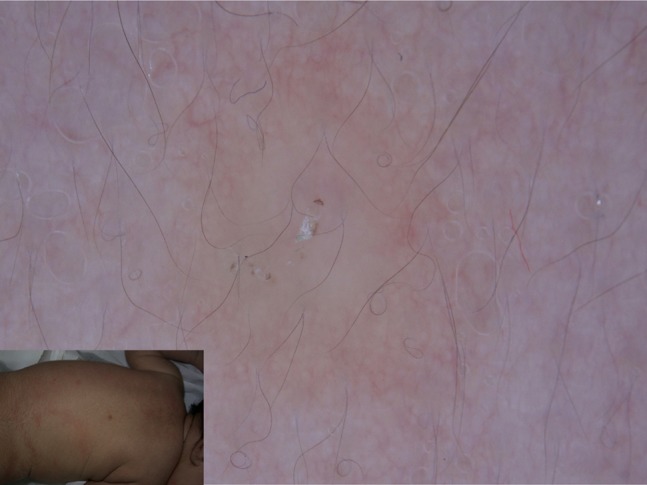
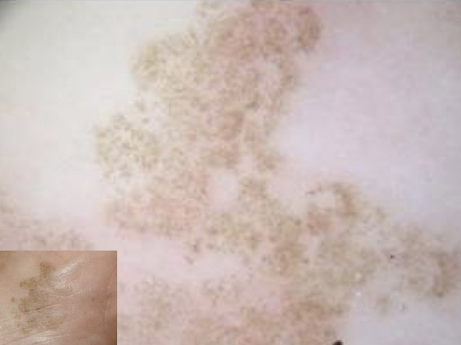
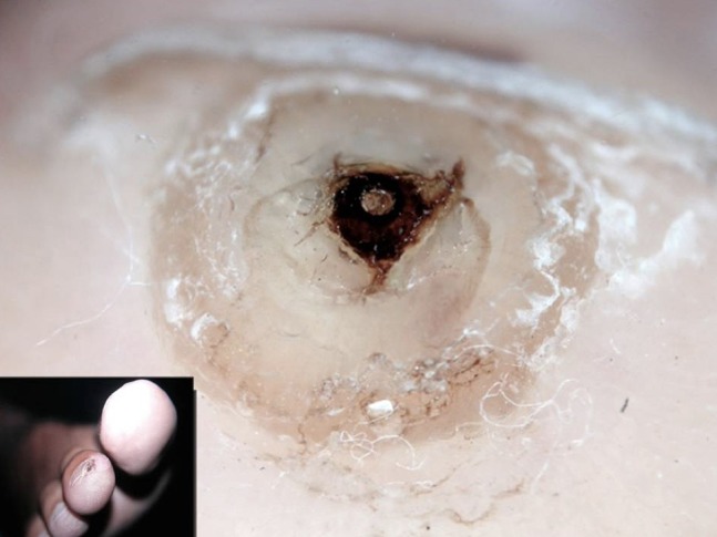
Nowadays, dermoscopy is a global worldwide diffuse diagnostic tool supporting clinicians in their daily hard task of correct orientation among dermatological diseases. Born to be an instrument for early diagnosis of skin cancer, the dermatoscope is now considered the dermatologist's stethoscope, as it can be routinely used to support diagnosis in general dermatology, so spreading its utility in cutaneous inflammatory and infectious diseases, as adjuvant and not substitute to histology and potassium hydroxide examination. As concerns the latter, plenty of papers have been published since the first description of dermoscopic findings of scabies. The aim of this review is to give the clinician a practical approach to dermoscopic parameters of cutaneous infectious diseases with a focus on the latest updates in this topic.

Keywords: dermatoscopy; dermoscopy; entomodermoscopy; infections; infectious.


Conflict of interest statement

Competing interests: The author has no conflicts of interest to disclose.

Figures

Figure 1
Figure 1
Typical papillae in a common wart centered by hemorrhagic dots in turn surrounded by whitish halos.
Figure 2
Figure 2
Projected papillae in a filiform wart of beard area. Dotted vessels are detectable at the extremities of each papilla.
Figure 3
Figure 3
Dotted vessels on a whitish background found in flat warts of the scalp.
Figure 4
Figure 4
Plantar wart showing hemorrhagic dots corresponding to thrombosed vessels. Interruption of dermatoglyphics is clearly evident.
Figure 5
Figure 5
Mosaic pattern in anogenital warts of a child.
Figure 6
Figure 6
Typical dermoscopic appearance of molluscum contagiosum, showing a central pore on whitish amorphous structure surrounded by linear vessels.
Figure 7
Figure 7
The presence of central orifice is not mandatory in molluscum contagiosum as in this case.
Figure 8
Figure 8
Polylobular appearance of an “old” molluscum of the dorsum.
Figure 9
Figure 9
Diffuse comma hair in a patients affected by tinea capitis.
Figure 10
Figure 10
Tinea capitis showing different findings including comma hair, hair casts, dystrophic hairs, and zig-zag hair.
Figure 11
Figure 11
Corkscrew and barcode-like hair in tinea capitis.
Figure 12
Figure 12
Newly described vellus hair involvement in tinea corporis, identified by black dystrophic hair.
Figure 13
Figure 13
A challenging diagnosis of tinea incognito could be favored by dermoscopy showing vellus hair involvement and pustules.
Figure 14
Figure 14
Onychomycosis showing fringed proximal onycholytic border associated with longitudinal striae.
Figure 15
Figure 15
Ruin-like appearance of distal border of nail seen at dermoscopy.
Figure 16
Figure 16
Rare picture of pediculus capitis seen at dermoscopy.
Figure 17
Figure 17
Nits adherent to hair shafts highly diagnostic in pediculosis capitis.
Figure 18
Figure 18
A couple of Pthirus found on the pubis of young man. Note the scorpion-like appearance of blood-filled intestine.
Figure 19
Figure 19
Nits and lice adherent to eyelashes in a child affected by pthiriasis palpebrarum.
Figure 20
Figure 20
Delta-wing jet with contrail sign in scabies.
Figure 21
Figure 21
Grouped mite burrows configuring the “noodle sign” in crusted scabies.
Figure 22
Figure 22
Nodular scabies of newborn detected at dermoscopy.
Figure 23
Figure 23
Typical findings of tinea nigra showing weak pigmentation composed of thin bundles of spicules, arranged in parallel lines mainly in some peripheral areas.
Figure 24
Figure 24
Dermoscopy of tungiasis showing a homogeneous white area centered by a black pore.
Figure 25
Figure 25
A child affected by cutaneous leishmaniasis showing at dermoscopy yellow tears on a reddish orange background.
Figure 26
Figure 26
Segmented serpiginous appearance of larva migrans at dermoscopy.
Figure 27
Figure 27
Yellow-to-green discoloration of the nail at dermoscopic examination of nail plate.
Figure 28
Figure 28
Dermoscopic appearance of trichomycosis axillaris showing yellowish hair casts around axillary hairs.

Similar articles

Cited by

References

    1. Russo T, Piccolo V, Lallas A, Argenziano G. Recent advances in dermoscopy. F1000Res. 2016 Feb 17;5 doi: 10.12688/f1000research.7597.1. pii: F1000 Faculty Rev-184. - DOI - PMC - PubMed
    1. Zalaudek I, Lallas A, Moscarella E, Longo C, Soyer HP, Argenziano G. The dermatologist’s stethoscope—traditional and new applications of dermoscopy. Dermatol Pract Concept. 2013;3(2):67–71. - PMC - PubMed
    1. Argenziano G, Fabbrocini G, Delfino M. Epiluminescence microscopy: a new approach to in vivo detection of Sarcoptes scabiei. Arch Dermatol. 1997;133(6):751–753. - PubMed
    1. Errichetti E, Zalaudek I, Kittler H, et al. Standardization of dermoscopic terminology and basic dermoscopic parameters to evaluate in general dermatology (non-neoplastic dermatoses): an expert consensus on behalf of the International Dermoscopy Society. Br J Dermatol. 2019 May 11; doi: 10.1111/bjd.18125. Epub ahead of print. - DOI - PubMed
    1. Kelly SC, Purcell SM. Prevention of nosocomial infection during dermoscopy? Dermatol Surg. 2006;32(4):552–555. - PubMed