Review
. 2019 Dec 12:7:512.
doi: 10.3389/fped.2019.00512. eCollection 2019.

Tracheomalacia and Tracheobronchomalacia in Pediatrics: An Overview of Evaluation, Medical Management, and Surgical Treatment

Affiliations
Review

Tracheomalacia and Tracheobronchomalacia in Pediatrics: An Overview of Evaluation, Medical Management, and Surgical Treatment

Ali Kamran et al. Front Pediatr. .

Abstract

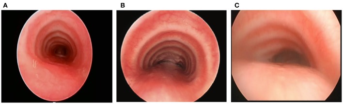
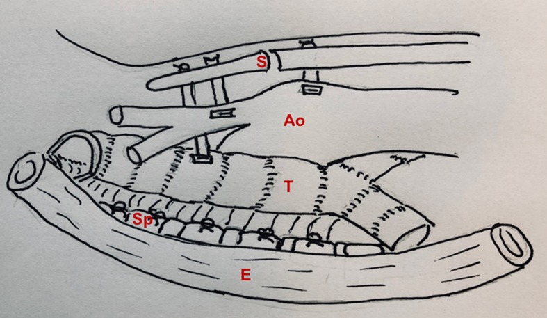
Tracheobronchomalacia (TBM) refers to airway collapse due to typically excessive posterior membrane intrusion and often associated with anterior cartilage compression. TBM occurs either in isolation or in association with other congenital or acquired conditions. Patients with TM typically present non-specific respiratory symptoms, ranging from noisy breathing with a typical barking cough to respiratory distress episodes to acute life-threatening events and recurrent and/or prolonged respiratory infections. There are no definitive standardized guidelines for the evaluation, diagnosis, and treatment of TBM; therefore, patients may be initially misdiagnosed and incorrectly treated. Although milder cases of TBM may become asymptomatic as the diameter of the airway enlarges with the child, in cases of severe TBM, more aggressive management is warranted. This article is an overview of the clinical presentation, evaluation, diagnosis, medical management, and surgical treatment options in pediatric tracheomalacia.

Keywords: aortopexy; tracheobronchomalacia; tracheobronchopexy; tracheomalacia; tracheopexy.


Figures

Figure 1
Figure 1
(A) Tracheal structure with normal C-shape rings. (B) U-shaped rings with a wider posterior membrane, demonstrating posterior intrusion. (C) Bow-shaped rings with a broad posterior membrane and severe posterior intrusion.
Figure 2
Figure 2
(A) Standardized form for documentation of 3-phase bronchoscopic findings. (B) Anatomic divisions of the trachea and mainstem bronchi to facilitate classification and description of tracheobronchomalacia. FOE, fiberoptic examination; AE, aryepiglottic; L cleft, laryngeal cleft; T1, trachea level 1; T2, trachea level 2; T3, trachea level 3; L1, left mainstem bronchus level 1; L2, left mainstem bronchus level 2; L3, left mainstem bronchus level 3; R1, right mainstem bronchus level 1; R2, right mainstem bronchus level 2; BI, bronchus intermedius; TEF, tracheoesophageal fistula; R, right-sided; L, left-sided; LMB, left mainstem bronchus; NL, normal; ELLIP, elliptical cricoid; U, U-shaped rings; BOW, bow-shaped rings [Illustration was adapted from Choi et al. (14) Copyright 2019 by American Medical Association].
Figure 3
Figure 3
Dynamic airway CT scan with 3-D reconstruction. (A) CT scan (cross-sectional view): partial collapse of the mid trachea (T) and dilated esophageal dilation (E) at end-inspiration. (B) CT (cross-sectional view): complete collapse of the mid trachea (arrow) at end-expiration. (C) 3-D reconstruction of the airways: partial collapse of the mid trachea at end-inspiration. (D) 3-D reconstruction of the airways: complete collapse of the mid trachea at end-expiration (arrow).
Figure 4
Figure 4
Anterior aortopexy. (A) Anterior collapse of the trachea, caused by compression of the aorta. (B) Anterior aortopexy with suspension of both the ascending aorta and the innominate artery to the posterior surface of the sternum using pledgeted horizontal mattress sutures. This image was illustrated by Dr. John Foker and published with his permission.
Figure 5
Figure 5
Posterior and anterior tracheopexy combined with anterior aortopexy. S, sternum; Ao, aorta; T, trachea; Sp, spine; E, esophagus.
Figure 6
Figure 6
Posterior descending aortopexy. (A) Anatomic relationships of the descending aorta to the esophagus and the spine. (B) Cross-sectional view: the esophagus is rotated to the right and the descending aorta is moved to the left and secured to the side of the spine as posteriorly as necessary to relieve posterior pressure off the left mainstem bronchus. (C) Descending aortopexy sutures are tied, relieving left mainstem posterior intrusion and compression between the descending aorta and the pulmonary artery.

References

    1. Baxter JD, Dunbar JS. Tracheomalacia. Ann Otol Rhinol Laryngol. (1963) 72:1013–23. 10.1177/000348946307200415 - DOI - PubMed
    1. Masters IB, Chang AB, Patterson L, Wainwright C, Buntain H, Dean BW, et al. Series of laryngomalacia, tracheomalacia, and bronchomalacia disorders and their associations with other conditions in children. Pediatr Pulmonol. (2002) 34:189–95. 10.1002/ppul.10156 - DOI - PubMed
    1. Masters IB, Zimmerman PV, Chang AB. Longitudinal quantification of growth and changes in primary tracheobronchomalacia sites in children. Pediatr Pulmonol. (2007) 42:906–13. 10.1002/ppul.20681 - DOI - PubMed
    1. Tan JZ, Ditchfield M, Freezer N. Tracheobronchomalacia in children: review of diagnosis and definition. Pediatr Radiol. (2012) 42:906–15. 10.1007/s00247-012-2367-5 - DOI - PubMed
    1. Carden KA, Boiselle PM, Waltz DA, Ernst A. Tracheomalacia and tracheobronchomalacia in children and adults: an in-depth review. Chest. (2005) 127:984–1005. 10.1378/chest.127.3.984 - DOI - PubMed

LinkOut - more resources