Review
. 2019 Dec 12:7:414.
doi: 10.3389/fbioe.2019.00414. eCollection 2019.

Cytotoxicity-Related Bioeffects Induced by Nanoparticles: The Role of Surface Chemistry

Affiliations
Review

Cytotoxicity-Related Bioeffects Induced by Nanoparticles: The Role of Surface Chemistry

Hainan Sun et al. Front Bioeng Biotechnol. .

Abstract

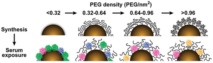
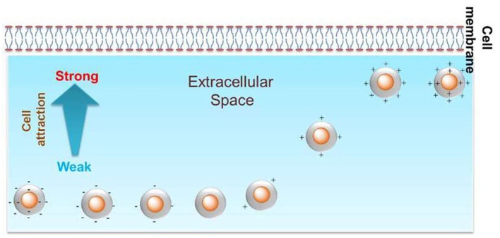
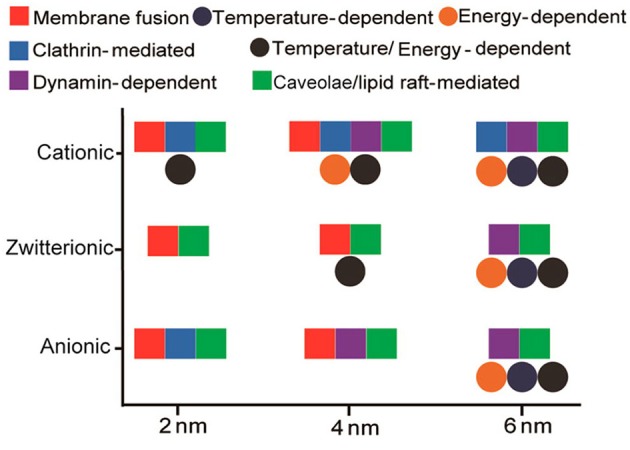
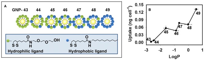
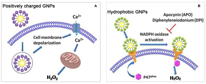
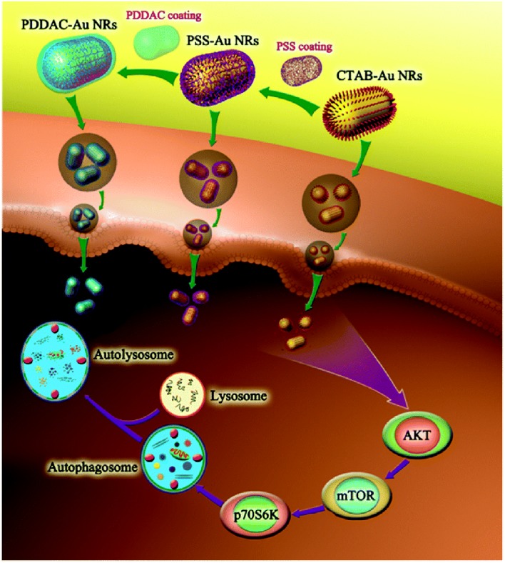
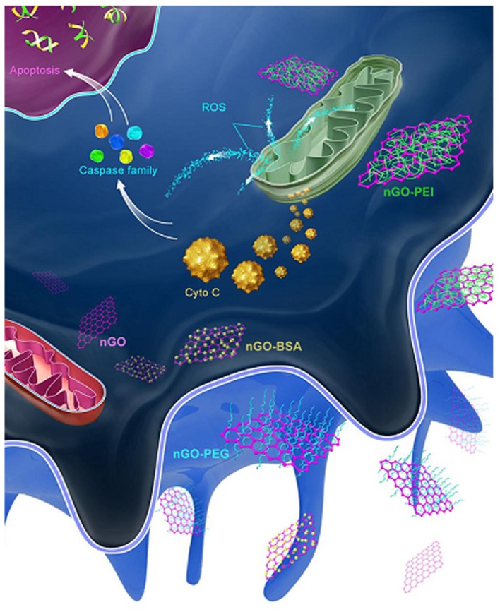
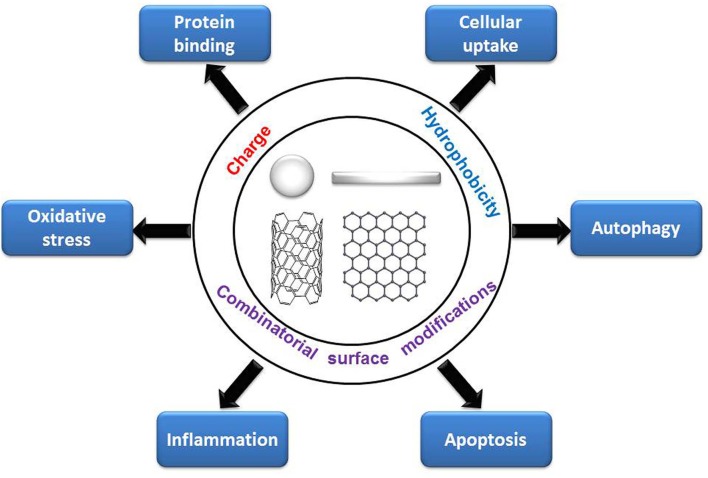
Nanoparticles (NPs) are widely used in a variety of fields, including those related to consumer products, architecture, energy, and biomedicine. Once they enter the human body, NPs contact proteins in the blood and interact with cells in organs, which may induce cytotoxicity. Among the various factors of NP surface chemistry, surface charges, hydrophobicity levels and combinatorial decorations are found to play key roles inregulating typical cytotoxicity-related bioeffects, including protein binding, cellular uptake, oxidative stress, autophagy, inflammation, and apoptosis. In this review, we summarize the recent progress made in directing the levels and molecular pathways of these cytotoxicity-related effects by the purposeful design of NP surface charge, hydrophobicity, and combinatorial decorations.

Keywords: PEG; charge; cytotoxicity; hydrophobicity; nanoparticles; surface chemistry.


Figures

Figure 1
Figure 1
The influence of PEG density on serum protein adsorption to gold nanoparticles. The top panel shows as-synthesized gold nanoparticles grafted with PEG at increasing density. As PEG density increases, PEG volume decreases as a result of PEG–PEG steric interactions. The lower panel illustrates how PEG density determines the amount and relative abundance of serum proteins adsorbed to the gold nanoparticle surface after serum exposure. Adapted with permission from Walkey et al. (2012).
Figure 2
Figure 2
Positive charge density could tune the cellular uptake level of GNPs in HeLa cells. Reproduced with permission from Su et al. (2012).
Figure 3
Figure 3
Interplay of size and surface functionality on the cellular uptake pathway of GNPs. Reproduced with permission from Jiang et al. (2015).
Figure 4
Figure 4
(A) GNP library with a continuous change in hydrophobicity. (B) Hydrophobicity regulates the cellular uptake level of GNPs in macrophages. Adapted with permission from Li et al. (2015a).
Figure 5
Figure 5
(A) Positively charged GNPs induce cell membrane depolarization and calcium channel opening, and stimulate mitochondria to generate intracellular oxidative stress. (B) Hydrophobic GNPs induce oxidative stress by perturbing NADPH oxidase. Adapted with permission from Sun et al. (2018).
Figure 6
Figure 6
CTAB-GNRs induce autophagy through mTOR-dependent pathway while PSS- and PDDAC-GNRs do not cause an obvious autophagy process. Reproduced with permission from Li et al. (2015b).
Figure 7
Figure 7
After being phagocytized, PEI-GO was more apt to interact with mitochondria and activate the apoptotic pathway. Reproduced with permission from Luo et al. (2015).
Figure 8
Figure 8
Regulation of cytotoxicity-related bioeffects by surface chemistry.

Similar articles

Cited by

References

    1. Abdelkhaliq A., Van Der Zande M., Punt A., Helsdingen R., Boeren S., Vervoort J. J. M., et al. . (2018). Impact of nanoparticle surface functionalization on the protein corona and cellular adhesion, uptake and transport. J. Nanobiotechnol. 16:70. 10.1186/s12951-018-0394-6 - DOI - PMC - PubMed
    1. Albanese A., Tang P. S., Chan W. C. (2012). The effect of nanoparticle size, shape, and surface chemistry on biological systems. Annu. Rev. Biomed. Eng. 14, 1–16. 10.1146/annurev-bioeng-071811-150124 - DOI - PubMed
    1. Aramesh M., Shimoni O., Ostrikov K., Prawer S., Cervenka J. (2015). Surface charge effects in protein adsorption on nanodiamonds. Nanoscale 7, 5726–5736. 10.1039/C5NR00250H - DOI - PubMed
    1. Asharani P. V., Low Kah Mun G., Hande M. P., Valiyaveettil S. (2009). Cytotoxicity and genotoxicity of silver nanoparticles in human cells. Acs Nano 3, 279–290. 10.1021/nn800596w - DOI - PubMed
    1. Ayala V., Herrera A. P., Latorre-Esteves M., Torres-Lugo M., Rinaldi C. (2013). Effect of surface charge on the colloidal stability and in vitro uptake of carboxymethyl dextran-coated iron oxide nanoparticles. J Nanopart. Res. 15:1874. 10.1007/s11051-013-1874-0 - DOI - PMC - PubMed