Review
. 2019 Dec 20:7:355.
doi: 10.3389/fcell.2019.00355. eCollection 2019.

Mitochondrial Involvement in Migration, Invasion and Metastasis

Affiliations
Review

Mitochondrial Involvement in Migration, Invasion and Metastasis

Tatiana V Denisenko et al. Front Cell Dev Biol. .

Abstract

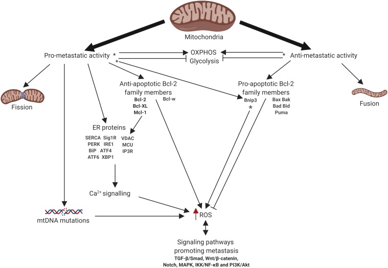
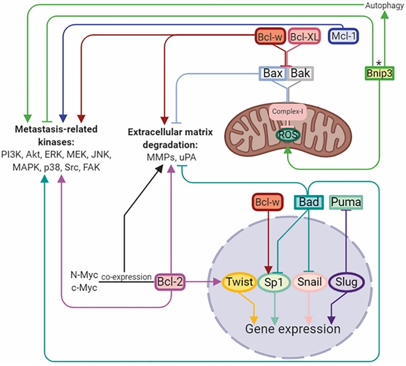
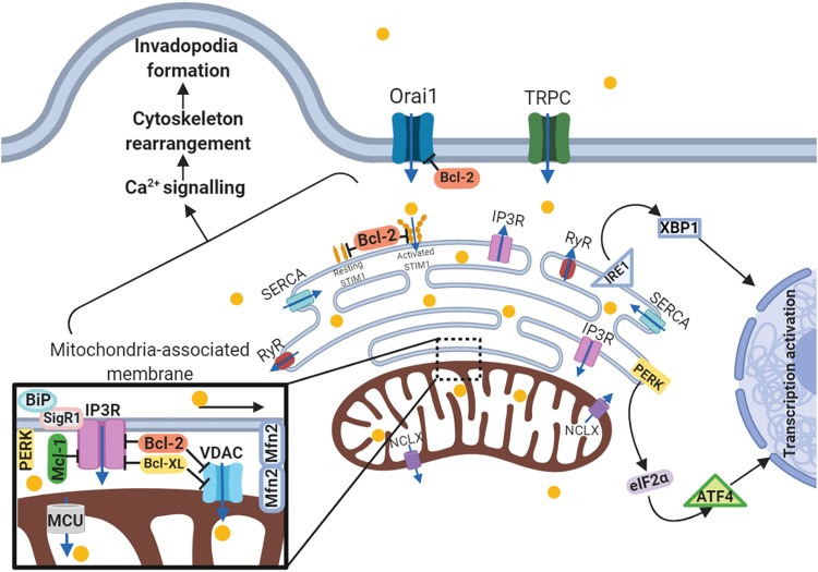
Mitochondria in addition to be a main cellular power station, are involved in the regulation of many physiological processes, such as generation of reactive oxygen species, metabolite production and the maintenance of the intracellular Ca2+ homeostasis. Almost 100 years ago Otto Warburg presented evidence for the role of mitochondria in the development of cancer. During the past 20 years mitochondrial involvement in programmed cell death regulation has been clarified. Moreover, it has been shown that mitochondria may act as a switchboard between various cell death modalities. Recently, accumulated data have pointed to the role of mitochondria in the metastatic dissemination of cancer cells. Here we summarize the modern knowledge concerning the contribution of mitochondria to the invasion and dissemination of tumor cells and the possible mechanisms behind that and attempts to target metastatic cancers involving mitochondria.

Keywords: cell death; invasion; metastasis; migration; mitochondria.


Figures

FIGURE 1
FIGURE 1
Schematic representation of mitochondrial involvement in metastasis. Arrows or blunt ends indicate activation or inhibition, respectively. Red arrow indicates increased level. - function depends on the tumor type. OXPHOS, oxidative phosphorylation; ER, endoplasmic reticulum; ROS, reactive oxygen species. For details, see text. Figure is created using BioRender.
FIGURE 2
FIGURE 2
Bcl-2 family members regulate metastasis by activation/inhibition of signaling kinases, matrix-degrading enzymes, and transcriptional factors. Arrows or blunt ends indicate activation or inhibition, respectively. Colored lines corresponds to each protein. - function depends on the tumor type. Bcl-2, B-cell lymphoma 2; Mcl-1, myeloid cell leukemia 1; Bcl-XL, B-cell lymphoma-extra large; Bcl-w, Bcl-2-like protein 2; Bnip3, BCL2/adenovirus E1B 19 kDa protein-interacting protein 3; Bax, Bcl-2-like protein 4; Bak, Bcl-2 homologous antagonist/killer; Bad, Bcl-2-associated death promoter; Puma, p53 upregulated modulator of apoptosis; Twist, class A basic helix-loop-helix protein 38; Sp1, specificity protein 1; Snail, zinc finger protein SNAI1; Slug, zinc finger protein SNAI2. For details, see text. Figure is created using BioRender.
FIGURE 3
FIGURE 3
Schematic representation of the link between ER-mitochondria network and motility of cancer cells. Arrows or blunt ends indicate activation or inhibition, respectively. Blue arrows indicate direction of Ca2+ current. Yellow circles – Ca2+. MAM, mitochondria-associated ER membrane; Orai1, calcium release-activated calcium channel protein 1; TRPC, transient receptor potential cation channel; SERCA, sarco/endoplasmic reticulum Ca2+-ATPase; STIM1, stromal interaction molecule 1; IP3R, inositol trisphosphate receptor; RyR, ryanodine receptor; IRE1, serine/threonine-protein kinase/endoribonuclease inositol-requiring enzyme 1; XBP1, X-box binding protein 1; PERK, protein kinase RNA-like endoplasmic reticulum kinase; eIF2α, eukaryotic translation initiation factor 2; ATF4, activating transcription factor 4; NCLX, mitochondrial Na+/Ca2+ exchanger; BiP, binding immunoglobulin protein; SigR1, sigma receptor 1; Mfn2, mitofusin2; VDAC, voltage-dependent anion-selective channel; MCU, mitochondrial calcium uniporter. For details, see text. Figure is created using BioRender.

References

    1. Adams J. M., Cory S. (2018). The BCL-2 arbiters of apoptosis and their growing role as cancer targets. Cell Death Differ. 25 27–36. 10.1038/cdd.2017.161 - DOI - PMC - PubMed
    1. Ahmad A., Aboukameel A., Kong D., Wang Z., Sethi S., Chen W., et al. (2011). Phosphoglucose Isomerase/Autocrine Motility factor mediates epithelial-mesenchymal transition regulated by miR-200 in breast cancer cells. Cancer Res. 71 3400–3409. 10.1158/0008-5472.CAN-10-0965 - DOI - PMC - PubMed
    1. Báthori G., Csordás G., Garcia-Perez C., Davies E., Hajnóczky G. (2006). Ca2+ -dependent control of the permeability properties of the mitochondrial outer membrane and voltage-dependent anion-selective channel (VDAC). J. Biol. Chem. 281 17347–17358. 10.1074/jbc.M600906200 - DOI - PubMed
    1. Beckner M. E., Gobbel G. T., Abounader R., Burovic F., Agostino N. R., Laterra J., et al. (2005). Glycolytic glioma cells with active glycogen synthase are sensitive to PTEN and inhibitors of PI3K and gluconeogenesis. Lab. Invest. 85 1457–1470. 10.1038/labinvest.3700355 - DOI - PubMed
    1. Ben-Kasus Nissim T., Zhang X., Elazar A., Roy S., Stolwijk J. A., Zhou Y., et al. (2017). Mitochondria control store-operated Ca2+ entry through Na + and redox signals. EMBO J. 36 797–815. 10.15252/embj.201592481 - DOI - PMC - PubMed