Review
. 2019 Dec:18:44-52.
doi: 10.1016/j.coisb.2019.10.008.

Modeling cell-cell communication for immune systems across space and time

Affiliations
Review

Modeling cell-cell communication for immune systems across space and time

Hirad Daneshpour et al. Curr Opin Syst Biol. 2019 Dec.

Abstract

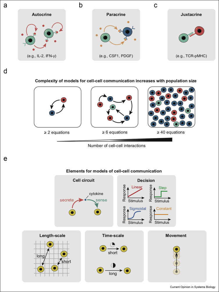
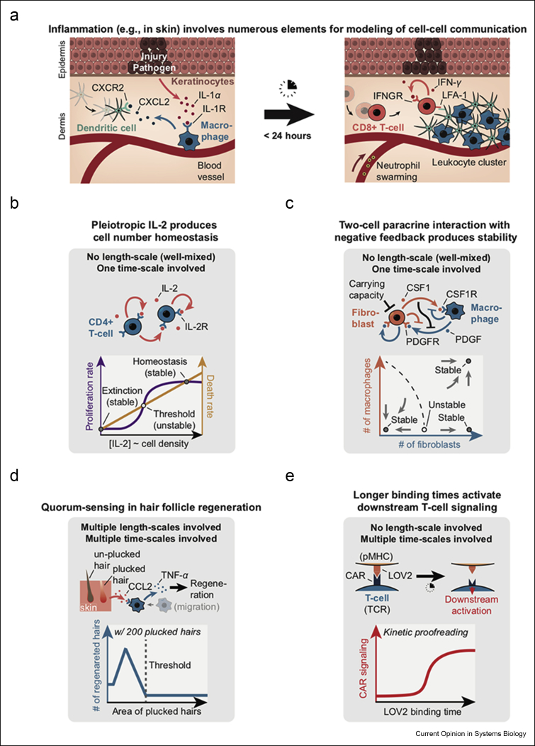
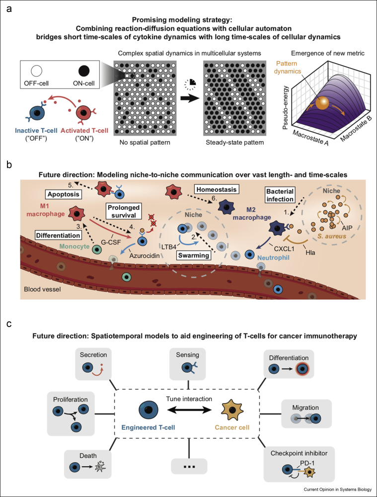
Communicating is crucial for cells to coordinate their behaviors. Immunological processes, involving diverse cytokines and cell types, are ideal for developing frameworks for modeling coordinated behaviors of cells. Here, we review recent studies that combine modeling and experiments to reveal how immune systems use autocrine, paracrine, and juxtacrine signals to achieve behaviors such as controlling population densities and hair regenerations. We explain that models are useful because one can computationally vary numerous parameters, in experimentally infeasible ways, to evaluate alternate immunological responses. For each model, we focus on the length-scales and time-scales involved and explain why integrating multiple length-scales and time-scales in a model remain challenging. We suggest promising modeling strategies for meeting this challenge and their practical consequences.

Keywords: Cellular automata; Cellular communication; Cytokines; Design principles; Immune systems; Modeling; Multicellular systems; Reaction–diffusion equations.


Figures

Figure 1
Figure 1
Ingredients for modeling cell–cell communication.(a) Autocrine signaling involves one cell type and cytokines such as IL-2 and IFN-γ. (b) Paracrine signaling involves at least two cell types, one that secretes a cytokine (e.g., CSF-1 and PDGF) without a cognate receptor and another cell type has the cognate receptor but does not secrete the cytokine. (c) Juxtacrine signaling involves at least two cell types communicating by a physical contact through a membrane-bound ligand (such as pMHC) and a receptor (such as TCR). (d) Models that describe communications among cells typically have as many equations as the number of cells involved. (e) Elements that enter a model for cell–cell communication. (Top left) Cell circuit that describes which cell secretes and which cell senses a cytokine; (Top right) Distinct responses to cytokines; (Bottom left) Distinct length-scales involved in cytokine-mediated communication; (Bottom middle) Distinct time-scales involved in cytokine-mediated communication; (Bottom right) cell motility.
Figure 2
Figure 2
Case studies for modeling cell–cell communication in immunological processes.(a) Schematic showing skin injury by invading pathogens. Left panel shows the immunological processes that occur soon after the skin injury, and the right panel shows how the skin injury is repaired [17]. (b) Population density of murine CD4+ T-cells controlled by their secreted IL-2 that they sense with the receptor (IL-2R). IL-2 simultaneously controls the proliferation rate (purple curve) and the death rate (orange curve) as shown in the graph. The graph shows two population densities that can be stably maintained (nearly zero and a value below a carrying capacity) and one that can be unstably maintained (“threshold” value) [18,19]. (c) Stable and robust maintenance of a ratio between two population densities (densities of fibroblasts and of macrophages). Fibroblasts secrete the autocrine and paracrine growth factor CSF1 and express the receptor, PDGFR, to sense the PDGF and the receptor, CSF1R, to sense the CSF1. Macrophages secrete the paracrine growth factor, PDGF, and express the receptor, CSF1R, to sense the CSF1. The graph shows three ratios of population densities that can be stably maintained and one that is an unstable, steady-state ratio [20,21]. (d) Regeneration of hair follicles on mouse skin by quorum-sensing. Plucked, distressed hair follicles secrete CCL2 which is sensed by M1 macrophages that are in turn recruited to the distressed follicles. Then, macrophages secrete TNF-α which then activates regeneration of the distressed hair follicles. Graph shows the number of regenerated hairs (blue curve) as a function of the skin area from which 200 hairs are plucked—only high density (small area) leads to appreciable regenerations [30]. (e) Kinetic proofreading as a mechanism to explain how a T-cell can distinguish between self and foreign peptides. Top cartoon shows a schematic of a recent experiment in which the binding time of the CAR (TCR) to LOV2 (pMHC) was optogenetically controlled. Graph shows the downstream activation in T-cell (CAR signaling) occurring only when the pMHC-TCR complex lives longer than a certain threshold duration [43].
Figure 3
Figure 3
Prospects for modeling cell–cell communication in immune systems.(a) A promising modeling strategy for reducing complexity—combining reaction–diffusion equations and cellular automata. Schematics here summarize two recent studies [46,47]. Two cell types (OFF-cell and ON-cell) communicate via an autocrine-signaling cytokine (bottom left shows a paracrine signal that one of the studies [47] also treats). Two fields of cells are shown. On the left is a disordered field of cells that, after some time, becomes more spatially organized (right field) because of cells coordinating their gene expressions through cytokine-mediated communications. (Rightmost picture): This self-organization dynamics can be quantitatively mapped to intuitive dynamics in which a ball (representing a field of cells) rolls down a Waddington-like landscape whose shape is determined by the various parameters in the model (e.g., secretion rate of the cytokines) [47]. (b) Schematic showing an example of a multistep process—niche-to-niche communication—of the type for which no suitable models exist yet because multiple length-scales and time-scales are involved. Picture shows a step-by-step process (following the numbers in order) that occurs after Staphylococcus aureus infects the skin [49,52]. (c) Various ingredients, shown in each box, that go into engineering T-cells (e.g., CAR-T) for cancer immunotherapy. Quantitative models will likely provide blueprints for better engineering CAR-T cells [53, 54, 55].

References

    1. Larkin J.W., Zhai X., Kikuchi K., Redford S., Prindle A., Liu J., Greenfield S., Walczak A.M., Garcia-Ojalvo J., Mugler A., Süel G.M. Signal percolation within a bacterial community. Cell Syst. 2018;7:1–9. - PMC - PubMed
    1. Altan-Bonnet G., Mukherjee R. Cytokine-mediated communication: a quantitative appraisal of immune complexity. Nat Rev Immunol. 2019;19:205–217. - PMC - PubMed
    1. Antonioli L., Blandizzi C., Pacher P., Guilliams M., Haskó G. Rethinking communication in the immune system: the quorum sensing concept. Trends Immunol. 2019;40:88–97. - PMC - PubMed
    1. Francis K., Palsson B.O. Effective intercellular communication distances are determined by the relative time constants for cyto/chemokine secretion and diffusion. Proc Natl Acad Sci. 1997;94:12258–12262. - PMC - PubMed
    2. This study mathematically shows how a single cell secreting cytokines or chemokines could achieve various length-scales and time-scales of communication based on physical and chemical parameters.

    1. Shvartsman S.Y., Steven Wiley H., Deen W.M., Lauffenburger D.A. Spatial range of autocrine signaling: modeling and computational analysis. Biophys J. 2001;81:1854–1867. - PMC - PubMed
    2. This study combines Brownian motion theory, Monte Carlo simulations and reaction-diffusion models to show how the spatial range of cell-secreted autocrine signals can be tuned based on molecular and cellular parameters.

LinkOut - more resources