. 2020 Apr 14;30(4):2070-2082.
doi: 10.1093/cercor/bhz223.

Active Sleep Promotes Coherent Oscillatory Activity in the Cortico-Hippocampal System of Infant Rats

Affiliations

Active Sleep Promotes Coherent Oscillatory Activity in the Cortico-Hippocampal System of Infant Rats

Carlos Del Rio-Bermudez et al. Cereb Cortex. .

Abstract

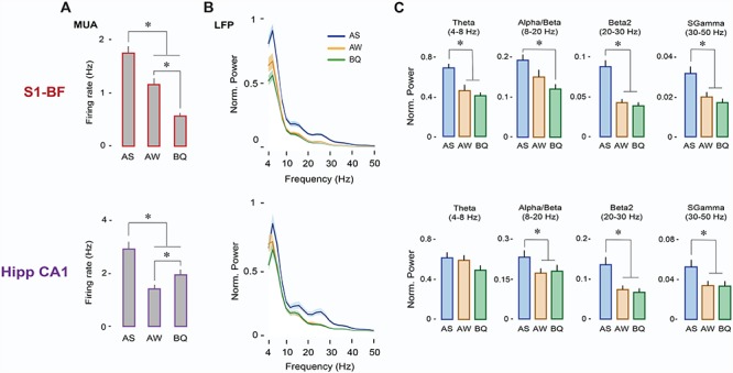
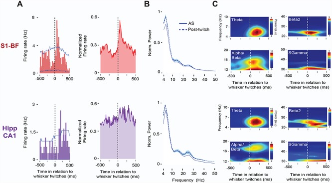
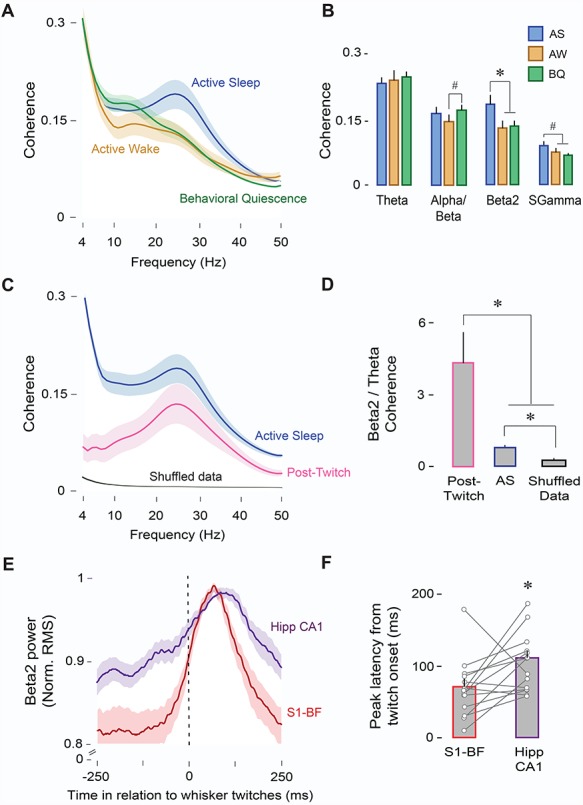
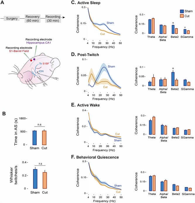
Active sleep (AS) provides a unique developmental context for synchronizing neural activity within and between cortical and subcortical structures. In week-old rats, sensory feedback from myoclonic twitches, the phasic motor activity that characterizes AS, promotes coherent theta oscillations (4-8 Hz) in the hippocampus and red nucleus, a midbrain motor structure. Sensory feedback from twitches also triggers rhythmic activity in sensorimotor cortex in the form of spindle bursts, which are brief oscillatory events composed of rhythmic components in the theta, alpha/beta (8-20 Hz), and beta2 (20-30 Hz) bands. Here we ask whether one or more of these spindle-burst components are communicated from sensorimotor cortex to hippocampus. By recording simultaneously from whisker barrel cortex and dorsal hippocampus in 8-day-old rats, we show that AS, but not other behavioral states, promotes cortico-hippocampal coherence specifically in the beta2 band. By cutting the infraorbital nerve to prevent the conveyance of sensory feedback from whisker twitches, cortical-hippocampal beta2 coherence during AS was substantially reduced. These results demonstrate the necessity of sensory input, particularly during AS, for coordinating rhythmic activity between these two developing forebrain structures.

Keywords: REM sleep; barrel cortex; development; functional connectivity; myoclonic twitching.


Figures

Figure 1
Figure 1
Neural activity in the cortico-hippocampal system across behavioral states at P8. (A) Illustration depicting simultaneous electrode placements in the S1 barrel field (S1-BF) and CA1 region of hippocampus and, at right, corresponding CO-stained coronal sections for a representative pup. DG: dentate gyrus; CPU: caudate-putamen. (B) Representative data in a P8 rat showing manually scored sleep and wake behavior (wake movements: horizontal red line; twitches: red ticks), raw LFP, time-frequency spectrograms, and unit activity (S1-BF, red traces; Hipp CA1, purple traces), and whisker and forelimb EMGs.
Figure 2
Figure 2
Neural activity in S1-BF and Hipp CA1 is expressed maximally during AS. (A) Mean (+SE) firing rates during AS, AW, and BQ for neurons in S1-BF (n = 117 units, top) and Hipp-CA1 (n = 90 units, bottom). * denotes significant difference from other states (P < 0.001). (B) Mean (±SE) LFP power spectra for S1-BF (top) and Hipp CA1 (bottom) during AS (blue), AW (orange), and BQ (green). (C) Mean LFP power (+SE) in S1-BF (n = 14 LFPs, top) and Hipp CA1 (n = 14 LFPs, bottom) across frequency bands and behavioral states (AS, AW, BQ). * denotes significant difference from AW and/or BQ (P < 0.005).
Figure 3
Figure 3
Whisker twitches during AS drive neural activity in S1-BF and Hipp CA1. (A) Left: Representative perievent histograms (10-ms bins) for spike activity in relation to whisker twitches in S1-BF (red, top) and Hipp CA1 (purple, bottom) in a P8 rat. Vertical dashed lines indicate twitch onset. Upper and lower acceptance bands (P < 0.05 for each band) are indicated by blue lines. Right: Same as at left but for normalized pooled data across those units in S1-BF (n = 32) and Hipp CA1 (n = 10) that exhibited significant twitch-related activity (P < 0.05). (B) Mean LFP power spectra (+SE) for AS (solid line) and post-twitch periods (500-ms window, dashed line) for S1-BF (n = 14, top) and Hipp CA1 (n = 14, bottom) from the same P8 rat. (C) Representative twitch-triggered time-frequency spectrograms for S1-BF (top) and Hipp CA1 (bottom) from the same P8 rat. Vertical dashed lines in spectrograms denote whisker twitch onset.
Figure 4
Figure 4
AS enables oscillatory coupling between S1-BF and Hipp CA1. (A) Mean LFP-LFP coherence spectra between S1-BF and Hipp CA1 (n = 14 pups, 14 LFP pairs) during AS (blue), AW (orange), and BQ (green). Shaded area indicates SE. (B) Mean (+ SE) LFP-LFP coherence values across frequency bands and behavioral states (AS, AW, and BQ). * denotes significant difference (P < 0.01). # denotes significant difference (P < 0.05). (C) Mean LFP-LFP coherence spectra between S1-BF and Hipp CA1 (n = 14 pups, 14 LFP pairs) during AS (blue), post-twitch periods (500-ms window, pink), and shuffled data (black). Shaded area indicates SE. (D) Mean beta2/theta ratios for coherence values during AS, post-twitch periods, and shuffled data. * denotes significant difference (P < 0.05). (E) Mean normalized twitch-triggered LFP power (beta2; 20–30 Hz; root mean square) pooled across subjects (14 pups, 14 LFPs) for S1-BF (red) and Hipp CA1 (purple). Shaded area indicates SE. Vertical line denotes whisker twitch onset. ( f ) Mean (+ SE) peak latency in (E) for S1-BF (red) and Hipp CA1 (purple). Vertical lines depict latency data from individual pups. * denotes significant difference (P < 0.05).
Figure 5
Figure 5
Transection of the ION decreases oscillatory coupling between S1-BF and Hipp CA1 in a state-dependent manner. (A) Experimental timeline and illustration depicting ION transections in a P8 rat. (B) Mean (+SE) time spent in AS (top) and whisker twitching rates (bottom) in the Sham (n = 6 pups, blue) and Cut (n = 6 pups, orange) experimental groups. n.s: not significant. (C) Left: Mean LFP-LFP coherence spectra between S1-BF and Hipp CA1 during AS in the Sham (n = 6 pups, 6 LFP pairs; blue line) and Cut (n = 6 pups, 6 LFP pairs; orange line) groups. Shaded area indicates SE. Right: Mean (+ SE) LFP-LFP coherence values across frequency bands in the Sham (blue) and Cut (orange) groups. (D) Same as in (C) but for post-twitch activity during AS (post-twitch window: 500 ms). (E) Same as in (C) but for AW. ( f ) Same as in (C) but for BQ.

References

    1. Abel T, Havekes R, Saletin JM, Walker MP. 2013. Sleep, plasticity and memory from molecules to whole-brain networks. Curr Biol. 23:R774–R788. - PMC - PubMed
    1. Akhmetshina D, Nasretdinov A, Zakharov A, Valeeva G, Khazipov R. 2016. The nature of the sensory input to the neonatal rat barrel cortex. J Neurosci. 36:9922–9932. - PMC - PubMed
    1. Alhbeck J, Song L, Chini M, Bitzenhofer SH, Hanganu-Opatz IL. 2018. Glutamatergic drive along the septo-temporal axis of hippocampus boosts prelimbic oscillations in the neontal mouse. elife. 7:e33158. - PMC - PubMed
    1. Amarasingham A, Harrison MT, Hatsopoulos NG, Geman S. 2012. Conditional modeling and the jitter method of spike re-sampling. J Neurophysiol. 107:517–531. - PMC - PubMed
    1. An S, Kilb W, Luhmann HJ. 2014. Sensory-evoked and spontaneous gamma and spindle bursts in neonatal rat motor cortex. J Neurosci. 34:10870–10883. - PMC - PubMed

Publication types