. 2020 Mar;21(3):1172-1180.
doi: 10.3892/mmr.2020.10918. Epub 2020 Jan 8.

Neural stem cell transplantation improves learning and memory by protecting cholinergic neurons and restoring synaptic impairment in an amyloid precursor protein/presenilin 1 transgenic mouse model of Alzheimer's disease

Affiliations

Neural stem cell transplantation improves learning and memory by protecting cholinergic neurons and restoring synaptic impairment in an amyloid precursor protein/presenilin 1 transgenic mouse model of Alzheimer's disease

Qing Zhu et al. Mol Med Rep. 2020 Mar.

Abstract

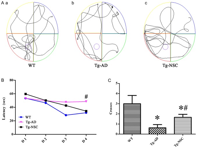
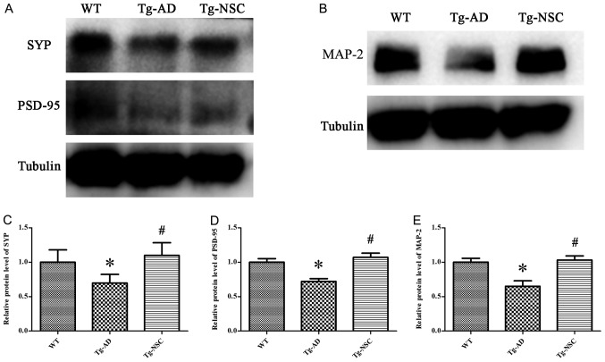
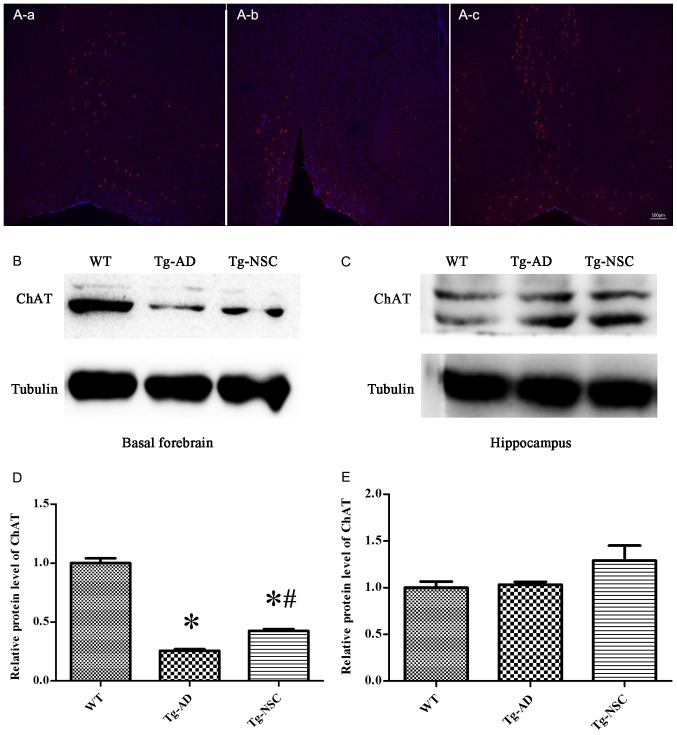
Alzheimer's disease (AD) is the most prevalent age‑related neurodegenerative disorder. It is featured by the progressive accumulation of β‑amyloid (Aβ) plaques and neurofibrillary tangles. This can eventually lead to a decrease of cholinergic neurons in the basal forebrain. Stem cell transplantation is an effective treatment for neurodegenerative diseases. Previous studies have revealed that different types of stem or progenitor cells can mitigate cognition impairment in different Alzheimer's disease mouse models. However, understanding the underlying mechanisms of neural stem cell (NSC) therapies for AD requires further investigation. In the present study, the effects and the underlying mechanisms of the treatment of AD by NSCs are reported. The latter were labelled with the enhanced green fluorescent protein (EGFP) prior to implantation into the bilateral hippocampus of an amyloid precursor protein (APP)/presenilin 1 (PS1) transgenic (Tg) mouse model of AD. It was observed that the number of basal forebrain cholinergic neurons was restored and the expression of choline acetyltransferase (ChAT) protein was increased. Moreover, the levels of synaptophysin (SYP), postsynaptic density protein 95 (PSD‑95) and microtubule‑associated protein (MAP‑2) were significantly increased in the hippocampus of NSC‑treated AD mice. Notably, spatial learning and memory were both improved after transplantation of NSCs. In conclusion, the present study revealed that NSC transplantation improved learning and memory functions in an AD mouse model. This treatment allowed repairing of basal forebrain cholinergic neurons and increased the expression of the cognition‑related proteins SYP, PSD‑95 and MAP‑2 in the hippocampus.

Keywords: alzheimer's disease; neural stem cell; cholinergic neurons; synapse; transplantation.


Figures

Figure 1.
Figure 1.
Mouse EGFP-NSCs have the ability of self-renewal and multi-differentiation in vitro. (A) Cultured NSCs were amassed as neurospheres and co-expressed EGFP (green) and Nestin (red). Subpart A-d is a merge image, shows DAPI (blue fluorescence, A-a) + EGFP (green fluorescence, A-b) + Nestin (red fluorescence, A-c). (B) NSCs differentiated into astrocyte, expression of GFAP-positive markers (red). Subpart B-c is a merge image, shows DAPI (blue fluorescence, B-a) + GFAP (red fluorescence, B-b). (C) NSCs differentiated into neurons, expression of Tuj1-positive markers (red). Subpart C-c is a merge image, shows DAPI (blue fluorescence, C-a) + Tuj1 (red fluorescence, C-b). Scale bar, 100 µm. EGFP, enhanced green fluorescent protein; NSCs, neural stem cells.
Figure 2.
Figure 2.
The fate of NSCs two weeks after transplantation to the hippocampus. (A) Engrafted cells migrated to ambient regions including the corpus callosum, adjacent cortex, and deeper in the hippocampus. A-a shows EGFP-NSCs migrated to the corpus callosum and the adjacent cortex (as indicated by the arrow). A-b shows the cells migrated in the hippocampus (the arrows show EGFP-NSCs). (B) Engrafted cells differentiated into GFAP + astrocytes (as indicated by the arrow); subpart B-a is the injection site, B-b is the partial magnification of the marked area in B-a. (C) Certain engrafted cells differentiated into DCX + neurons (as indicated by the arrow). NSCs, neural stem cells.
Figure 3.
Figure 3.
MWM test performances two weeks after NSC transplantation. (A) Representative movement traces from 3 groups on the day of exploration. The Tg-mice exhibited more dispersed paths, suggesting memory impairments; the Tg-NSC and WT groups had slightly concentrated swimming paths. A-a, A-b and A-c show the WT, Tg-AD and Tg-NSc groups, respectively. (B) Time latency in a navigation test. All animals exhibited improvements in finding the platform. (C) Crossing times of the original platform. *P<0.05 compared with WT group; #P<0.05 compared with Tg-AD group. MWM, Morris water-maze; NSC, neural stem cell; Tg, transgenic; AD, Alzheimer's disease; WT, wild-type.
Figure 4.
Figure 4.
Cognitive-related protein expression in the hippocampus. (A and B) The protein levels of SYP, PSD-95 and MAP-2 in the hippocampus after NSC transplantation. (C-E) Relative levels of SYP, PSD-95 and MAP-2 protein. *P<0.05 compared with WT group; #P<0.05 compared with Tg-AD group. SYP, synaptophysin; PSD-95, postsynaptic density protein 95; MAP-2, microtubule-associated protein; NSC, neural stem cell; WT, wild-type; Tg, transgenic; AD, Alzheimer's disease.
Figure 5.
Figure 5.
Cholinergic neurons in the basal forebrain. (A) Confocal display the ChAT-positive neurons, (A-a) WT group; (A-b) Tg-AD group; (A-c) Tg-NSC group. (B and C) ChAT protein expression in the basal forebrain and hippocampus. (D and E) Relative levels of ChAT protein in the basal forebrain and hippocampus. *P<0.05 compared with WT group; #P<0.05 compared with Tg-AD group. ChAT, choline acetyltransferase; WT, wild-type; Tg, transgenic; AD, Alzheimer's disease; NSC, neural stem cell.

References

    1. Anand R, Gill KD, Mahdi AA. Therapeutics of Alzheimer's disease: Past, present and future. Neuropharmacology. 2014;76:27–50. doi: 10.1016/j.neuropharm.2013.07.004. - DOI - PubMed
    1. Cattaneo A, Calissano P. Nerve growth factor and Alzheimer's disease: New facts for an old hypothesis. Mol Neurobiol. 2012;46:588–604. doi: 10.1007/s12035-012-8310-9. - DOI - PubMed
    1. Small G, Bullock R. Defining optimal treatment with cholinesterase inhibitors in Alzheimer's disease. Alzheimers Dement. 2011;7:177–184. doi: 10.1016/j.jalz.2010.03.016. - DOI - PubMed
    1. Teng YD. Functional multipotency of stem cells: Biological traits gleaned from neural progeny studies. Semin Cell Dev Biol. 2019;pii:S1084–S9521. (Epub ahead of print) - PubMed
    1. Li XY, Bao XJ, Wang RZ. Potential of neural stem cell-based therapies for Alzheimer's disease. J Neurosci Res. 2015;93:1313–1324. doi: 10.1002/jnr.23555. - DOI - PubMed

MeSH terms