. 2020 Feb;32(7):e1905914.
doi: 10.1002/adma.201905914. Epub 2020 Jan 10.

Ultrasound-Triggered Enzymatic Gelation

Affiliations

Ultrasound-Triggered Enzymatic Gelation

Valeria Nele et al. Adv Mater. 2020 Feb.

Abstract

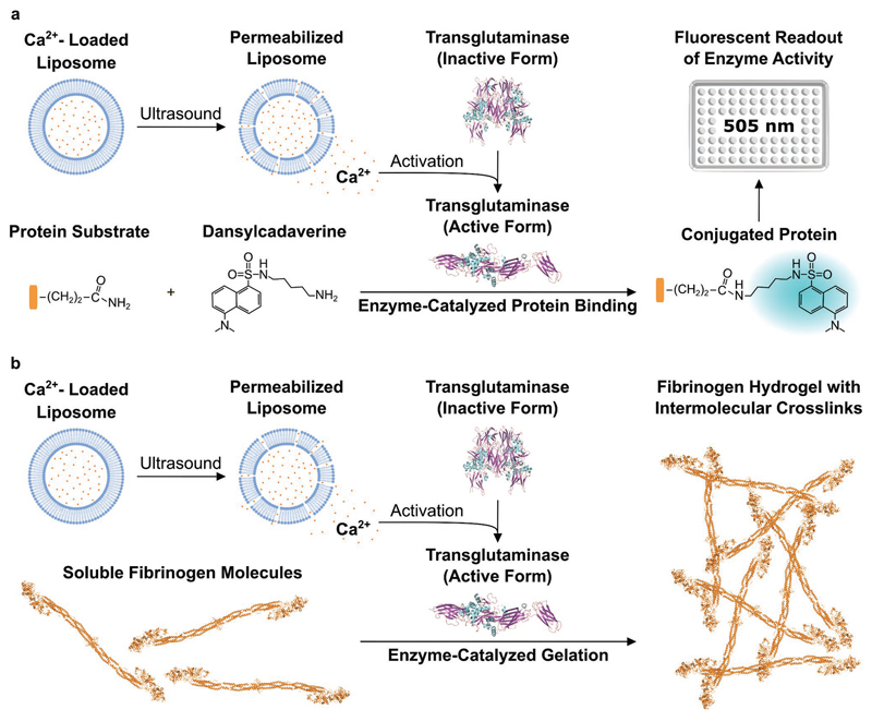
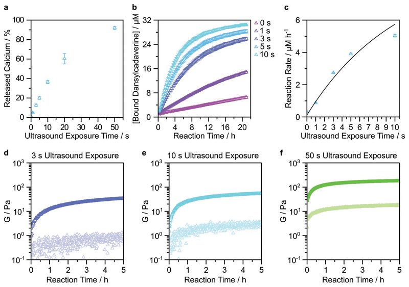
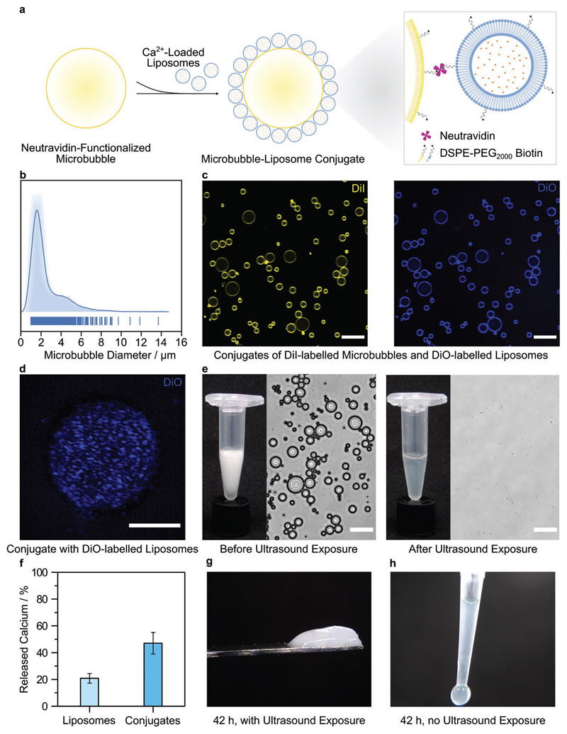
Hydrogels are formed using various triggers, including light irradiation, pH adjustment, heating, cooling, or chemical addition. Here, a new method for forming hydrogels is introduced: ultrasound-triggered enzymatic gelation. Specifically, ultrasound is used as a stimulus to liberate liposomal calcium ions, which then trigger the enzymatic activity of transglutaminase. The activated enzyme catalyzes the formation of fibrinogen hydrogels through covalent intermolecular crosslinking. The catalysis and gelation processes are monitored in real time and both the enzyme kinetics and final hydrogel properties are controlled by varying the initial ultrasound exposure time. This technology is extended to microbubble-liposome conjugates, which exhibit a stronger response to the applied acoustic field and are also used for ultrasound-triggered enzymatic hydrogelation. To the best of the knowledge, these results are the first instance in which ultrasound is used as a trigger for either enzyme catalysis or enzymatic hydrogelation. This approach is highly versatile and can be readily applied to different ion-dependent enzymes or gelation systems. Moreover, this work paves the way for the use of ultrasound as a remote trigger for in vivo hydrogelation.

Keywords: enzymes; hydrogels; liposomes; microbubbles; ultrasound.


Conflict of interest statement

Conflict of Interest

V.N., J.P.K.A., C.E.S., and M.M.S. are inventors on a patent application related to the technology described in this paper.

Figures

Figure 1
Figure 1. Schematic of ultrasound-triggered enzyme catalysis and hydrogelation.
a) Ultrasound is applied to calcium-loaded liposomes in order to liberate calcium ions and activate transglutaminase. The active transglutaminase then catalyzes isopeptide bond formation between a protein substrate and dansylcadaverine. This conjugation produces a shift in the maximum fluorescence emission wavelength of dansylcadaverine and an increase in fluorescence intensity at 505 nm. b) A similar ultrasound-triggered process is used to catalyze the crosslinking of soluble fibrinogen molecules. In this scenario, intermolecular crosslinking is used to generate fibrinogen hydrogels. The graphics for the structures of inactive and active transglutaminase and of fibrinogen were adapted from the Protein Data Bank (PDB) and processed with Visual Molecular Dynamics (VMD) software. Inactive transglutaminase PDB ID: 1kv3; active transglutaminase PDB ID: 2q3z; fibrinogen PDB ID: 3ghg.[30] The graphics for the 96-well plate were adapted from the Servier Medical Art website.
Figure 2
Figure 2. Ultrasound-triggered enzyme catalysis and hydrogelation using calcium-loaded liposomes.
a) Calcium-loaded liposomes were exposed to ultrasound for 0–50 s, with the released calcium quantified using an o-CPC assay. Data shown are the mean and standard deviation from four technical replicates across two batches of liposomes. b) The enzymatically catalyzed conversion of dansylcadaverine was measured after calcium-loaded liposomes were exposed to ultrasound for 0–10 s. Data shown are the mean and standard deviation from three technical replicates from one batch of liposomes. c) The rate of dansylcadaverine conversion was measured as a function of ultrasound exposure. Data shown are the mean and standard deviation, fitted to an asymptotic regression model (R2 = 0.95). d–f) The transglutaminase-catalyzed hydrogelation of fibrinogen was measured using timesweep rheology after the application of ultrasound for 3 s (d), 10 s (e), or 50 s (f). The final concentrations of fibrinogen and transglutaminase were 22.4 mg mL–1 and 5 × 10⁻⁶ m, respectively. Measurements were carried out at 1% strain and 1 rad s–1 at 25°C. G′ and G″ are shown with dark and light markers, respectively. Data shown for one technical repeat. For unexposed controls, see Figure S8 in the Supporting Information.
Figure 3
Figure 3. Ultrasound-triggered hydrogelation using calcium-loaded microbubble–liposome conjugates.
a) Schematic of the microbubble–liposome conjugation process, not to scale. b) Average-shifted histogram showing the diameter distribution of the microbubbles, as determined by image analysis of 890 microbubbles. The average diameter was measured as 2.5 ± 1.6 μm (mean ± standard deviation). c) Confocal fluorescence microscopy showing conjugates with fluorescence from both 1,1′-dioctadecyl-3,3,3′,3′-tetramethylindocarbocyanine (DiI)-labeled microbubbles (yellow) and 3,3′-dioctadecyloxacarbocyanine perchlorate (DiO)-labeled liposomes (blue). Scale bar: 20 μm. d) Super-resolution z-projection of DiO-labeled liposomes (blue) conjugated to the surface of a microbubble, obtained using structured illumination microscopy. Scale bar: 3 μm. e) Camera images and bright-field microscopy showing a turbid solution of intact microbubble–liposome conjugates before ultrasound exposure and a clear solution with no observable conjugates after ultrasound exposure (20 kHz, 25% duty cycle, 20% amplitude, 5 s). Scale bar: 20 μm. f) The percentage of calcium ions released from dose-matched liposomes and microbubble–liposome conjugates after ultrasound exposure (20 kHz, 25% duty cycle, 20% amplitude, 5 s) was measured using an o-CPC assay. Data are shown as mean and standard deviation of six technical replicates from the same batch of sonicated liposomes or microbubble–liposome conjugates. g) Image of a fibrinogen hydrogel formed after exposing calcium-loaded microbubble–liposome conjugates to ultrasound for 5 s. h) Image of an unexposed control, which remained liquid after 42 h.

References

    1. Seliktar D. Science. 2012;336:1124. - PubMed
    1. Annabi N, Tamayol A, Uquillas JA, Akbari M, Bertassoni LE, Cha C, Camci-Unal G, Dokmeci MR, Peppas NA, Khademhosseini A. Adv Mater. 2013;26:85. - PMC - PubMed
    1. Li J, Mooney DJ. Nat Rev Mater. 2016;1 16071. - PMC - PubMed
    1. Cianchetti M, Laschi C, Menciassi A, Dario P. Nat Rev Mater. 2018;3:143.
    1. Yuk H, Lu B, Zhao X. Chem Soc Rev. 2019;48:1642. - PubMed

LinkOut - more resources