. 2020 Jan 10;15(1):e0227386.
doi: 10.1371/journal.pone.0227386. eCollection 2020.

Immunopathogenesis of canine chronic ulcerative stomatitis

Affiliations

Immunopathogenesis of canine chronic ulcerative stomatitis

J G Anderson et al. PLoS One. .

Abstract

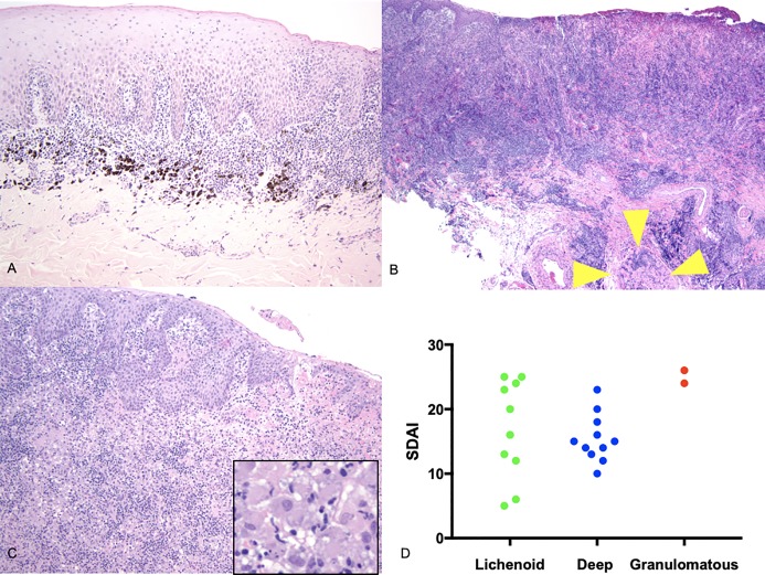
Canine Chronic Ulcerative Stomatitis is a spontaneously occurring inflammatory disease of the oral mucosa. An immune-mediated pathogenesis is suspected though not yet proven. We have recently reported on the clinical and histologic features, and identification of select leukocyte cell populations within the lesion. A clinical and histologic similarity to oral lichen planus of people was proposed. In the present study, these initial observations are extended by examining lesions from 24 dogs with clinical evidence of chronic ulcerative stomatitis. Because dogs with chronic ulcerative stomatitis often have concurrent periodontal disease, we wondered if dental plaque/biofilm may be a common instigator of inflammation in both lesions. We hypothesized that dogs with chronic ulcerative stomatitis would exhibit a spectrum of pathologic changes and phenotype of infiltrating leukocytes that would inform lesion pathogenesis and that these changes would differ from inflammatory phenotypes in periodontitis. Previously we identified chronic ulcerative stomatitis lesions to be rich in FoxP3+ and IL17+ cells. As such, we suspect that these leukocytes play an important role in lesion pathogenesis. The current study confirms the presence of moderate to large numbers of FoxP3+ T cells and IL17+ cells in all ulcerative stomatitis lesions using confocal immunofluorescence. Interestingly, the majority of IL17+ cells were determined to be non-T cells and IL17+ cell frequencies were negatively correlated with severity on the clinical scoring system. Three histologic subtypes of ulcerative stomatitis were determined; lichenoid, deep stomatitis and granulomatous. Periodontitis lesions, like stomatitis lesions, were B cell and plasma cell rich, but otherwise differed from the stomatitis lesions. Direct immunofluorescence results did not support an autoantibody-mediated autoimmune disease process. This investigation contributes to the body of literature regarding leukocyte involvement in canine idiopathic inflammatory disease pathogenesis.


Conflict of interest statement

The authors have declared that no competing interests exist.

Figures

Fig 1
Fig 1. CCUS sample distribution.
Fig 2
Fig 2. Histologic subtypes of CCUS.
A-Lichenoid stomatitis-CASE G 100x; B-Deep stomatitis-CASE V 40x (yellow 445 arrowheads-inflamed skeletal muscle); C-Granulomatous stomatitis-CASE U 100x (inset-446 histiocytic inflammation, 200x) D-Clinical score (CUSDAI) versus CCUS histologic subtype.
Fig 3
Fig 3. Leukocyte subsets- CCUS versus periodontitis controls.
Fig 4
Fig 4. T helper subset distribution in CCUS lesions.
A -Normal canine oral mucosa illustrates very few CD3+ T cells (white arrows) that are distributed at the mucosal–submucosal interface (A, stitched 20x tile image); B -Co-labeling of CD3 (green) and FoxP3 (red) reveals abundant FoxP3+ T cells that are noted with the submucosa of CCUS lesions; C-IL17 (red) and CD3(green) co-staining reveals marked infiltration of sub-epithelial and mucosal tissues with CD3+T cells and IL17 producing cells. Note that most of the IL17 producing cells (i.e. red) are not double labeled with CD3 (i.e. green) indicating that IL17 producing cells are primarily non-T cells. D—Close up (630x) image illustrates a single positive, IL17, cell (white arrow), a single positive CD3 T cell (white arrow head) and a group of double positive (i.e. IL17/CD3) IL17 producing T cells (asterisk). E—No significant differences were identified in the frequencies of CD3+/FoxP3+ cells, CD3+ cells, IL17+ cells and DP positive cells between the three histologic variants.
Fig 5
Fig 5. T cell and IL17 producing cell subsets in CCUS histologic types.
A-There was no statistically significant difference in the number of infiltrating T cells; B-IL17+ cells; C and D-percent double positive (IL17+/CD3+); and E-Treg cells. F-A negative correlation was found between the numbers of IL17+ cells per 20x field and the stomatitis disease activity index.
Fig 6
Fig 6
A-C Direct Immunofluorescence in health and CCUS, IgG. A-DIF–tissue-bound IgG–using gingival mucosa from a healthy dog (A) or dog with CUS (B, C); B-A band of IgG positive cells in the lamina propria below the mucosal epithelium (green) with scattered IgG positive cells in deeper layers of the lamina propria; C-In rare dogs, in addition to the IgG positive mononuclear cells in the lamina propria, a faint and patchy basement membrane zone deposit could be appreciated. Blue nuclear staining = DAPI.
Fig 7
Fig 7
A-D Direct Immunofluorescence in health and CCUS, IgM and IgA. DIF–tissue-bound IgM (A-B) and IgA(C-D)–using gingival mucosa from a healthy dog (A, C) or dog with CUS (B, D). Positively stained mononuclear cells in the lamina propria and within mucosal epithelium depicted in green. Blue nuclear staining = DAPI.

References

    1. Anderson JG, Peralta S, Kol A, Kass PH, Murphy B. Clinical and Histopathologic Characterization of Canine Chronic Ulcerative Stomatitis. Vet Pathol. 2017;54(3):511–9. Epub 2017/01/24. 10.1177/0300985816688754 . - DOI - PubMed
    1. Tesmer LA, Lundy SK, Sarkar S, Fox DA. Th17 cells in human disease. Immunol Rev. 2008;223:87–113. Epub 2008/07/11. 10.1111/j.1600-065X.2008.00628.x - DOI - PMC - PubMed
    1. Gaffen SL, Hajishengallis G. A new inflammatory cytokine on the block: re-thinking periodontal disease and the Th1/Th2 paradigm in the context of Th17 cells and IL-17. J Dent Res. 2008;87(9):817–28. Epub 2008/08/23. 10.1177/154405910808700908 - DOI - PMC - PubMed
    1. Mailer RKW. Alternative Splicing of FOXP3-Virtue and Vice. Front Immunol. 2018;9:530 Epub 2018/03/30. 10.3389/fimmu.2018.00530 - DOI - PMC - PubMed
    1. Chellappa S, Hugenschmidt H, Hagness M, Line PD, Labori KJ, Wiedswang G, et al. Regulatory T cells that co-express RORgammat and FOXP3 are pro-inflammatory and immunosuppressive and expand in human pancreatic cancer. Oncoimmunology. 2016;5(4):e1102828 Epub 2016/05/04. 10.1080/2162402X.2015.1102828 - DOI - PMC - PubMed

Publication types

MeSH terms