Review
. 2020 Dec 1;1862(12):183175.
doi: 10.1016/j.bbamem.2020.183175. Epub 2020 Jan 7.

VGLUT substrates and inhibitors: A computational viewpoint

Affiliations
Review

VGLUT substrates and inhibitors: A computational viewpoint

Charles M Thompson et al. Biochim Biophys Acta Biomembr. .

Abstract

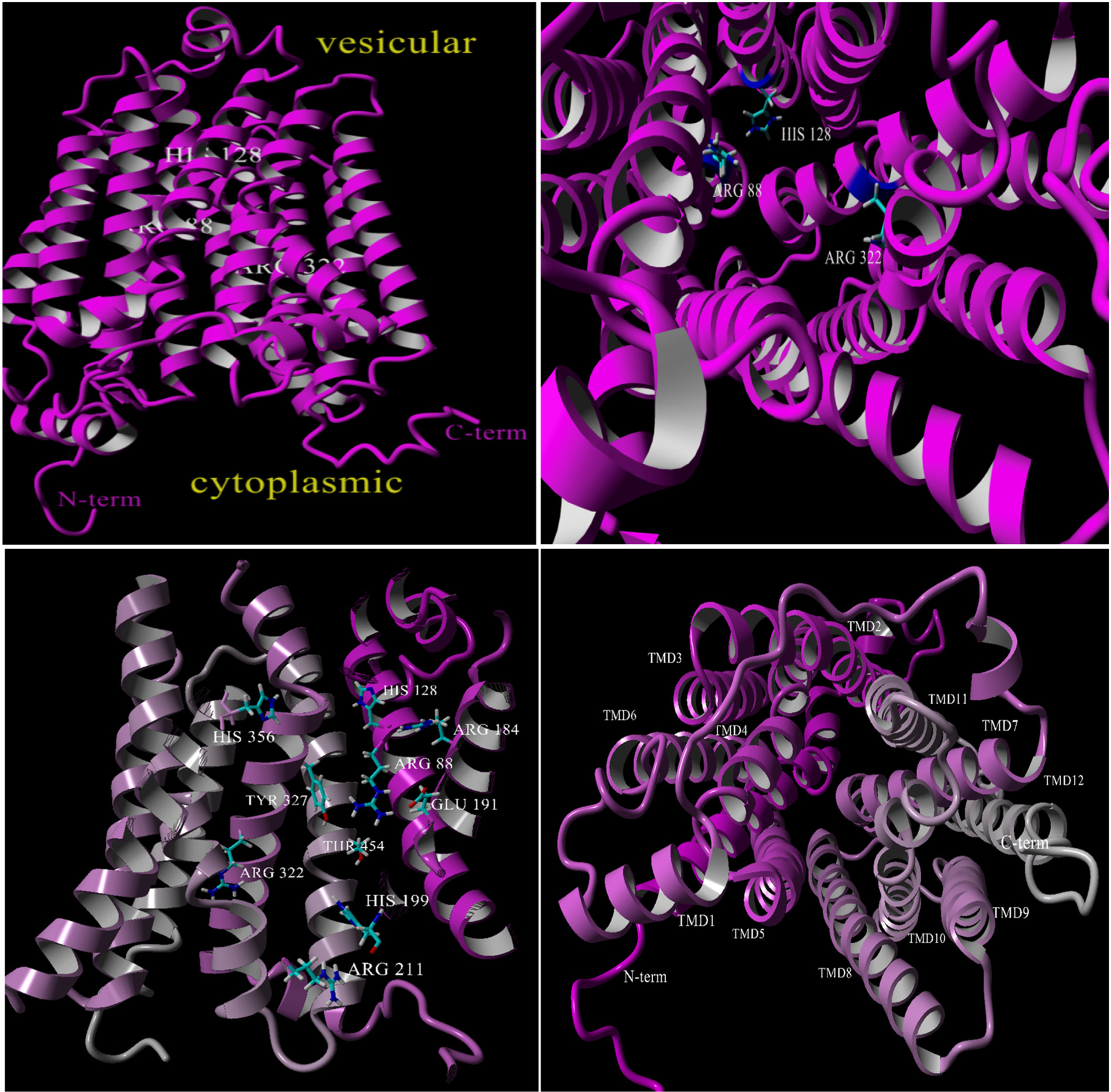
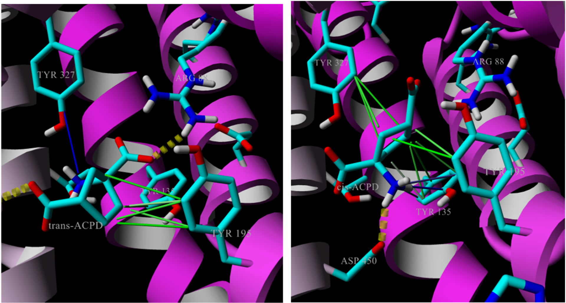
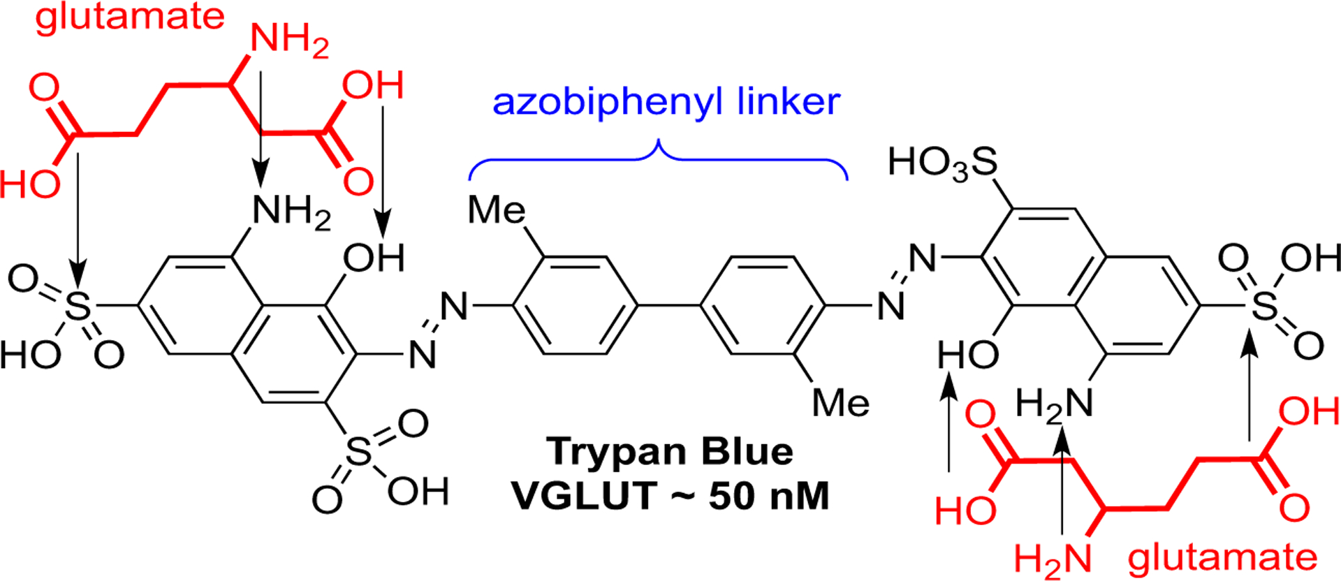
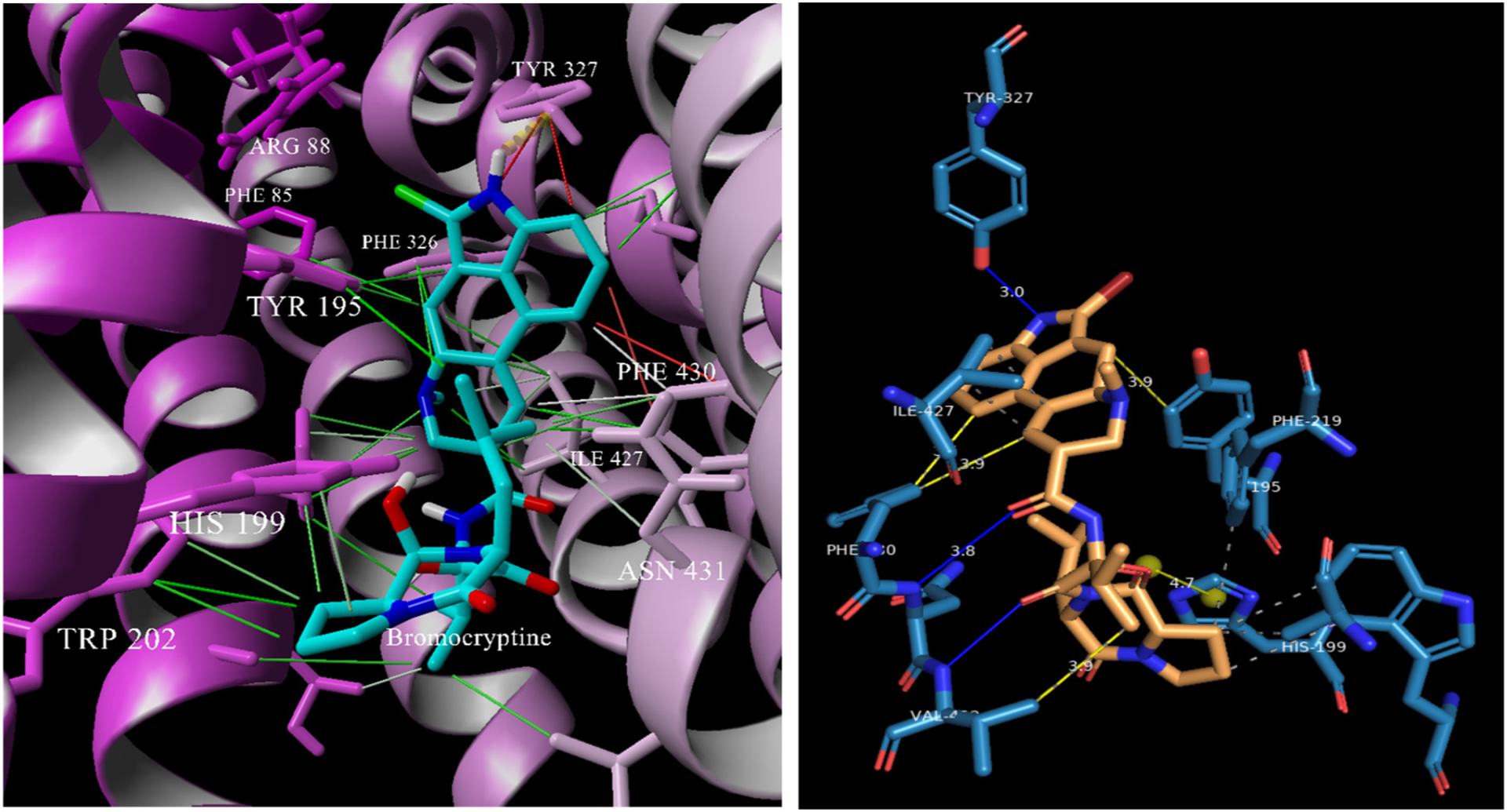
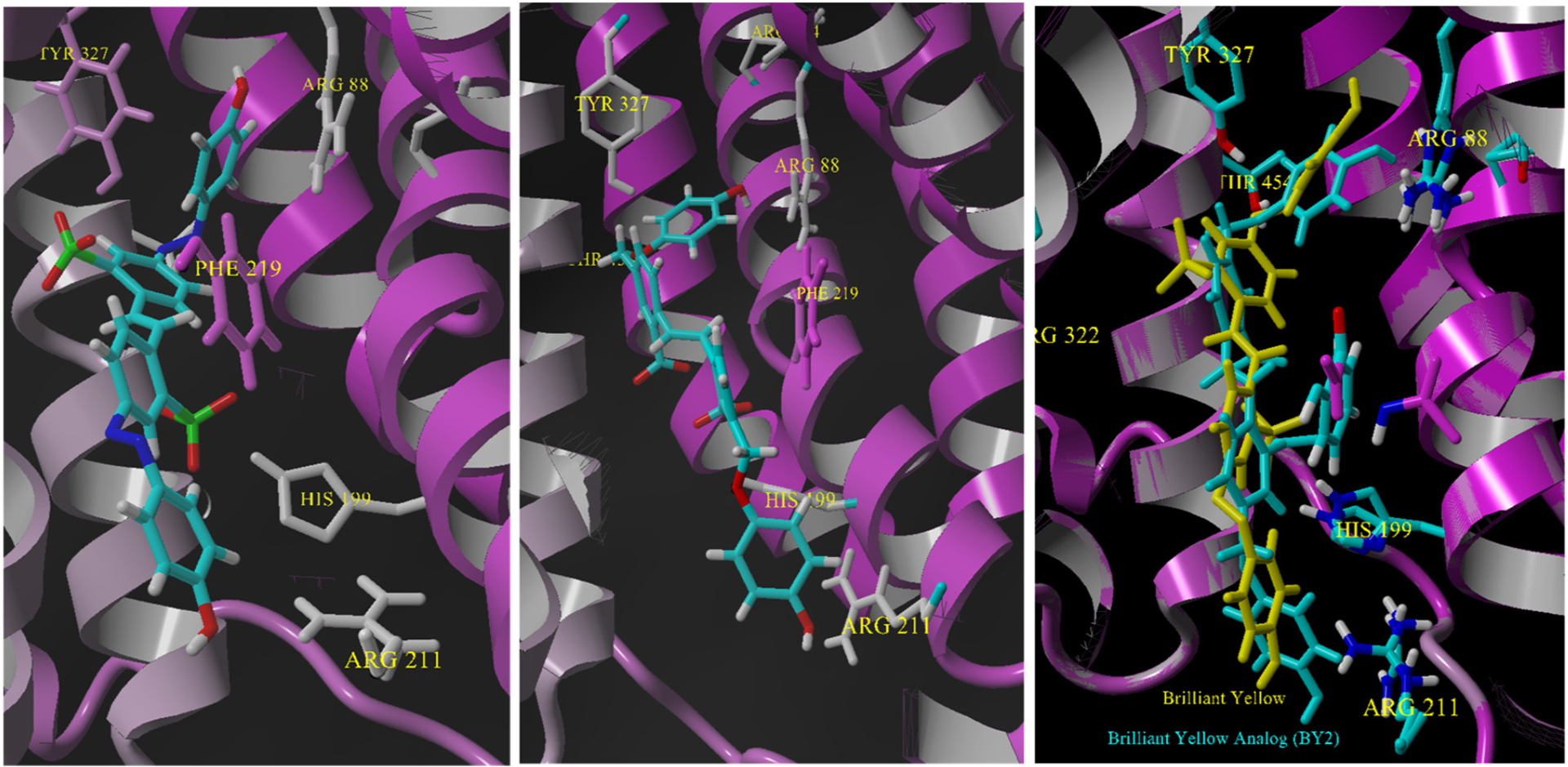
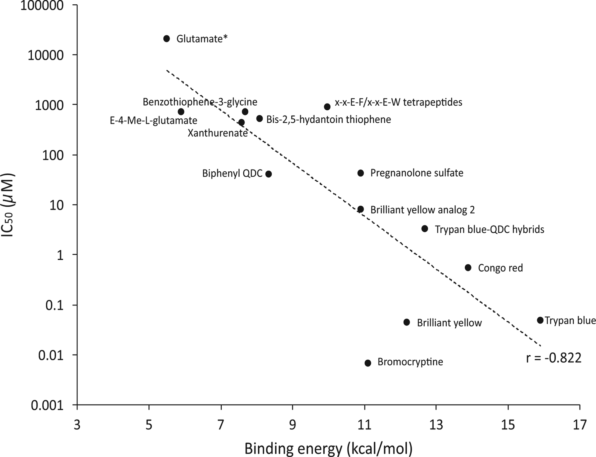
The vesicular glutamate transporters (VGLUTs) bind and move glutamate (Glu) from the cytosol into the lumen of synaptic vesicles using a H+-electrochemical gradient (ΔpH and Δψ) generated by the vesicular H+-ATPase. VGLUTs show very low Glu binding and to date, no three-dimensional structure has been elucidated. Prior studies have attempted to identify the key residues involved in binding VGLUT substrates and inhibitors using homology models and docking experiments. Recently, the inward and outward oriented crystal structures of d-galactonate transporter (DgoT) emerged as possible structure templates for VGLUT. In this review, a new homology model for VGLUT2 based on DgoT has been developed and used to conduct docking experiments to identify and differentiate residues and binding orientations involved in ligand interactions. This review describes small molecule-ligand interactions including docking using a VGLUT2 homology model derived from DgoT.

Keywords: Docking; Glutamate; Homology models; Inhibitor; Substrate; Vesicular glutamate transporter.


Conflict of interest statement

Declaration of competing interest The authors declare that they have no known competing financial interests or personal relationships that could have appeared to influence the work reported in this paper.

Figures

Fig. 1.
Fig. 1.
Left: Glu release, recycling and receptor action. Right: Glu transport into vesicles and inhibitor blocking Glu uptake.
Fig. 2.
Fig. 2.
Top left: VGLUT2 homology model based on DgoT (magenta) as a side view. Top right: VGLUT2 homology model view from cytoplasmic side showing the position of R88, H128 and R322 for clarity. Bottom left: VGLUT2 (cutaway) showing previously identified residues and as included in docking studies. Bottom right: assignment of the VGLUT2-DgoT homology model transmembrane domains (TMDs).
Fig. 3.
Fig. 3.
Structures of substrates and non-substrates.
Fig. 4.
Fig. 4.
L-Glutamate (left) and D-glutamate (right) docked into VGLUT2 homology model showing nearby interactions.
Fig. 5.
Fig. 5.
Trans-ACPD (left) and cis-ACPD (right) docked into VGLUT2 homology model showing nearby interactions. Trans-ACPD orients similarly to L-Glu with charged amino toward Y327 and carboxylate interaction with R88 whereas the amino of cis-ACPD faces toward Y135/D450. S323 and Q319 not shown.
Fig. 6.
Fig. 6.
Left: Docking image of E-4-methylglutamate binding to VGLUT2. Right: Key interactions between E-4-methyl glutamate and nearby residues along with distances. Binding to R88/Y327 is similar to that found for the natural substrate L-Glu.
Fig. 7.
Fig. 7.
Left: Overlay of the substrates glutamate, E4MeGlu and trans-ACPD in VGLUT2. Right: Overlay of L-aspartate, D-glutamate, and cis-ACPD non-substrate binding modes in VGLUT2. Cation-pi interactions in blue; hydrophobic interactions in green; gold dotted cylinders are salt bridges and hydrogen bonds.
Fig. 8.
Fig. 8.
Structures of representative VGLUT inhibitors.
Fig. 9.
Fig. 9.
Overlay of glutamates onto matching functional groups of Trypan Blue.
Fig. 10.
Fig. 10.
Left: Bromocriptine docked in VGLUT2 showing an array of interactions with residues identified as binding the substrate Glu. Right: A 2D ligand map showing the interactions and distances in the absence of protein.
Fig. 11.
Fig. 11.
Left: Docking of TB in VGLUT2 showing the span length of the inhibitor in a pose entering from the cytosolic side. Center: TB in a transparent VDW surface docked in VGLUT2. Right: two-dimensional schematic of TB (in orange) without protein showing the residue interactions (salt-bridges or H-bonding shown as cyan bars).
Fig. 12.
Fig. 12.
Left: Docking of CR in VGLUT2 homology model. Right: two-dimensional ligand-protein interaction map (PLIP; [69]) showing the key residues that interact.
Fig. 13.
Fig. 13.
Left: docking of Brilliant Yellow. Center: Docking of Brilliant Yellow analog 2 (BYA2). Right: an overlay of the two structures in VGLUT2 using the Mustang program [42] in YASARA.
Fig. 14.
Fig. 14.
Left: xanthurenic acid docked in VGLUT2 near the substrate residues. Right: The key residues that bind xanthurenic acid without protein.
Fig. 15.
Fig. 15.
Left: Pose of 6-(4-biphenyl)QDC docked in VGLUT in which the 2-carboxylic acid faces R88 and Y327. Right: Ligand-interaction map showing the residues and distances for binding.
Fig. 16.
Fig. 16.
Left: Rose Bengal lactone (cyan; closed) and Rose Bengal carboxylic acid (yellow; open) docked near the cytosolic entry regions of the VGLUT2 model. Right: Expansion of the docking pose showing interactions of RB with H199, P196, M296 and L293.
Fig. 17.
Fig. 17.
Structure of biliverdin.
Fig. 18.
Fig. 18.
Plot of IC50 values of competitive inhibitors versus their calculated binding energies. Position of the substrate glutamate shown but not included in the correlation. Rose Bengal was not included as a non-competitive inhibitor. (For interpretation of the references to colour in this figure legend, the reader is referred to the web version of this article.)
Scheme 1.
Scheme 1.
LEFT: mVGLUT2 (top line) and DGOT (lower line) sequence similarity alignment using T-Coffee (score 891/1000) [40]. Legend: inside or cytoplasmic domains = yellow; outside or lumenal domains = blue; helical membrane regions = pink. Single, fully conserved residue = asterisk(*); conservation between groups of strongly similar properties = colon(:); conservation between groups of weakly similar properties = period(.).

References

    1. Zhang FX, Ge SN, Dong YL, Shi J, Feng YP, Li Y, Li YQ, Li JL, Vesicular glutamate transporter isoforms: the essential players in the somatosensory systems, Prog. Neurobiol 171 (2018) 72–89. - PubMed
    1. Omote H, Miyaji T, Juge N, Moriyama Y, Vesicular neurotransmitter transporter: bioenergetics and regulation of glutamate transport, Biochemistry 50 (2011) 5558–5565. - PubMed
    1. El Mestikawy S, Wallen-Mackenzie A, Fortin GM, Descarries L, Trudeau LE, From glutamate co-release to vesicular synergy: vesicular glutamate transporters, Nat. Rev. Neurosci 12 (2011) 204–216. - PubMed
    1. Liguz-Lecznar M, Skangiel-Kramska J, Vesicular glutamate transporters (VGLUTs): the three musketeers of glutamatergic system, Acta Neurobiol. Exp. (Wars) 67 (2007) 207–218. - PubMed
    1. Takamori S, VGLUTs: ‘exciting’ times for glutamatergic research? Neurosci. Res 55 (2006) 343–351. - PubMed

Publication types

MeSH terms

Substances

LinkOut - more resources