Review
. 2020 Mar:186:111207.
doi: 10.1016/j.mad.2020.111207. Epub 2020 Jan 7.

Mitophagy and DNA damage signaling in human aging

Affiliations
Review

Mitophagy and DNA damage signaling in human aging

Mansi Babbar et al. Mech Ageing Dev. 2020 Mar.

Abstract

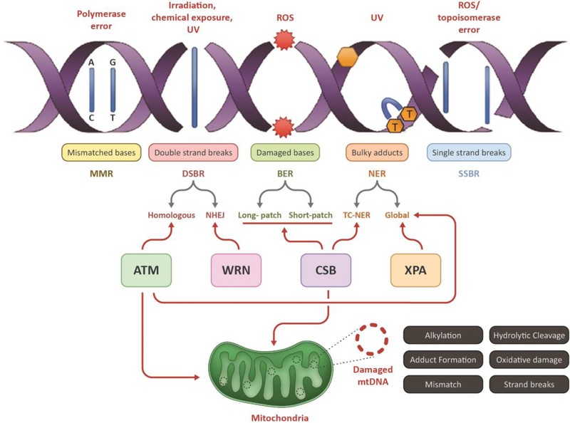
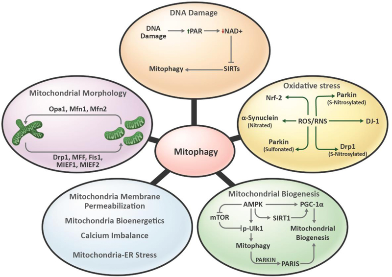
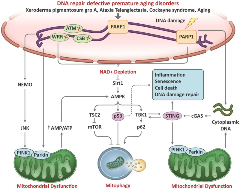
Aging is associated with multiple human pathologies. In the past few years mitochondrial homeostasis has been well correlated with age-related disorders and longevity. Mitochondrial homeostasis involves generation, biogenesis and removal of dysfunctional mitochondria via mitophagy. Mitophagy is regulated by various mitochondrial and extra-mitochondrial factors including morphology, oxidative stress and DNA damage. For decades, DNA damage and inefficient DNA repair have been considered as major determinants for age-related disorders. Although defects in DNA damage recognition and repair and mitophagy are well documented to be major factors in age-associated diseases, interactivity between these is poorly understood. Mitophagy efficiency decreases with age leading to accumulation of dysfunctional mitochondria enhancing the severity of age-related disorders including neurodegenerative diseases, inflammatory diseases, cancer, diabetes and many more. Therefore, mitophagy is being targeted for intervention in age-associated disorders. NAD+ supplementation has emerged as one intervention to target both defective DNA repair and mitophagy. In this review, we discuss the molecular signaling pathways involved in regulation of DNA damage and repair and of mitophagy, and we highlight the opportunities for clinical interventions targeting these processes to improve the quality of life during aging.

Keywords: Aging; DNA damage; DNA repair; Mitochondria; Mitophagy.


Figures

Figure 1:
Figure 1:. Schematic representation of different types of nuclear DNA damages and its associated repair pathways:
This figure depicts the major type of nuclear DNAdamages (by stressors) and repair pathways involved in eukaryotic cells including damage recognition, lesion excision;, processing, synthesis of new bases and ligation (left panel). The major repair processes include the mismatch DNA repair (MMR), double strand break repair (DSBR), base excision repair (BER), nucleotide excision repair (NER) and single-strand breaks repair (SSBR). Key players of each step include damage recognition by proteins like glycosylases, lesion excision by nucleases, synthesis of new bases by polymerases and ligation of nicks by ligases. Double strand breaks (DSB) on DNA caused by stressors like UV and ionizing radiation are specifically recognized and repaired by the players of the DSB repair (DSBR) pathway, that includes either non-homologous end joining (NHEJ) or homologous recombination (HR), depending on the cell-type and stages of cell cycle involved.
Figure 2:
Figure 2:. Schematic representation of autophagy, mitophagy and proteins involved.
(A) Autophagy involves removal of unnecessary or dysfunctional cellular components like proteins, lipids or organelles. Broadly, autophagy is classified as 1. Macroautophagy- It is the most extensively studied form of autophagy for bulk degradation of cellular components. It involves formation of phagophore, a double membrane structure around the targeted components. Autophagosome formed around the target merges with the lysosome leading to formation of autophagolysosomes to enable degradation of engulfed contents. Micro autophagy or chaperone-mediated autophagy involves direct uptake of targeted contents into lysosomes via lysosomal invagination or binding of chaperon tagged proteins to lysosomal-associated membrane protein LAMP-2A. (B) and (C) Mitophagy is a type of macroautophagy targeting mitochondria specifically. (C) Mitophagy receptors can tag mitochondria for phagophore formation in ubiquitin-dependent, LIR-motif protein-dependent and lipid-mediated pathway. The ubiquitin-dependent pathway involves ubiquitination of OMM proteins to form poly ubiquitin chains with the help of E3 ligases and deubiquitinating enzymes. The polyubiquitin chains are detected by NDP52, OPTN, NBR1 and p62 which contains both UBD and LIR motifs. These proteins aid in interaction of ubiquitinated mitochondria to LC3 containing phagophore. LIR-motif protein-dependent pathway involves regulation of expression and modification of LIR-motif containing OMM proteins. Proteins like NIX, BNIP3, FUNDC1 helps with the interaction of targeted-mitochondria to LC3-containing phagophore. Lastly, lipid-mediated mitophagy involves accumulation of LC3 interacting lipids including cardiolipin and ceramide on OMM, tagging mitochondria for phagophore interaction.
Figure 3:
Figure 3:. Mitochondrial factors regulating mitophagy.
Mitochondrial homeostasis plays an important role in regulation of mitophagy. DNA damage, including mtDNA damage, causes increase in PARP activity and thereby reduces cellular NAD+ pools. This further reduces the activity of NAD+-dependent proteins like SIRT1 and thereby mitophagy. Oxidative stress modulates activities of various mitophagy-related proteins. For instance, RNS regulates nitrosylation of Parkin, Drp1, α-synuclein and mitophagy. Mitochondrial biogenesis helps in maintaining mitochondrial content along with mitophagy. Protein like AMPK coordinates the biogenesis and mitophagy balance which is important for maintaining mitochondrial homeostasis. Mitochondrial frgmentation regulated by fission proteins including Drp1, MFF, Fis1, MIEF1and MIEF2 helps in segregation of defective mitochondria from healthy mitochondria and aids in mitophagy. Mitochondrial biogenesis, energetics, calcium imbalance and mitochondrial-ER stress regulates mitophagy.
Figure 4:
Figure 4:. Schematic representation of the cross talk between DNA damage to mitophagy.
Compromised DNA repair downregulates mitophagy in aging-related disorders such as Ataxia telangiectasia, Werner syndrome, Cockayne syndrome, and Alzheimer’s disease. When DNA breaks occur, the DNA break sensor PARP1 detects break sites and initiates DNA repair signaling through generation of PAR, a process called PARylation, which consumes NAD+. A decrease in NAD+ and an increase upon DNA damage, nuclear SIRT1 can be activated to facilitate DNA repair. After lethal levels of nuclear DNA damage, SIRT1 is inhibited by the DNA damage response, leading to p53 acetylation and cell death. Loss of SIRT1 activity decreases the stimulation of mitophagy through peroxisome proliferator-activated receptor-γ co-activator 1α (PGC1α)- and AMP-activated kinase (AMPK)-dependent pathways. SIRT1 regulates mitochondrial biogenesis and mitophagy through deacetylation of PGC1α, and it also interacts with AMPK to regulate mitophagy. In response to DNA damage, ATM is auto phosphorylated within an MRN multiprotein complex that binds DSBs. Activated ATM initiates a pathway that results in activation of AMPK and activates the mTORC1 inhibitor protein tuberous sclerosis complex 2 (TSC2), thereby further activating autophagy. AMPK also positively regulates PGC1α activity, which stimulates mitochondrial biogenesis. AMPK phosphorylates and activates p53, which propagates pro-apoptotic signals. p53 also has been well characterized in the response to genotoxic stimuli, in which it transcriptionally activates pro-apoptotic proteins such as BAX and p21 while simultaneously inhibiting the ULK1-containing autophagy-initiating complex. In addition, p53 activation may lead to decreased expression of parkin and decreased activation of parkin–PINK1-mediated mitophagy. Damaged DNAfrom nuclear or cytoplasm release into the cytosol. Aberrant localization of DNA in the cytosol activates the cGAS-cGAMP-STING pathway, leading to enhanced inflammatory gene expression. Activation of the cGAS-cGAMP-STING pathway may stimulate autophagy/mitophagy via a cGAS/beclin-1 interaction–dependent mechanism. The cGAS-cGAMP-STING pathway also promotes autophagy/mitophagy by TBK1-dependent phosphorylation and activation of receptors OPTN and p62 (SQSTM). PINK/Parkin-induced mitophagy inhibits cGAS-cGAMP-STING signaling and innate immunity by mitophagy-mediated mtDNAclearance.
Figure 5:
Figure 5:. Human pathologies and the organs affected by mitophagy.
Deregulation in mitophagy is linked with various diseases. Mitophagy-associated diseases may affect single organs (like cardiac myopathy, pulmonary disease, fatty liver disease, muscle atrophy) or multiple organs (like cancer, aging, diabetes, inflammatory disease).

References

    1. Pitt JN and Kaeberlein M, Why is aging conserved and what can we do about it? PLoS Biol, 2015. 13(4): p. e1002131. - PMC - PubMed
    1. Cohen AA, Aging across the tree of life: The importance of a comparative perspective for the use of animal models in aging. Biochim Biophys Acta Mol Basis Dis, 2018. 1864(9 Pt A): p. 26802689. - PubMed
    1. Gensler HL and Bernstein H, DNA damage as the primary cause of aging. Q Rev Biol, 1981. 56(3): p. 279–303. - PubMed
    1. Stewart JB and Chinnery PF, The dynamics of mitochondrial DNA heteroplasmy: implications for human health and disease. Nat Rev Genet, 2015. 16(9): p. 530–42. - PubMed
    1. Ashrafi G. and Schwarz TL, The pathways of mitophagy for quality control and clearance of mitochondria. Cell Death Differ, 2013. 20(1): p. 31–42. - PMC - PubMed

Publication types