. 2020 Apr:133:115224.
doi: 10.1016/j.bone.2020.115224. Epub 2020 Jan 8.

Early-onset Paget's disease of bone in a Mexican family caused by a novel tandem duplication (77dup27) in TNFRSF11A that encodes RANK

Affiliations

Early-onset Paget's disease of bone in a Mexican family caused by a novel tandem duplication (77dup27) in TNFRSF11A that encodes RANK

Sean J Iwamoto et al. Bone. 2020 Apr.

Abstract

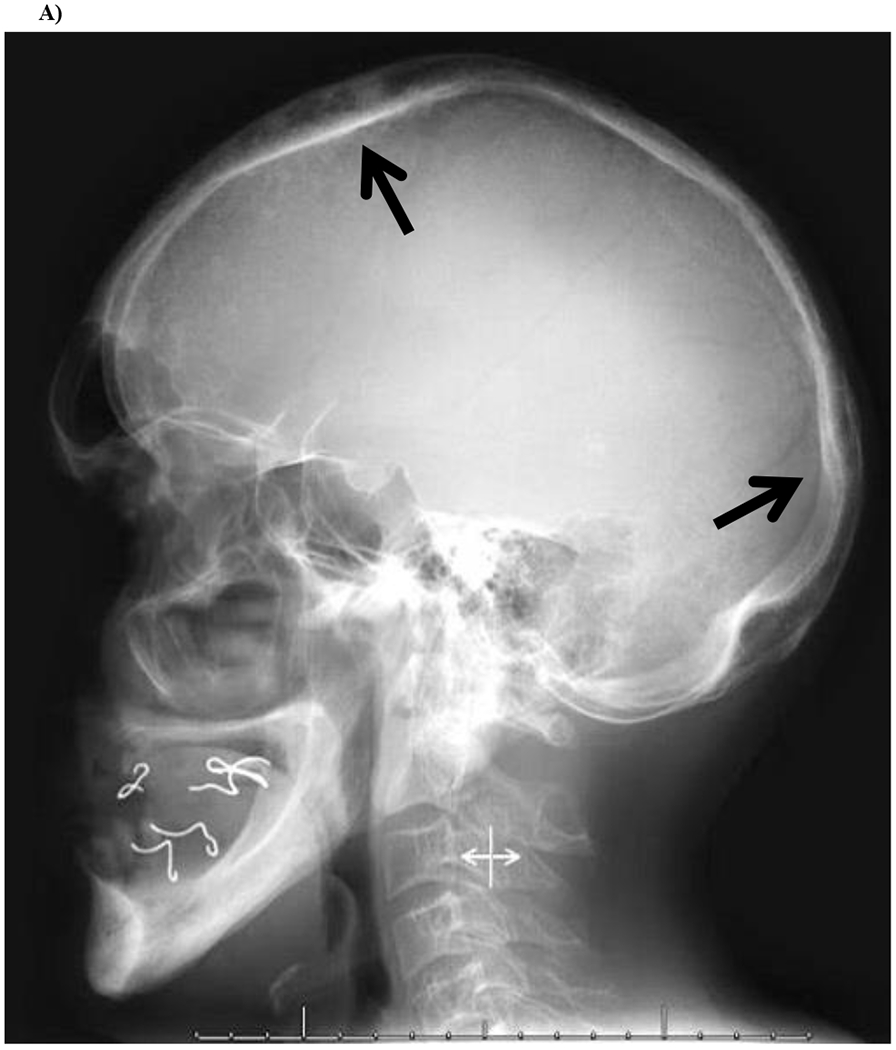
Four heterozygous in-frame tandem duplications of different lengths in TNFRSF11A, the gene that encodes receptor activator of nuclear factor κB (RANK), constitutively activate RANK and lead to high turnover skeletal disease. Each duplication elongates the signal peptide of RANK. The 18-base pair (bp) duplication at position 84 (84dup18) causes familial expansile osteolysis (FEO), the 15-bp duplication at position 84 (84dup15) causes expansile skeletal hyperphosphatasia (ESH), the 12-bp duplication at position 90 (90dup12) causes panostotic expansile bone disease (PEBD), and the 27-bp duplication causes early-onset Paget's disease of bone (PDB2). The severity of the associated skeletal disease seems inversely related to the duplication's length. Additional 15- and 18-bp duplications of TNFRSF11A fit this pattern. Herein, we delineate the skeletal disease of a middle-aged man of Mexican descent who we found to harbor a novel 27-bp tandem duplication at position 77 (77dup27) of TNFRSF11A. His disorder shares features, particularly hand involvement, with the single Japanese (75dup27) and Chinese (78dup27) kindreds with PDB2 (PDB2Jpn and PDB2Chn, respectively). However, his distinct hearing loss developed later in adulthood compared to the other 27-bp families. He reported no morbidities during childhood, but in his late 20s developed unexplained tooth loss, low-trauma fractures, post-operative hypercalcemia, and painless enlargement of his fingers. Biochemical studies showed elevated serum alkaline phosphatase (ALP), bone-specific ALP, C-telopeptide, and osteocalcin consistent with rapid bone remodeling. Radiologic imaging revealed remarkably lucent bones with vertebral compression fractures, calvarial lucencies, and thinned long bone cortices. DXA showed extremely low bone mineral density. His disorder genetically and phenotypically fits best with PDB2 and can be called PDB2Mex.

Keywords: Bone markers; Bone turnover; Deafness; Early Paget's disease of bone; Fracturing; HIV infection; Hypercalcemia; Hyperphosphatasemia; NF-κB; Osteoclast; RANK; RANKL; TNFRSF11A; Tooth loss.


Conflict of interest statement

Declaration of competing interest None.

Figures

Figure 1:
Figure 1:
77dup27 Family Pedigree
Figure 2:
Figure 2:
Propositus’ Enlarged Fingers
Figure 3:
Figure 3:. Radiographs of Propositus
A) Skull Lateral skull radiograph demonstrates slight calvarial expansion and rounded lucencies (arrows) in the frontal and occipital bones. There is also evidence that the propositus’ mouth was essentially edentulous. B) Spine Lateral radiograph of the thoracic spine is notable for multiple upper thoracic compression fractures with severe kyphosis. C) Right Tibia and Fibula AP radiograph of the right leg demonstrates enlargement and hyperostosis of the entire tibia and fibula, with cortical thickening and coarsening of the trabecular pattern. Similar features are noted in the distal femur and talus. D) Left Tibia and Fibula Lateral radiograph of the left leg demonstrates marked anterior bowing of the tibia (saber shin deformity) with cortical thickening, as well as central lucency of the medullary cavity and coarsened trabecular pattern. E) Pelvis and Femurs AP pelvic radiograph demonstrates healed bilateral femoral shaft fractures fixated with intramedullary nails. Note the thickened iliopectineal (arrow) and ilioischial (arrowhead) lines with lucent bones. F) Right Humerus AP radiograph of the right arm demonstrates a thickened cortex with coarse trabecular bone pattern, involving not only the humerus but also the proximal radius and ulna, similar to the findings in the propositus’ other long bones.
Figure 4:
Figure 4:. Whole Body Bone Scintigraphy
Anterior and posterior planar views from Tc-99m MDP bone scintigraphy demonstrate mild varus bowing of the lower extremity long bones, with abnormally increased tracer in the appendicular long bones, particularly the tibias.
Figure 5:
Figure 5:
Electropherograms showing RANK exon 1 duplication 77dup27 in the family members vs. control. The horizontal black line at the top of the control panel represents the duplicated 27 bp and the black arrows indicate the beginning of the duplication.
Figure 6:
Figure 6:. Duplications in RANK Causing FEO and Related Skeletal Diseases
RANK exon 1 tandem duplications causing high-turnover skeletal disease. RANK protein is shown at the top, with important domains indicated, and normal DNA sequence just below. PEBD: panostotic expansile bone disease; ESH: expansile skeletal hyperphosphatasia; FEO: familial expansile osteolysis; PDB2: early-onset Paget’s disease of bone. Red C: One base difference between the Chinese PDB2 mutation compared to the Original Japanese and Mexican mutations; however, this base difference did not change the amino acid sequence. Note: Mutation numbering (e.g. 84dup18) is based on Hughes et al. (2000) using cDNA GenBank: AF018253.1, with the first base of exon 1 as the number one position instead of the current convention of using the A of the ATG start codon as number one [6]. (For interpretation of the references to colour in this figure legend, the reader is referred to the web version of this article.)

References

    1. Martin TJ. Paracrine regulation of osteoclast formation and activity: milestones in discovery. J Musculoskelet Neuronal Interact. September 2004;4(3):243–53. - PubMed
    1. Boyce BF, Xing L. Functions of RANKL/RANK/OPG in bone modeling and remodeling. Archives of biochemistry and biophysics. May 15 2008;473(2): 139–46. 10.1016/j.abb.2008.03.018. - DOI - PMC - PubMed
    1. Albagha OME, Rojas J, Van’t Hof R, Dorin J, Ralston SH. A mouse model of early onset Paget’s disease of bone caused by an insertion mutation affecting the rank signal peptide. Calcified Tissue Int. 2007;80:S34–S.
    1. Park JH, Lee NK, Lee SY. Current Understanding of RANK Signaling in Osteoclast Differentiation and Maturation. Mol Cells. October 2017;40(10):706–13. 10.14348/molcells.2017.0225. - DOI - PMC - PubMed
    1. Martin TJ, Sims NA. RANKL/OPG; Critical role in bone physiology. Rev Endocr Metab Disord. June 2015; 16(2): 131–9. 10.1007/s11154-014-9308-6. - DOI - PubMed

Publication types

Substances