Review
. 2020 Jan;245(2):96-108.
doi: 10.1177/1535370219898141. Epub 2020 Jan 10.

Mechanisms of liver fibrosis and its role in liver cancer

Affiliations
Review

Mechanisms of liver fibrosis and its role in liver cancer

Debanjan Dhar et al. Exp Biol Med (Maywood). 2020 Jan.

Abstract

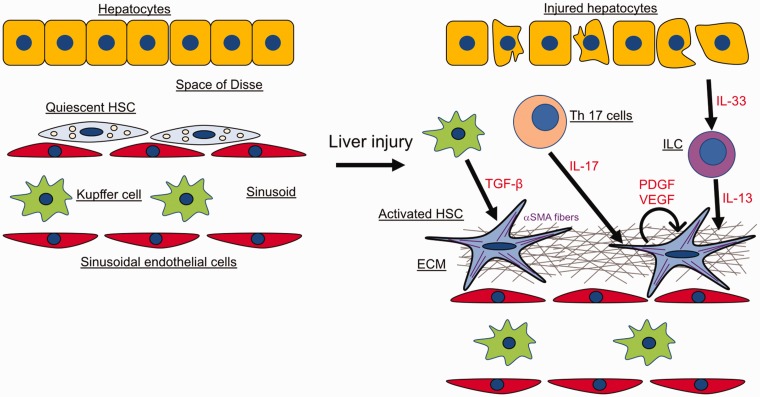
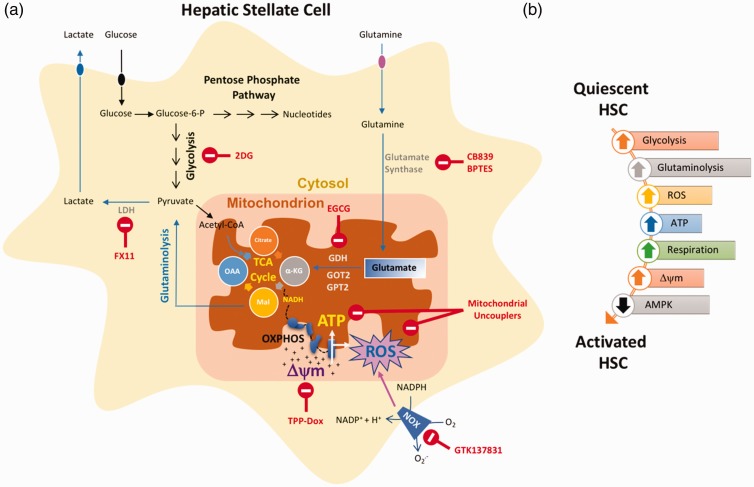
Hepatic fibrogenesis is a pathophysiological outcome of chronic liver injury hallmarked by excessive accumulation of extracellular matrix proteins. Fibrosis is a dynamic process that involves cross-talk between parenchymal cells (hepatocytes), hepatic stellate cells, sinusoidal endothelial cells and both resident and infiltrating immune cells. In this review, we focus on key cell-types that contribute to liver fibrosis, cytokines, and chemokines influencing this process and what it takes for fibrosis to regress. We discuss how mitochondria and metabolic changes in hepatic stellate cells modulate the fibrogenic process. We also briefly review how the presence of fibrosis affects development of hepatocellular carcinoma.

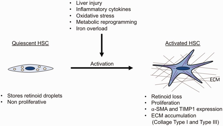
Impact statement: Advanced liver fibrosis results in cirrhosis, portal hypertension, and liver failure and often requires liver transplantation. Advanced liver fibrosis and cirrhosis are also major risk factors for hepatocellular carcinoma (HCC). Hepatic stellate cells (HSCs) play a pivotal role in the pathogenesis of liver fibrosis. In this review, we summarize the basic mechanisms that influence liver fibrosis development and how oxidative stress, mitochondrial dysfunction, and metabolic remodeling modulate HSC activation and indicate areas of potential therapeutic intervention.

Keywords: Liver fibrosis; PNPLA3; fibrosis regression; hepatic stellate cells; liver cancer; metabolic pathways; mitochondria.


Figures

Figure 1.
Figure 1.
Cell types in liver fibrosis. Quiescent HSCs in healthy livers are localized in the space of Disse. Following chronic liver damage, injured hepatocytes, immune cells and activated Kupffer cells release pro-fibrogenic molecules which activate HSCs. Activated HSCs upregulate ⍺-SMA expression, secrete growth factors, and produce large amounts of ECM. The figure depicts the different cell types and fibrogenic mediators involved in the fibrotic process and the crosstalk between them (see text for details).
Figure 2.
Figure 2.
Features of quiescent and activated HSCs. Quiescent HSCs store retinoid droplets, proliferate slowly, and express high levels of GFAP and LRAT. Upon liver injury, increase of inflammatory cytokines, ROS production, metabolic reprogramming or iron overload, HSCs become activated. Activated HSCs are characterized by loss of retinoid droplets, increased proliferation, and expression of fibrogenic markers (⍺-SMA, TIMP1, Lox and LoxL2). Moreover, activated HSCs produce ECM proteins such as collagen type I and collagen type III which are hallmarks of liver fibrosis.
Figure 3.
Figure 3.
Metabolic reprogramming of HSC. (a) Multiple aspects of glucose and glutamine metabolism are altered during HSC activation that can provide potential therapeutic avenues (indicated by red font). (b) A simplified depiction of metabolic pathways altered during HSC activation. Up arrows indicate upregulated pathway and down arrows mean downregulated. Δψm: Mitochondrial membrane potential; LDH: lactate dehydrogenase; GDH: glutamate dehydrogenase; GOT2: glutamic-oxaloacetic transaminase 2; GPT2: glutamate pyruvate transaminase 2.

References

    1. 1. Friedman SL. Liver fibrosis – from bench to bedside. J Hepatol 2003; 38(Suppl 1):S38–53 - PubMed
    1. Hernandez-Gea V, Friedman SL. Pathogenesis of liver fibrosis. Annu Rev Pathol 2011; 6:425–56 - PubMed
    1. Bataller R, Brenner DA. Liver fibrosis. J Clin Invest 2005; 115:209–18 - PMC - PubMed
    1. Gabele E, Brenner DA, Rippe RA. Liver fibrosis: signals leading to the amplification of the fibrogenic hepatic stellate cell. Front Biosci 2003; 8:d69–77 - PubMed
    1. Kisseleva T, Cong M, Paik Y, Scholten D, Jiang C, Benner C, Iwaisako K, Moore-Morris T, Scott B, Tsukamoto H, Evans SM, Dillmann W, Glass CK, Brenner DA. Myofibroblasts revert to an inactive phenotype during regression of liver fibrosis. Proc Natl Acad Sci U S A 2012; 109:9448–53 - PMC - PubMed

Publication types