. 2020 Jan 10;19(1):7.
doi: 10.1186/s12933-019-0980-4.

Sodium-glucose cotransporter 2 inhibitor Dapagliflozin attenuates diabetic cardiomyopathy

Affiliations

Sodium-glucose cotransporter 2 inhibitor Dapagliflozin attenuates diabetic cardiomyopathy

M Arow et al. Cardiovasc Diabetol. .

Abstract

Background: Diabetes mellitus type 2 (DM2) is a risk factor for developing heart failure but there is no specific therapy for diabetic heart disease. Sodium glucose transporter 2 inhibitors (SGLT2I) are recently developed diabetic drugs that primarily work on the kidney. Clinical data describing the cardiovascular benefits of SGLT2Is highlight the potential therapeutic benefit of these drugs in the prevention of cardiovascular events and heart failure. However, the underlying mechanism of protection remains unclear. We investigated the effect of Dapagliflozin-SGLT2I, on diabetic cardiomyopathy in a mouse model of DM2.

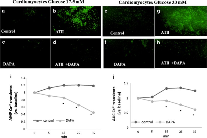
Methods: Cardiomyopathy was induced in diabetic mice (db/db) by subcutaneous infusion of angiotensin II (ATII) for 30 days using an osmotic pump. Dapagliflozin (1.5 mg/kg/day) was administered concomitantly in drinking water. Male homozygous, 12-14 weeks old WT or db/db mice (n = 4-8/group), were used for the experiments. Isolated cardiomyocytes were exposed to glucose (17.5-33 mM) and treated with Dapagliflozin in vitro. Intracellular calcium transients were measured using a fluorescent indicator indo-1.

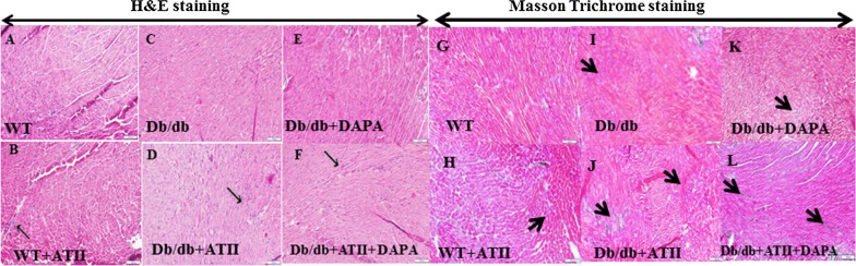
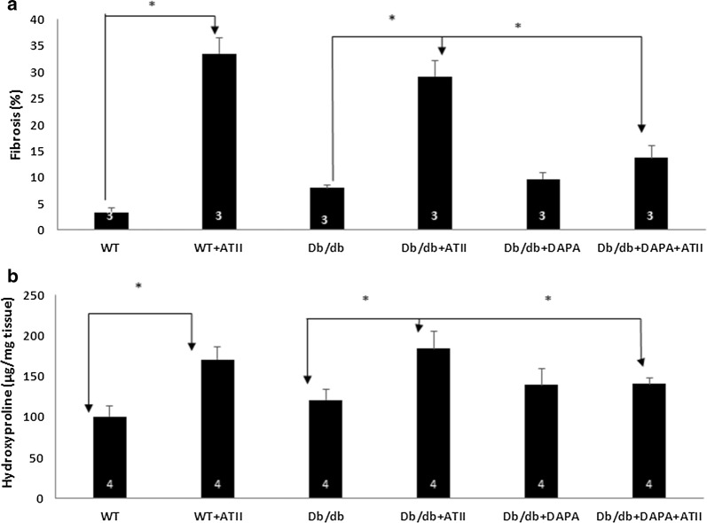
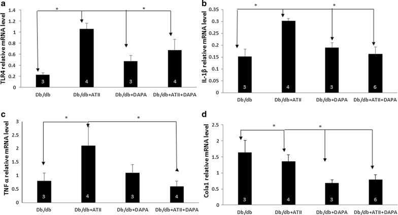
Results: Angiotensin II infusion induced cardiomyopathy in db/db mice, manifested by cardiac hypertrophy, myocardial fibrosis and inflammation (TNFα, TLR4). Dapagliflozin decreased blood glucose (874 ± 111 to 556 ± 57 mg/dl, p < 0.05). In addition it attenuated fibrosis and inflammation and increased the left ventricular fractional shortening in ATII treated db/db mice. In isolated cardiomyocytes Dapagliflozin decreased intracellular calcium transients, inflammation and ROS production. Finally, voltage-dependent L-type calcium channel (CACNA1C), the sodium-calcium exchanger (NCX) and the sodium-hydrogen exchanger 1 (NHE) membrane transporters expression was reduced following Dapagliflozin treatment.

Conclusion: Dapagliflozin was cardioprotective in ATII-stressed diabetic mice. It reduced oxygen radicals, as well the activity of membrane channels related to calcium transport. The cardioprotective effect manifested by decreased fibrosis, reduced inflammation and improved systolic function. The clinical implication of our results suggest a novel pharmacologic approach for the treatment of diabetic cardiomyopathy through modulation of ion homeostasis.

Keywords: Calcium transport fibrosis; Cardiomyocytes; Cardiomyopathy; Dapagliflozin; Diabetes mellitus type 2; Inflammation; ROS.


Conflict of interest statement

The authors declare that they have no competing interests.

Figures

Fig. 1
Fig. 1
H&E staining: wt/wt and db/db mice showed normal architecture with very well-shaped cardiomyocytes in the heart (a and c). ATII treatment increased the mononuclear inflammatory cells infiltration in left ventricle of heart (b and d, thin arrows). DAPA ameliorated the damage as seen by the organized architecture of the cardiomyocytes and reduction of mononuclear inflammatory cells (e and f), Scale bar = 100 µm. Masson trichrome staining: ATII induced fibrosis (blue staining-thick arrows) in both wt/wt and db/db mice (h vs g and j vs i). Tissues treated with DAPA were stained with normal pink color (l and k), Scale bar = 100 µm
Fig. 2
Fig. 2
a Quantification of fibrosis by Photoshop illustrates the significant reduction of fibrosis by DAPA. b Collagen content: the hydroxyproline assay was used. Measurement of hydroxyproline has been used to quantify collagen levels. ATII increases the collagen content in both wt/wt and db/db tissues (p < 0.05) while DAPA reduced it
Fig. 3
Fig. 3
PCR analysis revealed that treating db/db mice with ATII increases the inflammatory markers TLR4 and TNFα (a and c). Adding DAPA results in a significant reduction in TLR4, IL-1β and TNFα (ac). PCR analysis revealed that treating db/db mice with ATII increases the fibrosis marker Chola 1. However, adding DAPA causes a significant reduction in Chola 1 (d), (*p < 0.05)
Fig. 4
Fig. 4
DAPA alleviates oxidative stress. Neonatal cardiomyocytes were exposed to 17.5 mM and 33 mM glucose (ah). ROS was detected using a 2′, 7′-dichlorofluorescin diacetate (DCF-DA) reagent. Adding DAPA (5 μM), with/without ATII (1 μM) reduced the production of oxidative species in both glucose concentrations. i, j Adding DAPA to cardiomyocytes causes an immediate reduction in both amplitude (AMP) and area under the curve (AUC) of calcium transients. *p < 0.05
Fig. 5
Fig. 5
a Western blot for CACNA1C protein. b Densitometry analysis of CACNA1C normalized to β actin *p < 0.05. c Western blots for NCX1 protein. d Densitometry analysis of NCX1 normalized to β actin *p < 0.05. e Western blots for NHE1 protein. f Densitometry analysis of NHE1 normalized to β actin *p < 0.05
Fig. 6
Fig. 6
Summary of our understanding of the pathways accounting to improved cardiac function in diabetes in response to DAPA. The glucose lowering kidney targeted agent, DAPA, demonstrates direct cardiac effects by lowering myocardial [Na+]c through inhibition of NHE-1 and NCX and as well as Ca2+c through LTCC. The reduction in Ca2+ by DAPA appears to be directly correlated to the lowering of [Na+]c. The reduction in Ca2+ current by DAPA may partly explain the negative inotropic effects of DAPA in diabetic heart as seen by decreased hypertrophy better diastolic diameter following ATII treatment in one hand but augmented cell viability better mitochondrial function resulting less ROS production, reduced fibrosis and inflammation; all leading to better myocardial function on one hand and less cardiomyopathy markers on the other hand

Similar articles

Cited by

References

    1. Di Angelantonio E, Kaptoge S, Wormser D, Willeit P, Butterworth AS, Bansal N, O’Keeffe LM, Gao P, Wood AM, Burgess S, et al. Association of cardiometabolic multimorbidity with mortality. JAMA. 2015;314(1):52–60. doi: 10.1001/jama.2015.7008. - DOI - PMC - PubMed
    1. Boudina S, Abel ED. Diabetic cardiomyopathy, causes and effects. Rev Endocr Metab Disord. 2010;11(1):31–39. doi: 10.1007/s11154-010-9131-7. - DOI - PMC - PubMed
    1. Abel ED, O’Shea KM, Ramasamy R. Insulin resistance: metabolic mechanisms and consequences in the heart. Arterioscler Thromb Vasc Biol. 2012;32(9):2068–2076. doi: 10.1161/ATVBAHA.111.241984. - DOI - PMC - PubMed
    1. Bugger H, Abel ED. Molecular mechanisms of diabetic cardiomyopathy. Diabetologia. 2014;57(4):660–671. doi: 10.1007/s00125-014-3171-6. - DOI - PMC - PubMed
    1. Stratmann B, Tschoepe D. Heart in diabetes: not only a macrovascular disease. Diabetes Care. 2011;34(Suppl 2):S138–S144. doi: 10.2337/dc11-s208. - DOI - PMC - PubMed

Publication types

MeSH terms