. 2020 Jan 10;51(1):2.
doi: 10.1186/s13567-019-0733-0.

Precision cut lung slices: a novel versatile tool to examine host-pathogen interaction in the chicken lung

Affiliations

Precision cut lung slices: a novel versatile tool to examine host-pathogen interaction in the chicken lung

Karen Jane Bryson et al. Vet Res. .

Abstract

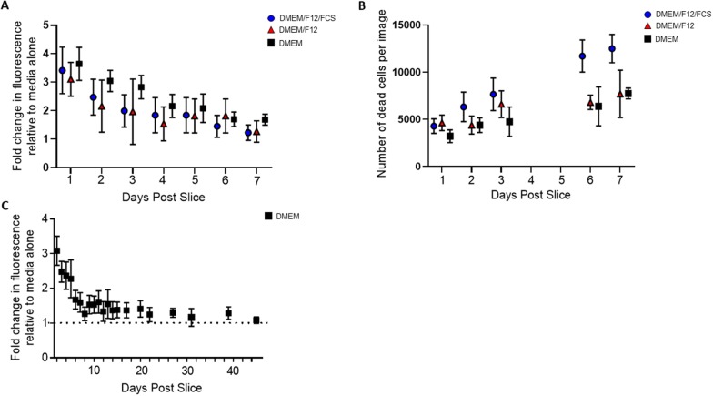
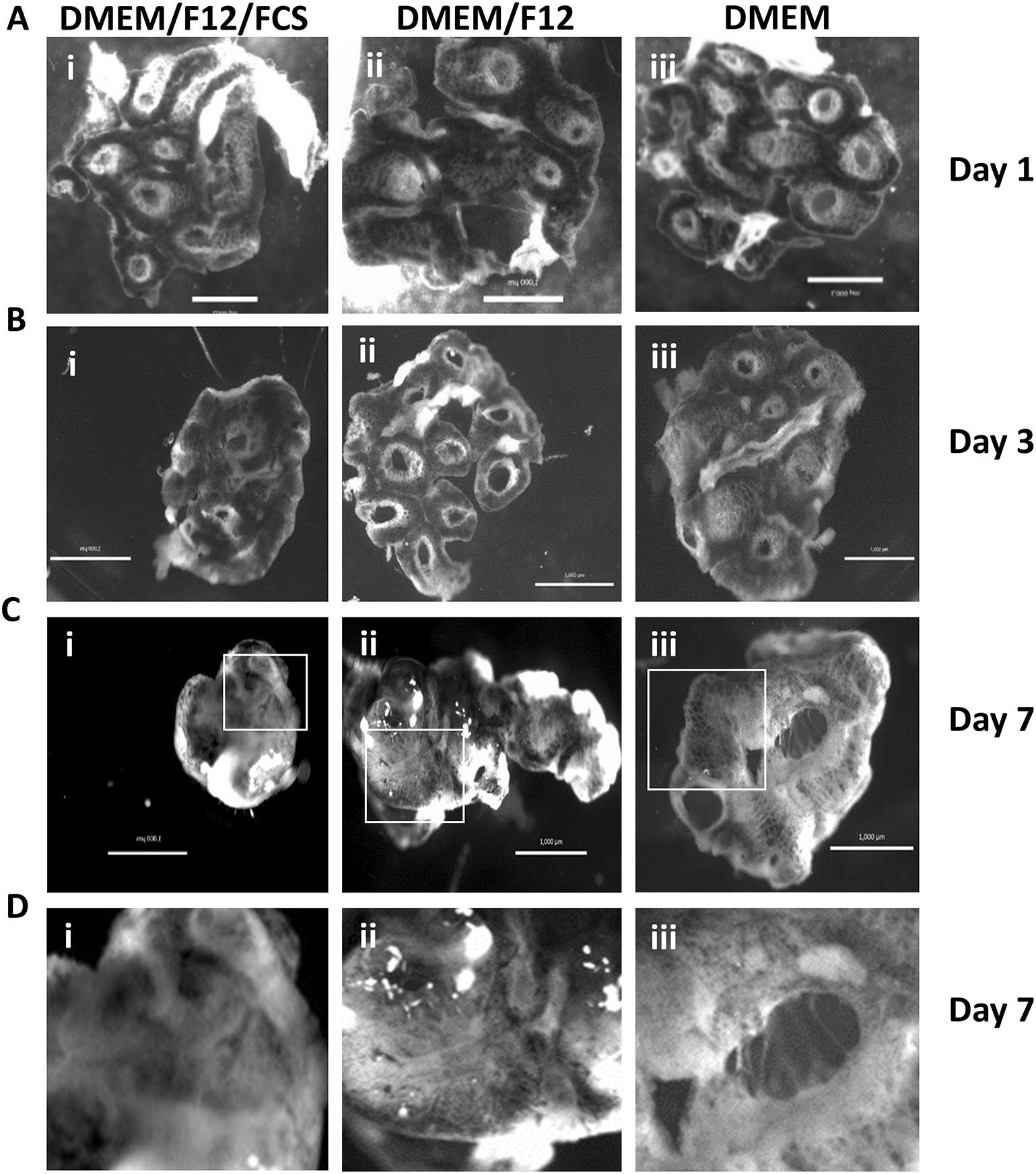
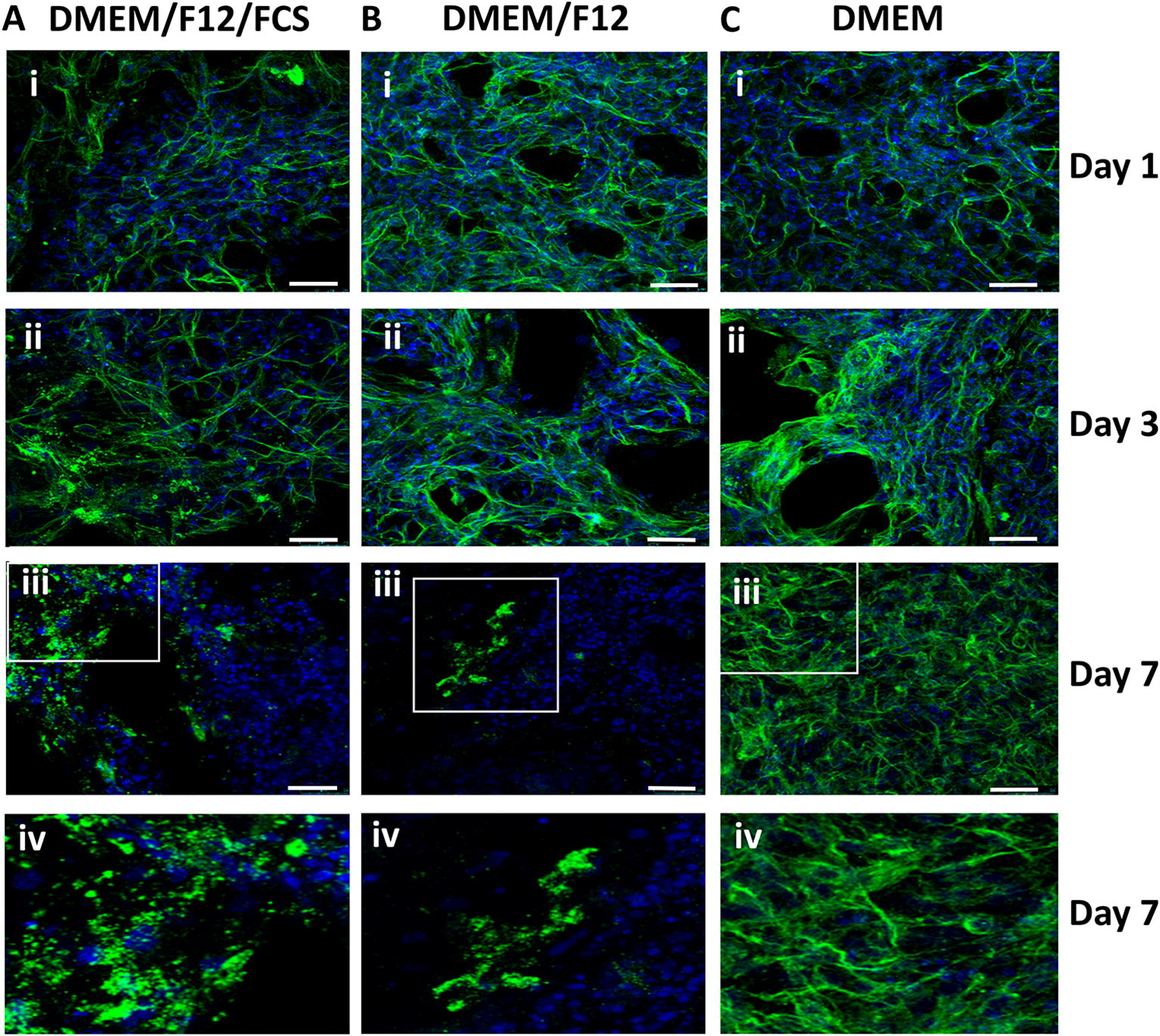
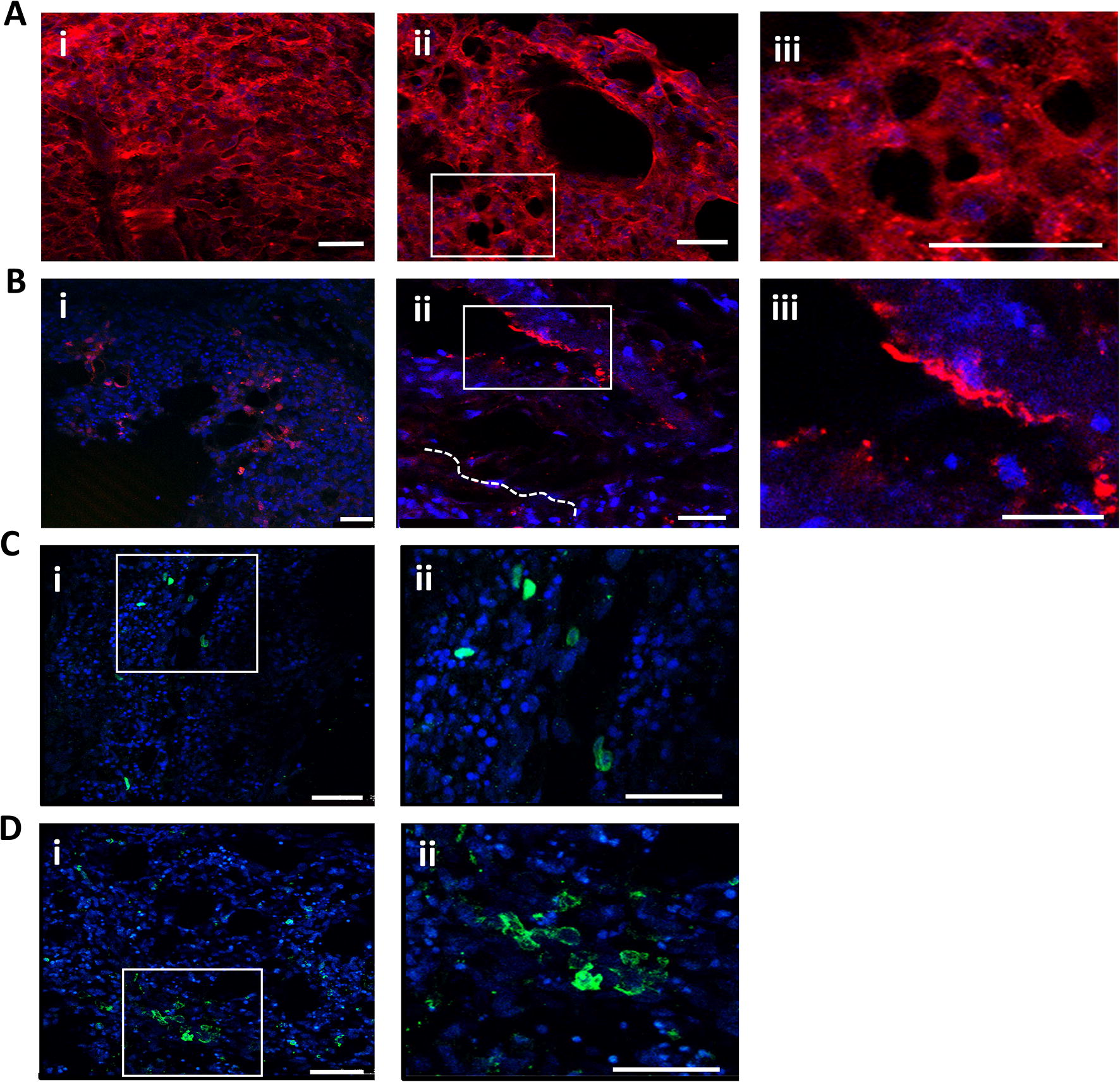
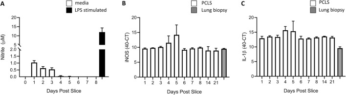
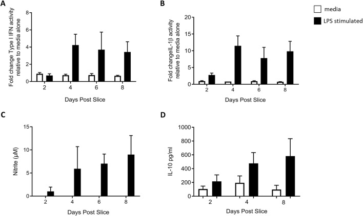
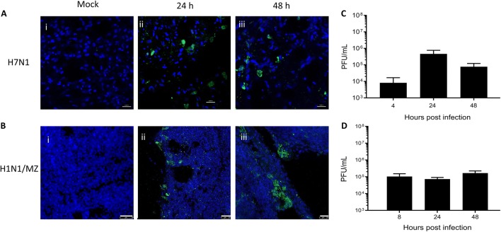
The avian respiratory tract is a common entry route for many pathogens and an important delivery route for vaccination in the poultry industry. Immune responses in the avian lung have mostly been studied in vivo due to the lack of robust, relevant in vitro and ex vivo models mimicking the microenvironment. Precision-cut lung slices (PCLS) have the major advantages of maintaining the 3-dimensional architecture of the lung and includes heterogeneous cell populations. PCLS have been obtained from a number of mammalian species and from chicken embryos. However, as the embryonic lung is physiologically undifferentiated and immunologically immature, it is less suitable to examine complex host-pathogen interactions including antimicrobial responses. Here we prepared PCLS from immunologically mature chicken lungs, tested different culture conditions, and found that serum supplementation has a detrimental effect on the quality of PCLS. Viable cells in PCLS remained present for ≥ 40 days, as determined by viability assays and sustained motility of fluorescent mononuclear phagocytic cells. The PCLS were responsive to lipopolysaccharide stimulation, which induced the release of nitric oxide, IL-1β, type I interferons and IL-10. Mononuclear phagocytes within the tissue maintained phagocytic activity, with live cell imaging capturing interactions with latex beads and an avian pathogenic Escherichia coli strain. Finally, the PCLS were also shown to be permissive to infection with low pathogenic avian influenza viruses. Taken together, immunologically mature chicken PCLS provide a suitable model to simulate live organ responsiveness and cell dynamics, which can be readily exploited to examine host-pathogen interactions and inflammatory responses.


Conflict of interest statement

The authors declare that they have no competing interests.

Figures

Figure 1
Figure 1
PCLS viability over time. PCLS viability was assessed by AlamarBlue assay (A and C) and Live/Dead staining (B). PCLS (500 μm) prepared from CSF1R-eGFP transgenic chickens were cultured for 7 days in either DMEM/F12/FCS (blue circle), DMEM/F12 (red triangle) or DMEM (black square) and viability was assessed using the AlamarBlue assay (A). n = 6–18 slices generated from 3 birds, data are represented as the mean ± SD. PCLS prepared from SPF PA12 chickens (200–300 μm) were cultured for 7 days in either DMEM/F12/FCS (blue circle), DMEM/F12 (red triangle) or DMEM (black square) and were stained with a Live/Dead kit and subsequently examined by epifluorescence microscopy (B). The number of dead cells were enumerated in images from 3 to 4 PCLS generated from 2 birds at different time-points using the Fiji-ImageJ image processing software. Data are expressed as the mean ± SD, per time-point of one representative experiment. n = 3–4 slices. The maintenance of cell viability in PCLS (500 μm) prepared from CSF1R-eGFP transgenic chickens was assessed in DMEM media by AlamarBlue assay (C). n = 16 slices generated from 2 birds, data are represented as the mean ± SD.
Figure 2
Figure 2
Structural integrity of PCLS. Bright field images were captured at 1 (A), 3 (B) and 7 (C and D) days post slice for PCLS (500 μm) prepared from CSF1R-eGFP transgenic chickens. The PCLS were cultured with DMEM/F12/FCS (i), DMEM/F12 (ii) or DMEM (iii). The white boxes (C, panels i, ii and iii) represent the area digitally enlarged (D, panels i, ii, iii respectively). Images were captured using the Zeiss Axio zoom V16. Images are representative of randomly selected PCLS from 3 individual birds. Scale bar = 1000 µm.
Figure 3
Figure 3
PCLS cytoskeleton structure under different media conditions. PCLS (200–300 μm) prepared from PA12 SPF chickens were cultured in DMEM/F12/FCS (A), DMEM/F12 (B), or DMEM (C) from 1 (i), 3 (ii) or 7 days (iii and iv) post slice. PCLS were fixed and β-tubulin filaments (green) and nuclei (blue) visualized. The white boxes (A, panel iii, B, panels iii and C iii) represent the area digitally enlarged (A, panel iv, B, panel iv, C, panel iv respectively). Images were captured using a Leica TCS P8 confocal microscope at ×630 magnification. Scale bars = 25 μm. Images are representative of 3 to 4 PCLS randomly selected from 2 birds per time point and condition.
Figure 4
Figure 4
Immunostaining of PCLS. PCLS (200–300 μm) prepared from PA12 SPF chickens 1 day post slice were stained with a panel of antibodies and examined by confocal microscopy. Nuclei are represented in blue (DAPI). Actin cytoskeleton staining using rhodamine phalloidin (red) (A). Longitudinal section revealing boundaries of capillaries from the parabronchi (A, panel i); Transversal section revealing a parabronchus and air capillaries in greater details (A, panels ii and iii). The white box (A, panels ii) represents the area digitally enlarged (A, panels iii). von Willebrand Factor staining (red) (B), a marker of endothelial cells. Overall view of vWF-stained endothelial cells within the lung parenchyma (B, panel i); Longitudinal section (B, panel ii) with a schematic drawing of capillary networks beneath the lung parenchyma (dotted lines). The white box (B, panel ii) represents the area digitally enlarged (B, panel iii). Capillary ends are observed scattered in the tissue (B, panel ii); a capillary ramification close to a parabronchus and a capillary end in greater detail (B, panel iii). Longitudinal section showing MCRL1-B+ phagocytes (green) (C) within the lung parenchyma (C, panels i and ii), especially close to the septae. The white box (C, panel i) represents the area digitally enlarged (C, panel ii). Transversal section showing CD45+ leucocytes (green) within the lung parenchyma (D, panels i and ii), scattered in the atria and septae. The white box (D, panel i) represent the area digitally enlarged (D, panel ii). Images were captured using a Leica TCS P8 confocal microscope. Scale bars = 75 μm (B, panel i) and 25 μm. Images are representative of 3 to 4 PCLS randomly selected from 2 birds per staining strategy.
Figure 5
Figure 5
Basal levels of inflammatory mediators in cultured PCLS. Nitrite produced by PCLS (500 μm) prepared from CSF1R-eGFP transgenic chickens was assessed in the supernatant over 8 days of culture by Griess assay (white) (A). The supernatant was harvested every 24 h. As positive control, on day 7 a number of PCLS were stimulated with LPS for 24 h (black) and the supernatant harvested on day 8. n = 6–10 slices generated from 4 individual birds. iNOS (B) and IL-1β (C) mRNA levels were assessed by qPCR in the PCLS over 21 days post slice and normalised to 28S expression (white). n = 6 slices generated from 4 individual birds. iNOS and IL-1β levels were also assessed in a fresh lung biopsy sample (grey) that had not been sliced into sections or cultured with DMEM. n = 3–4 slices generated from 3 individual birds. All data are represented as the mean ± SD. *p < 0.05 all time points compared.
Figure 6
Figure 6
PCLS responses to LPS. PCLS (500 μm) prepared from from CSF1R-eGFP transgenic chickens and cultured in the presence (black) or absence (white) of LPS for 24 h on either day 1, 3, 5 or 7 post slice and supernatants harvested on day 2, 4, 6 and 8 post slice. IL-1β (A) and type I IFNs (B) production were measured in the supernatants using luciferase-based Mx- or NFκB-reporter bioassays. Results are expressed as fold change in luciferase activity relative to unstimulated PCLS. n = 3 slices generated from 2 individual birds. Nitrite produced by PCLS over 8 days of culture in response to LPS for 24 h was assessed in the supernatant by Griess assay (C). n = 6 slices generated from 4 individual birds. The concentration of IL-10 was assessed in PCLS supernatant by ELISA on day 2, 4 and 8 post slice (D). n = 6 slices generated from 4 individual birds. Data are represented as the mean ± SD.
Figure 7
Figure 7
PCLS are susceptible to low pathogenic avian influenza virus strains. PCLS (500 μm) were generated from Hy-Line conventional (A) and PCLS (200–300 μm) were generated from PA12 SPF (B) chickens, infected with H7N1 (A) and H1N1 (B) LPAI virus strains, and stained for the viral proteins NP (A) and NS1 (B). Representative images showing uninfected PCLS (i) and virus-infected cells (green) within the PCLS at 24 (ii) and 48 (iii) hours post-infection. The nuclei are represented in blue. Images were captured by confocal microscopy at ×400 magnification. Scale bar = 10 µm (A) and 25 µm (B). n = 8 slices generated from 2 individual birds. Viral titre of supernatants harvested from H7N1-infected PCLS at 4, 24 and 48 h post-infection (C) and H1N1-infected PCLS at 8, 24 and 48 h post-infection (D). n = 8 slices generated from 2 individual birds, data are represented as the mean ± SD.

References

    1. Parrish AR, Gandolfi AJ, Brendel K. Precision-cut tissue slices: applications in pharmacology and toxicology. Life Sci. 1995;57:1887–1901. doi: 10.1016/0024-3205(95)02176-J. - DOI - PubMed
    1. Siminski JT, Kavanagh TJ, Chi E, Raghu G. Long-term maintenance of mature pulmonary parenchyma cultured in serum-free conditions. Am J Physiol. 1992;262:L105–110. - PubMed
    1. Ressmeyer AR, Larsson AK, Vollmer E, Dahlen SE, Uhlig S, Martin C. Characterisation of guinea pig precision-cut lung slices: comparison with human tissues. Eur Respir J. 2006;28:603–611. doi: 10.1183/09031936.06.00004206. - DOI - PubMed
    1. Rosales Gerpe MC, van Vloten JP, Santry LA, de Jong J, Mould RC, Pelin A, Bell JC, Bridle BW, Wootton SK. Use of precision-cut lung slices as an ex vivo tool for evaluating viruses and viral vectors for gene and oncolytic therapy. Mol Ther Methods Clin Dev. 2018;10:245–256. doi: 10.1016/j.omtm.2018.07.010. - DOI - PMC - PubMed
    1. Meng F, Wu NH, Nerlich A, Herrler G, Valentin-Weigand P, Seitz M. Dynamic virus-bacterium interactions in a porcine precision-cut lung slice coinfection model: swine influenza virus paves the way for Streptococcus suis infection in a two-step process. Infect Immun. 2015;83:2806–2815. doi: 10.1128/IAI.00171-15. - DOI - PMC - PubMed

Publication types

Substances

LinkOut - more resources