. 2020 Mar 4;105(5):855-866.e5.
doi: 10.1016/j.neuron.2019.12.014. Epub 2020 Jan 7.

Astrocyte Unfolded Protein Response Induces a Specific Reactivity State that Causes Non-Cell-Autonomous Neuronal Degeneration

Affiliations

Astrocyte Unfolded Protein Response Induces a Specific Reactivity State that Causes Non-Cell-Autonomous Neuronal Degeneration

Heather L Smith et al. Neuron. .

Abstract

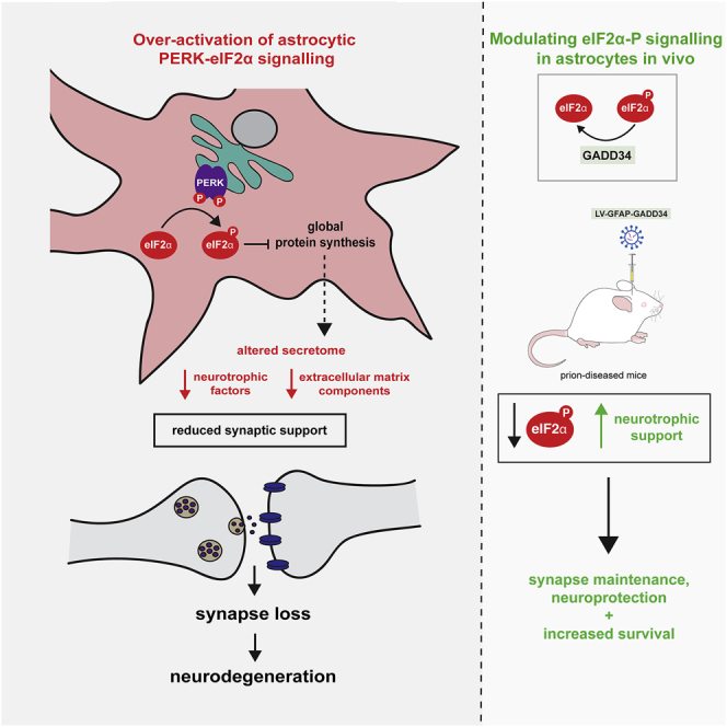
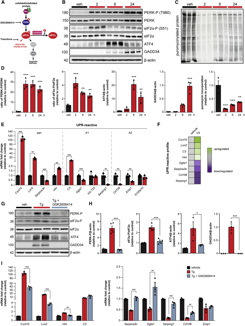
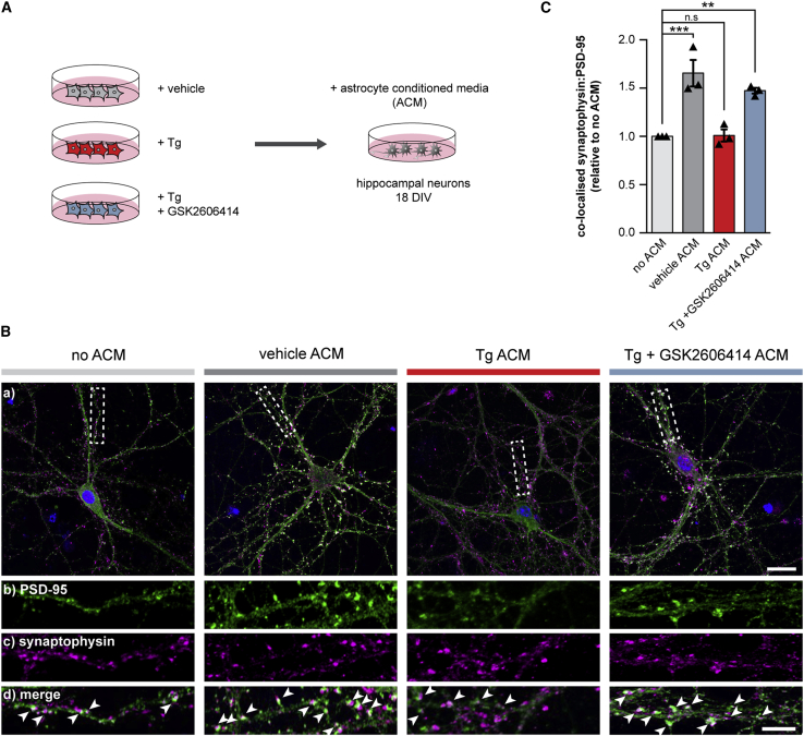
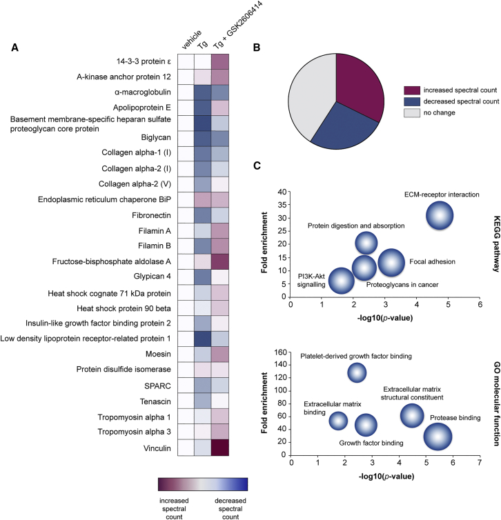
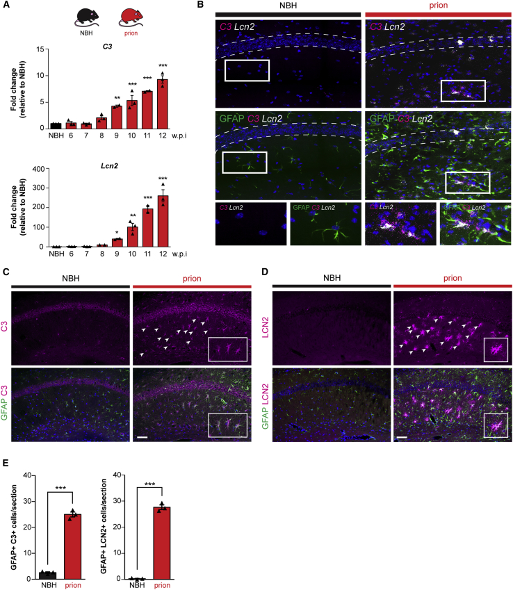
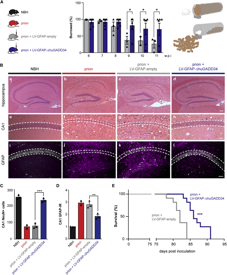
Recent interest in astrocyte activation states has raised the fundamental question of how these cells, normally essential for synapse and neuronal maintenance, become pathogenic. Here, we show that activation of the unfolded protein response (UPR), specifically phosphorylated protein kinase R-like endoplasmic reticulum (ER) kinase (PERK-P) signaling-a pathway that is widely dysregulated in neurodegenerative diseases-generates a distinct reactivity state in astrocytes that alters the astrocytic secretome, leading to loss of synaptogenic function in vitro. Further, we establish that the same PERK-P-dependent astrocyte reactivity state is harmful to neurons in vivo in mice with prion neurodegeneration. Critically, targeting this signaling exclusively in astrocytes during prion disease is alone sufficient to prevent neuronal loss and significantly prolongs survival. Thus, the astrocyte reactivity state resulting from UPR over-activation is a distinct pathogenic mechanism that can by itself be effectively targeted for neuroprotection.

Keywords: LCN2; PERK signalling; astrocyte reactivity state; astrocytes; neurodegeneration; neuroprotection; secretome; synapse; translational neuroscience; unfolded protein response.


Conflict of interest statement

Declaration of Interests The authors declare no competing interests. O.J.F. is now an employee of AstraZeneca.

Figures

None
Graphical abstract
Figure 1
Figure 1
PERK-eIF2α Signaling Induces a Reactive Phenotype in Primary Cultured Astrocytes (A) Schematic of the PERK branch of the UPR, including the sites of action of the PERK branch inhibitors, GSK2606414 and trazodone. (B) Western blots showing PERK-eIF2α signaling in primary astrocytes treated with 300 nM thapsigargin (Tg) for 2, 6, or 24 h. (C) Primary astrocytes show reduced protein synthesis rates in the presence of Tg, as measured by the incorporation of puromycin into nascent proteins. (D) Quantification of western blots. (E) qPCR analysis of astrocyte reactivity markers revealed an altered profile on Tg treatment. (F) Astrocyte reactivity markers that characterize the UPR-reactive profile. (G) PERK-eIF2α signaling is significantly reduced in primary astrocytes cultured in the presence of Tg and 5 μM GSK2606414. (H) Western blots quantified. (I) GSK2606414 significantly blunts the reactivity profile of Tg-stressed astrocytes. All bar graphs show mean ± SEM. p < 0.05; ∗∗p < 0.01; ∗∗∗p < 0.001; one-way ANOVA. n = 3 biological replicates.
Figure 2
Figure 2
UPR-Reactive Astrocytes Fail to Promote Synaptogenesis In Vitro (A) Schematic illustrating the generation of astrocyte-conditioned media (ACM). Primary astrocytes were treated with vehicle, 300 nM Tg, or 300 nM Tg and 5 μM GSK2606414. Astrocytes were washed 24 h post-treatment and incubated with fresh neuron media for a further 24 h. Conditioned media were transferred to primary hippocampal neurons at 18 days in vitro (DIV). (B) Representative images of primary hippocampal neurons grown without ACM or with vehicle, Tg, or Tg and GSK2606414 ACM. Neurons were immunostained with the pre-synaptic marker, synaptophysin (magenta) and the post-synaptic marker, PSD-95 (green). Arrows highlight co-localized synaptophysin and PSD-95 puncta. Scale bars, (a) 25 μm; (b–d) 10 μm. (C) Relative number of synapses normalized to the no ACM condition. Tg ACM failed to promote synaptogenesis, whereas Tg and GSK2606414 ACM retained its synaptogenic properties. 10 neurons were counted per condition, per biological replicate (n = 3). Bar graph shows mean normalized synapse number ± SEM. ∗∗p < 0.01; ∗∗∗p < 0.001; n.s., non-significant; one-way ANOVA.
Figure 3
Figure 3
UPR-Reactive Astrocytes Have an Altered Secretome The proteome of conditioned media from vehicle-, Tg-, or Tg and GSK2606414-treated astrocytes was analyzed by LC/MS. (A) Heatmap showing normalized spectral counts of the 25 most abundant proteins detected in the conditioned media of vehicle-treated astrocytes. 10 of the 25 proteins showed reduced spectral counts on Tg treatment. These changes were largely reversed by PERK inhibition. (B) Pie chart showing the percentage of proteins that exhibited an increase, decrease, or no change in spectral counts on Tg treatment. (C) Bubble plots showing KEGG and GO functional analysis of the proteins that displayed reduced spectral counts on Tg treatment, as determined using DAVID. Terms relating to the extracellular matrix and cell adhesion were significantly enriched upon PERK activation (p < 0.0001). Size of bubble represents protein count. LC/MS was performed on conditioned media from 3 biological replicates.
Figure 4
Figure 4
UPR-Reactive Astrocytes Upregulate C3 and LCN2 In Vivo (A) qPCR analysis of hippocampal C3 and Lcn2 mRNA levels across the prion time course. p < 0.05; ∗∗p < 0.01; ∗∗∗p < 0.001; n = 3 mice per time point. (B) RNA scope showing the localization of C3 (magenta) and Lcn2 (white) to GFAP+ astrocytes (green) in the hippocampus of prion-diseased mice at 10 w.p.i. (C and D) Immunostaining of C3 (C) and LCN2 (D) in the hippocampus of prion-diseased mice at 10 w.p.i. Scale bars, 100 μm. (E) Prion-diseased mice showed a significant increase in the number of C3+/GFAP+ and LCN2+/GFAP+ astrocytes. Bar graphs show mean ± SEM; ∗∗∗p < 0.001; Student’s t test. n = 3 mice.
Figure 5
Figure 5
Genetic Modulation of Astrocytic PERK-eIF2α Signaling Ameliorates the UPR-Reactivity State In Vivo (A) Prion-inoculated mice were injected with lentivirus at 5 w.p.i., prior to synapse loss. The astrocytic expression of ΔhuGADD34 significantly reduced the mRNA levels of the UPR-reactivity markers C3 and Lcn2 at 10 w.p.i., as analyzed by qPCR. ∗∗p < 0.01; n = 5 mice per condition. (B) RNA scope also revealed a reduction in C3 and Lcn2 upon the expression of ΔhuGADD34. (C and D) Protein levels of C3 (C) and LCN2 (D) were similarly reduced by astrocytic ΔhuGADD34, as determined by immunohistochemistry. Scale bars, 100 μm. (E) Bar graphs represent quantification of C3+ and LCN2+ astrocytes treated with ΔhuGADD34 or empty virus. Bar graphs show mean ± SEM; ∗∗∗p < 0.001; Student’s t test. n = 3 mice.
Figure 6
Figure 6
Targeting Astrocytic PERK-eIF2α Signaling Is Neuroprotective in Prion-Diseased Mice (A) Prion-infected mice received hippocampal injections of LV-GFAP-empty or LV-GFAP-ΔhuGADD34 at 5 w.p.i. The overexpression of ΔhuGADD34 prevented the decline in burrowing behavior at 9, 10, and 11 w.p.i. (B) Representative images of hematoxylin-and-eosin-stained hippocampus from normal brain homogenate (NBH) and prion-inoculated mice injected with LV-GFAP-empty or LV-GFAP-ΔhuGADD34. Scale bars, (a–d) 200 μm; (e–j) 50 μm. (C) Astrocytic expression of ΔhuGADD34 resulted in profound neuroprotection. (D) The levels of GFAP were also reduced. (E) Targeting astrocytic PERK-eIF2α significantly prolonged survival. LV-GFAP-empty = 6 mice; LV-GFAP-ΔhuGADD34 = 11 mice. ∗∗∗p < 0.001; Mantel-Cox test.

Comment in

  • Withdrawn support.
    Lewis S. Lewis S. Nat Rev Neurosci. 2020 Mar;21(3):119. doi: 10.1038/s41583-020-0268-4. Nat Rev Neurosci. 2020. PMID: 31996802 No abstract available.

References

    1. Allen N.J., Bennett M.L., Foo L.C., Wang G.X., Chakraborty C., Smith S.J., Barres B.A. Astrocyte glypicans 4 and 6 promote formation of excitatory synapses via GluA1 AMPA receptors. Nature. 2012;486:410–414. - PMC - PubMed
    1. Axten J.M., Medina J.R., Feng Y., Shu A., Romeril S.P., Grant S.W., Li W.H., Heerding D.A., Minthorn E., Mencken T. Discovery of 7-methyl-5-(1-{[3-(trifluoromethyl)phenyl]acetyl}-2,3-dihydro-1H-indol-5-yl)-7H-pyrrolo[2,3-d]pyrimidin-4-amine (GSK2606414), a potent and selective first-in-class inhibitor of protein kinase R (PKR)-like endoplasmic reticulum kinase (PERK) J. Med. Chem. 2012;55:7193–7207. - PubMed
    1. Bastide A., Peretti D., Knight J.R., Grosso S., Spriggs R.V., Pichon X., Sbarrato T., Roobol A., Roobol J., Vito D. RTN3 is a novel cold-induced protein and mediates neuroprotective effects of RBM3. Curr. Biol. 2017;27:638–650. - PMC - PubMed
    1. Celardo I., Costa A.C., Lehmann S., Jones C., Wood N., Mencacci N.E., Mallucci G.R., Loh S.H., Martins L.M. Mitofusin-mediated ER stress triggers neurodegeneration in pink1/parkin models of Parkinson’s disease. Cell Death Dis. 2016;7:e2271. - PMC - PubMed
    1. Christopherson K.S., Ullian E.M., Stokes C.C., Mullowney C.E., Hell J.W., Agah A., Lawler J., Mosher D.F., Bornstein P., Barres B.A. Thrombospondins are astrocyte-secreted proteins that promote CNS synaptogenesis. Cell. 2005;120:421–433. - PubMed

Publication types

MeSH terms