. 2020 Feb 15;204(4):763-774.
doi: 10.4049/jimmunol.1901086. Epub 2020 Jan 10.

Pyruvate Kinase M2 Promotes Expression of Proinflammatory Mediators in House Dust Mite-Induced Allergic Airways Disease

Affiliations

Pyruvate Kinase M2 Promotes Expression of Proinflammatory Mediators in House Dust Mite-Induced Allergic Airways Disease

Cheryl van de Wetering et al. J Immunol. .

Abstract

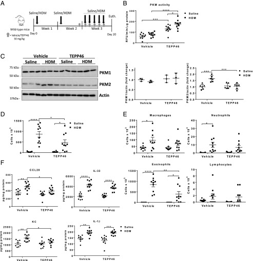
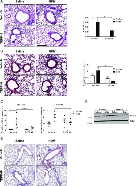
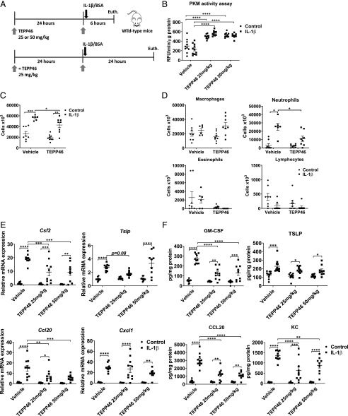
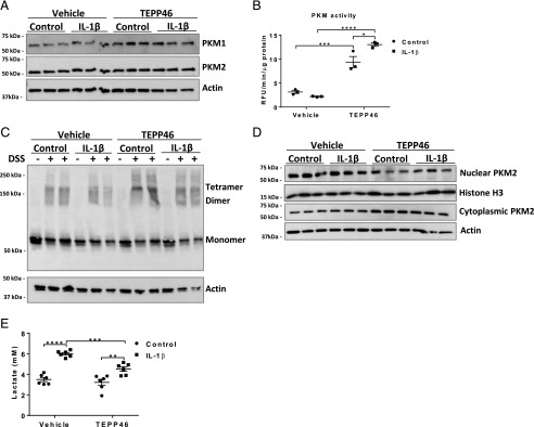
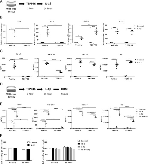
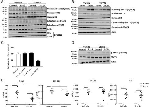
Asthma is a chronic disorder characterized by inflammation, mucus metaplasia, airway remodeling, and hyperresponsiveness. We recently showed that IL-1-induced glycolytic reprogramming contributes to allergic airway disease using a murine house dust mite model. Moreover, levels of pyruvate kinase M2 (PKM2) were increased in this model as well as in nasal epithelial cells from asthmatics as compared with healthy controls. Although the tetramer form of PKM2 converts phosphoenolpyruvate to pyruvate, the dimeric form of PKM2 has alternative, nonglycolysis functions as a transcriptional coactivator to enhance the transcription of several proinflammatory cytokines. In the current study, we examined the impact of PKM2 on the pathogenesis of house dust mite-induced allergic airways disease in C57BL/6NJ mice. We report, in this study, that activation of PKM2, using the small molecule activator, TEPP46, augmented PKM activity in lung tissues and attenuated airway eosinophils, mucus metaplasia, and subepithelial collagen. TEPP46 attenuated IL-1β-mediated airway inflammation and expression of proinflammatory mediators. Exposure to TEPP46 strongly decreased the IL-1β-mediated increases in thymic stromal lymphopoietin (TSLP) and GM-CSF in primary tracheal epithelial cells isolated from C57BL/6NJ mice. We also demonstrate that IL-1β-mediated increases in nuclear phospho-STAT3 were decreased by TEPP46. Finally, STAT3 inhibition attenuated the IL-1β-induced release of TSLP and GM-CSF, suggesting that the ability of PKM2 to phosphorylate STAT3 contributes to its proinflammatory function. Collectively, these results demonstrate that the glycolysis-inactive form of PKM2 plays a crucial role in the pathogenesis of allergic airways disease by increasing IL-1β-induced proinflammatory signaling, in part, through phosphorylation of STAT3.


Figures

FIGURE 1.
FIGURE 1.
Activation of PKM2 by TEPP46 attenuates proinflammatory cytokines in mice with HDM-induced allergic airway disease. (A) Schematic depicting the exposure regimen. Mice were sensitized twice with 10 μg of HDM or saline on days 1 and 8. Mice were treated with 50 mg/kg TEPP46 i.p. daily, starting on day 14. Mice were challenged with HDM on days 15–19 and euthanized 24 h after the final HDM challenge. (B) Assessment of PKM activity in lung tissue homogenates. (C) Representative Western blots and quantification for total PKM1 and PKM2 levels. β-Actin; loading control (n = 3–6 per group). (D and E) Total and differential cell counts in BAL fluid. (F) Measurements of CCL20, IL-33, KC, and IL-1β in lung tissue homogenates by ELISA. (A, B, and D–F) n = 8–10 per group. *p < 0.05, **p < 0.01, ***p < 0.001, ****p < 0.0001 (two-way ANOVA).
FIGURE 2.
FIGURE 2.
Activation of PKM2 by TEPP46 attenuates mucus metaplasia, subepithelial collagen, and markers of airway remodeling in mice with HDM-induced allergic airway disease. (A) Assessment and quantification of mucus metaplasia by Periodic acid–Schiff (PAS) staining intensity and (B) collagen deposition by Masson’s trichrome staining. Scale bar, 200 μm. (C) mRNA expression of Muc5AC, and Col1a1, normalized to Ppia. (D) Representative Western blots for SMA levels and the loading control β-actin. (E) Assessment of α-SMA staining around large airways (n = 5 per group). Scale bar, 300 μm. *p < 0.05, ***p < 0.001 (two-way ANOVA).
FIGURE 3.
FIGURE 3.
PKM2 activation attenuates the release of proinflammatory cytokines following intranasal administration of IL-1β. (A) Schematic depicting the pretreatment with 25 or 50 mg/kg TEPP46 prior to intranasal administration of 1 μg of IL-1β for either 6 or 24 h. The total cell count and cell differentials in the BAL fluid reflect 24 h after IL-1β treatment, the other results shown are obtained 6 h post–IL-1β. (B) Assessment of PKM activity in lung tissue homogenates. (C and D) Total and differential cell counts in BAL fluid. (E) mRNA expression of proinflammatory cytokine genes in lung tissue homogenates. Results were normalized to the housekeeping gene Ppia. (F) Levels of proinflammatory mediators TSLP, GM-CSF, KC, and CCL20 in lung tissue homogenates by ELISA (n = 8–11 per group). *p < 0.05, **p < 0.01, ***p < 0.001, ****p < 0.0001 (two-way ANOVA).
FIGURE 4.
FIGURE 4.
TEPP46 augments PKM activity and PKM2’s cytosolic presence and attenuates IL-1β–mediated lactate secretion in primary MTE cells. MTE cells were treated with 100 μM TEPP46 for 1 h prior to stimulation with 10 ng/ml IL-1β for 24 h. (A) Representative Western blot of total PKM1 and PKM2 levels and β-actin. (B) PKM activity assay in MTE cells and (C) representative Western blot for tetrameric, dimeric, and monomeric PKM2 and the loading control β-actin. MTE cells were incubated in the presence or absence (first lane of each condition) of the disuccinimidyl suberate (DSS) cross-linker to evaluate the formation of the isoforms of PKM2. (D) Representative Western blots of nuclear and cytosolic extracts of PKM2 (n = 3 per group). (E) Lactate levels in supernatants of MTE cells. Experiments were performed at least three times. *p < 0.05, **p < 0.01, ***p < 0.001, ****p < 0.0001 (two-way ANOVA).
FIGURE 5.
FIGURE 5.
Activation of PKM2 attenuates IL-1β–mediated proinflammatory responses in primary MTE cells and the release of proinflammatory mediators following subsequent exposure to HDM. (A) Schematic depicting the pretreatment with 100 μM TEPP46, followed by stimulation of 10 ng/ml IL-1β for 24 h. (B) mRNA expression of Tslp, Csf2, Cxcl1, and Ccl20 in MTE cells. Ppia is used as housekeeping gene. (C) Proinflammatory cytokine mediators TSLP, GM-CSF, KC, and CCL20 in cell culture supernatants of MTE cells were detected by ELISA. (D) Schematic depicting the pretreatment with 100 μM TEPP46, followed by stimulation of 10 ng/ml IL-1β for 24 h. Media was replaced and exposed to HDM (50 μg/ml) for an additional 2 h. (E) Proinflammatory cytokine mediators TSLP, GM-CSF, KC, and CCL20 in cell culture supernatants of MTE cells. (F) Cell survival was evaluated by Crystal Violet staining (left) and Calcein AM Assay (right) in MTE cells (n = 3–6 per group). Experiments were performed at least three times. *p < 0.05, **p < 0.01, ***p < 0.001, ****p < 0.0001 analyzed (two-way ANOVA).
FIGURE 6.
FIGURE 6.
PKM2-mediated phosphorylation of STAT3 contributes to IL-1β–mediated proinflammatory signaling in epithelial cells. (A) Representative Western blots of total and phosphorylated STAT3 in nuclear and cytosolic extracts from MTE cells and total IKKε levels in whole-cell lysates. (B) Representative Western blots of total and phosphorylated STAT3 in nuclear and cytosolic extracts from HDM- or saline-treated lung tissues. (C) Impact of Stattic on survival of MTE cells was evaluated by a Calcein AM Assay. (D) Representative Western blots of total and phosphorylated STAT3 in whole-cell lysates from control or IL-1β–stimulated MTE cells pretreated with Stattic or vehicle control. (E) Proinflammatory mediators TSLP, GM-CSF, KC, and CCL20 in cell culture supernatants of MTE cells after treatment for 1 h with 0.5 μM Stattic, followed by stimulation of 10 ng/ml IL-1β for 24 h. Experiments were conducted at least three times. **p < 0.01, ***p < 0.001, ****p < 0.0001 analyzed (two-way ANOVA).

References

    1. Fahy J. V. 2015. Type 2 inflammation in asthma--present in most, absent in many. Nat. Rev. Immunol. 15: 57–65. - PMC - PubMed
    1. Domblides C., Lartigue L., Faustin B. 2018. Metabolic stress in the immune function of T cells, macrophages and dendritic cells. Cells 7: 68. - PMC - PubMed
    1. Qian X., Aboushousha R., van de Wetering C., Chia S. B., Amiel E., Schneider R. W., van der Velden J. L. J., Lahue K. G., Hoagland D. A., Casey D. T., et al. 2018. IL-1/inhibitory κB kinase ε-induced glycolysis augment epithelial effector function and promote allergic airways disease. J. Allergy Clin. Immunol. 142: 435–450.e10. - PMC - PubMed
    1. Doherty J. R., Cleveland J. L. 2013. Targeting lactate metabolism for cancer therapeutics. J. Clin. Invest. 123: 3685–3692. - PMC - PubMed
    1. Ward P. S., Thompson C. B. 2012. Metabolic reprogramming: a cancer hallmark even warburg did not anticipate. Cancer Cell 21: 297–308. - PMC - PubMed

Publication types