. 2020 Feb 14;295(7):1915-1925.
doi: 10.1074/jbc.RA119.012078. Epub 2020 Jan 10.

Single-molecule imaging analysis reveals the mechanism of a high-catalytic-activity mutant of chitinase A from Serratia marcescens

Affiliations

Single-molecule imaging analysis reveals the mechanism of a high-catalytic-activity mutant of chitinase A from Serratia marcescens

Akasit Visootsat et al. J Biol Chem. .

Abstract

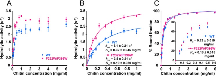
Chitin degradation is important for biomass conversion and has potential applications for agriculture, biotechnology, and the pharmaceutical industry. Chitinase A from the Gram-negative bacterium Serratia marcescens (SmChiA) is a processive enzyme that hydrolyzes crystalline chitin as it moves linearly along the substrate surface. In a previous study, the catalytic activity of SmChiA against crystalline chitin was found to increase after the tryptophan substitution of two phenylalanine residues (F232W and F396W), located at the entrance and exit of the substrate binding cleft of the catalytic domain, respectively. However, the mechanism underlying this high catalytic activity remains elusive. In this study, single-molecule fluorescence imaging and high-speed atomic force microscopy were applied to understand the mechanism of this high-catalytic-activity mutant. A reaction scheme including processive catalysis was used to reproduce the properties of SmChiA WT and F232W/F396W, in which all of the kinetic parameters were experimentally determined. High activity of F232W/F396W mutant was caused by a high processivity and a low dissociation rate constant after productive binding. The turnover numbers for both WT and F232W/F396W, determined by the biochemical analysis, were well-replicated using the kinetic parameters obtained from single-molecule imaging analysis, indicating the validity of the reaction scheme. Furthermore, alignment of amino acid sequences of 258 SmChiA-like proteins revealed that tryptophan, not phenylalanine, is the predominant amino acid at the corresponding positions (Phe-232 and Phe-396 for SmChiA). Our study will be helpful for understanding the kinetic mechanisms and further improvement of crystalline chitin hydrolytic activity of SmChiA mutants.

Keywords: Serratia marcescens; biomass conversion; biotechnology; chitin; chitin degradation; chitinase; high-speed atomic force microscopy; processivity; single-molecule biophysics; single-molecule fluorescence imaging.


Conflict of interest statement

The authors declare that they have no conflicts of interest with the contents of this article

Figures

Figure 1.
Figure 1.
Schematic model of SmChiA. A, crystal structure of SmChiA complexed with (GlcNAc)7 (sphere model) and crystalline chitin (stick model). The schematic shows the CD and CBD. Phe-232 and Phe-396 (sphere model, colored with cyan) are located at the entrance and exit of the chitin-binding cleft, respectively. B and C, structural superimposition to compare the aromatic residues inside the chitin-binding cleft of SmChiA (blue and side chain highlighted with cyan; PDB entry 1CTN) and OfChi-h bound with (GlcNAc)7 (pink; PDB entry 5GQB) in side view (B) and bottom view (C). The red boxes indicate the two different aromatic residues in SmChiA and OfChi-h. The numbers (−6 to +2) represent the chitin-binding subsites of SmChiA according to the standard nomenclature (38, 39). The red dashed lines in B and C show the position of the bond cleavage.
Figure 2.
Figure 2.
Biochemical analysis. A, hydrolytic activity of SmChiA WT (blue) and F232W/F396W (pink) at various concentrations of crystalline chitin (0–6 mg/ml). B, same as A at a low concentration range (0–1 mg/ml). The data points were fitted with the Michaelis–Menten equation to estimate kcat and Km of WT and F232W/F396W. Hydrolytic activity was measured in 50 mm sodium phosphate (pH 6.0) at 25 °C. C, the bound fraction of WT and F232W/F396W at various concentrations of crystalline chitin (0–6 mg/ml). Inset, the low chitin concentration range (<0.6 mg/ml). The amount of free enzymes was used to calculate the bound fraction percentage. The distribution was fitted with the Langmuir equation to estimate the dissociation constant (Kd). Error bars, S.D. of the sextupled (A and B) or triplicate (C) measurements.
Figure 3.
Figure 3.
Comparison of the distribution of kon and koff of SmChiA WT (blue) and F232W/F396W (pink) obtained by single-molecule fluorescence imaging analysis. Top, the distributions of kon were fitted with the double Gaussian function. The first peak corresponds to the kon against the single crystalline chitin microfibril. The sample number (N) in the distribution of kon represents the number of crystalline chitins observed. Bottom, the distributions of koff were fitted with the double-exponential decay function. A slow dissociation is associated with the binding of the enzyme to the hydrophobic crystalline chitin surfaces, whereas the fast dissociation is associated with the binding of the enzyme to the hydrophilic crystalline chitin surfaces. The sample number (N) in the distribution of koff represents the number of SmChiA molecules observed.
Figure 4.
Figure 4.
Comparison of the distribution of ktr, run length, and moving time of WT (blue) and F232W/F396W (pink) obtained by HS-AFM. Left, the distributions of ktr were fitted with the single Gaussian function. The processive catalysis rate constant (kpc) was calculated by dividing ktr with the step size (product size: 1.04 nm). Center and right, the distributions of run length and moving time were fitted with the single-exponential decay function. The processivity (P) was estimated by dividing the run length by the step size. The inverse of the moving time was determined as the productive dissociation rate constant (koffP), because all of the molecules analyzed by HS-AFM were moving molecules. The N and N chitin represent the number of SmChiA molecules and chitin microfibrils, respectively. The first bins (gray bars) of the run length were not included for fitting, because precise measurement of short run length was difficult.
Figure 5.
Figure 5.
Reaction scheme of the processive catalysis of SmChiA described with the kinetic parameters obtained experimentally. The green, blue, and red arrows denote the productive binding/dissociation, nonproductive binding/dissociation, and processive catalysis (hydrolysis cycle), respectively. E, enzyme (SmChiA); S, substrate (crystalline chitin); ES, enzyme-substrate complex after productive binding; ES, enzyme-substrate complex after nonproductive binding; P, product (chitobiose).
Figure 6.
Figure 6.
Comparison of the aromatic amino acid residues responsible for the binding to the crystalline chitin surface and chain sliding into the chitin binding cleft. The amino acid sequences of 258 SmChiA-like proteins were aligned and visualized by Clustal-Omega and WebLogo3. The aromatic amino acids of SmChiA are shown in cyan. The alignment shows the conservation of several aromatic amino acid residues involved in chitin binding. The structural alignment image was constructed by superimposing the crystal structure of SmChiA (PDB entry 1CTN; blue and side chain highlighted with cyan), OfChi-h (PDB entry 5GQB; pink), and VhChiA (PDB entry 3B8S; orange) using PyMOL software. The side chains of aromatic amino acid residues are shown as stick models.

References

    1. Langner T., and Göhre V. (2016) Fungal chitinases: function, regulation, and potential roles in plant/pathogen interactions. Curr. Genet. 62, 243–254 10.1007/s00294-015-0530-x - DOI - PubMed
    1. Merzendorfer H., and Zimoch L. (2003) Chitin metabolism in insects: structure, function and regulation of chitin synthases and chitinases. J. Exp. Biol. 206, 4393–4412 10.1242/jeb.00709 - DOI - PubMed
    1. Hamed I., Özogul F., and Regenstein J. M. (2016) Industrial applications of crustacean by-products (chitin, chitosan, and chitooligosaccharides): a review. Trends Food Sci. Technol. 48, 40–50 10.1016/j.tifs.2015.11.007 - DOI
    1. Songsiriritthigul C., Pantoom S., Aguda A. H., Robinson R. C., and Suginta W. (2008) Crystal structures of Vibrio harveyi chitinase A complexed with chitooligosaccharides: implications for the catalytic mechanism. J. Struct. Biol. 162, 491–499 10.1016/j.jsb.2008.03.008 - DOI - PubMed
    1. Yan N., and Chen X. (2015) Sustainability: don't waste seafood waste. Nature 524, 155–157 10.1038/524155a - DOI - PubMed

Publication types

MeSH terms

LinkOut - more resources