Comment
. 2020 May 1;26(9):2163-2175.
doi: 10.1158/1078-0432.CCR-19-2840. Epub 2020 Jan 10.

Proteomic Analysis of CSF from Patients with Leptomeningeal Melanoma Metastases Identifies Signatures Associated with Disease Progression and Therapeutic Resistance

Affiliations
Comment

Proteomic Analysis of CSF from Patients with Leptomeningeal Melanoma Metastases Identifies Signatures Associated with Disease Progression and Therapeutic Resistance

Inna Smalley et al. Clin Cancer Res. .

Abstract

Purpose: The development of leptomeningeal melanoma metastases (LMM) is a rare and devastating complication of the late-stage disease, for which no effective treatments exist. Here, we performed a multi-omics analysis of the cerebrospinal fluid (CSF) from patients with LMM to determine how the leptomeningeal microenvironment shapes the biology and therapeutic responses of melanoma cells.

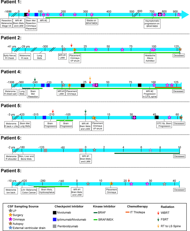
Experimental design: A total of 45 serial CSF samples were collected from 16 patients, 8 of these with confirmed LMM. Of those with LMM, 7 had poor survival (<4 months) and one was an extraordinary responder (still alive with survival >35 months). CSF samples were analyzed by mass spectrometry and incubated with melanoma cells that were subjected to RNA sequencing (RNA-seq) analysis. Functional assays were performed to validate the pathways identified.

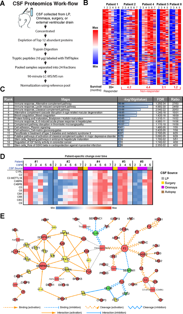
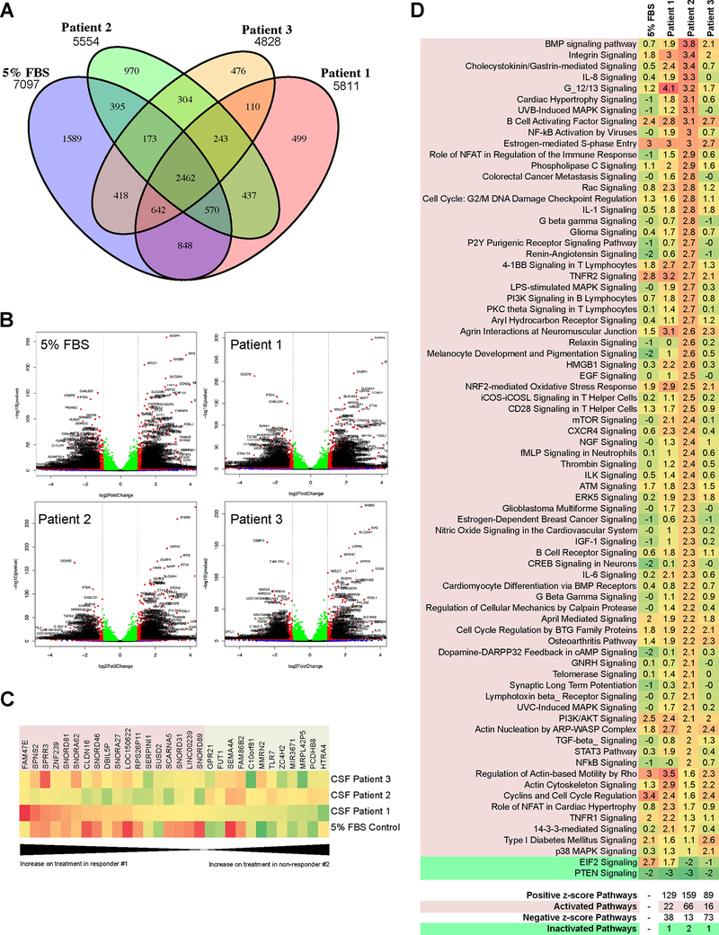
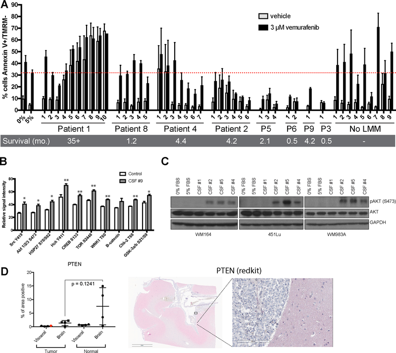
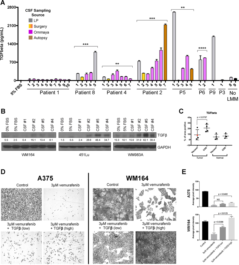
Results: Mass spectrometry analyses showed the CSF of most patients with LMM to be enriched for pathways involved in innate immunity, protease-mediated damage, and IGF-related signaling. All of these were anticorrelated in the extraordinary responder. RNA-seq analysis showed CSF to induce PI3K/AKT, integrin, B-cell activation, S-phase entry, TNFR2, TGFβ, and oxidative stress responses in the melanoma cells. ELISA assays confirmed that TGFβ expression increased in the CSF of patients progressing with LMM. CSF from poorly responding patients conferred tolerance to BRAF inhibitor therapy in apoptosis assays.

Conclusions: These analyses identified proteomic/transcriptional signatures in the CSF of patients who succumbed to LMM. We further showed that the CSF from patients with LMM has the potential to modulate BRAF inhibitor responses and may contribute to drug resistance.See related commentary by Glitza Oliva and Tawbi, p. 2083.


Conflict of interest statement

Conflicts of interest: Peter Forsyth serves on the advisory board for Novocure, BTG, Inovio, AbbVie Inc., Ziopharm, and Tocagen, outside the submitted work. All other authors have nothing to disclose.

Figures

Figure 1.
Figure 1.. Clinical timelines of leptomeningeal patient cohort.
Each timeline shows the unique course of patient’s disease and treatment and provides the clinical context for each CSF sampling. The time is indicated in days unless otherwise stated and is framed around the day of LMM diagnosis (day 0). Treatments include checkpoint inhibitors, kinase inhibitors and radiation therapies. CSF cytology results indicate counts for WBCs, RBCs and protein as well as the cytology finding when available (not done/negative, atypical, suspicious or positive). A star denotes CSF sampling, and the star color indicates CSF source.
Figure 2.
Figure 2.. Proteomic analysis of serial CSF specimens from leptomeningeal melanoma metastasis patients shows protein signatures associated with poor prognosis.
A. Workflow schematic showing the overall approach for CSF preparation and analysis. B. Heatmap showing LMM-specific protein signatures that change significantly over patient treatment time and are most anti-correlated between responder and non-responders. C. Pathway enrichment analysis of data shown in B illustrates components of the complement pathway, adhesion signaling and IGF activation pathways to be enriched for in CSF of poor responders. D. A closer look at the complement signatures identified using proteomics analysis of CSF in melanoma LMM patients (from panel B). Heatmap shows the protein level changes specific to each patient’s clinical timeline. E. Experimentally consistent literature network shows the major signaling mediators upregulated in CSF of non-responders.
Figure 3.
Figure 3.. RNAseq analysis shows LMM-derived CSF to elicit transcriptional modulation of melanoma cell response to BRAF inhibition.
A. Venn diagram showing number of genes significantly altered in WM164 melanoma cell line on BRAF inhibitor treatment in media supplemented with FBS (normal culturing conditions) or CSF-derived from Patients 1–3 (3 μM vemurafenib, 8 hours). B. Volcano plots showing significant upregulation and downregulation of genes when WM164 are treated with a BRAF inhibitor in media supplemented with FBS or CSF derived from Patients 1–3. C. Heatmap shows 30 of the mRNAs that were significantly altered (≥4-fold change in expression with treatment, padj <= 0.02) in the WM164 melanoma cell line following BRAF inhibitor treatment in the presence of patient CSF were also anti-correlated between Patient #1 (exceptional responder) and #2 (poor responder). mRNAs highlighted red increase during treatment in the presence of CSF from Patient #1, and the mRNAs highlighted green decrease. The opposite trend is observed for CSF from Patient #2. D. Heatmap of IPA pathway activation analysis comparing WM164 treated with BRAF inhibitor in context of media supplemented with FBS or different LMM patient-derived CSF. Pathways are considered to be activated when z-score is above 2.0 and inactivated when the z-score is below −2.0.
Figure 4.
Figure 4.. CSF protects melanoma cells from BRAF inhibitor therapy.
A. Flow cytometry-based apoptosis assay showing the percentage of cells undergoing apoptosis (Annexin V positive, TMRM negative) in WM164 treated with BRAF inhibitor in context of serum-free media, media supplemented with FBS or different patient-derived and commercially purchased CSF (72 hours). Serial specimens from individual patients are labeled in chronological order, numbered relative to their clinical timelines (Figure 1). No LMM CSF includes CSF obtained from 8 individual cancer patients who did not have leptomeningeal metastasis (#1–8) and a commercially available pooled CSF healthy individuals (#9). B. Kinase array showing the top 10 kinases activated in WM164 melanoma cell line treated with CSF from Patient #9 or serum-free media for 8 hours (p<0.05*; p<0.01**). C. Western blot analysis of lysates from WM164, 451Lu and WM983A cell lines treated with patient CSF for 8 hours confirms the increase in AKT activation. D. Quantitative image analysis of PTEN staining by IHC on tissue sections from various CNS sites of disease and other visceral organs (site of primary shown in red dot) obtained at autopsy from a patient with leptomeningeal metastases (left). Representative example of staining localization (right).
Figure 5.
Figure 5.. High TGF-β1 in CSF of patients with LMM promotes drug resistance.
A. ELISA assay showing levels of TGF-β1 in RPMI media supplemented 5% FBS and CSF specimens from patients with LMM, a cancer patient without LMM (No LMM #8) and commercially available healthy CSF (No LMM #9). Six out of seven poor responders show elevated levels of TGFβ1 in CSF compared to responder or none-LMM controls. Bar colors correspond to CSF collection source, p-values are denoted for a t-test comparing the average level of TGFβ1 over all time points for a patient compared to Patient #1 extraordinary responder (p<0.01**; p<0.001***; p<0.0001 ****). B. Western Blot analysis of lysates from WM164, 451Lu and WM983A cell lines treated with patient CSF for 8 hours confirms the increase in TGFβ (~45kDa). Quantification of TGF-β bands represent the mean pixel intensity above background. C. IHC analysis of TGF-β1 in autopsy specimens from different sites of disease in CNS and other visceral organs (site of primary shown in red dot) shows an elevated level of TGF-β1 staining in the tumor cells residing in CNS compared to tumors at visceral sites. D. Long term colony formation assays of A375 and WM164 melanoma cells treated with vemurafenib (3 μM) in the presence of absence of recombinant human TGF-β1 (high = 200pg/ml, low = 1ng/ml) for two weeks. E. Quantification of results shown in D. Data represents the average pixel intensity (inverse image) of 3 fields of view from each treatment condition.

Comment in

Comment on

References

    1. Smalley KS, Fedorenko IV, Kenchappa RS, Sahebjam S, Forsyth PA. Managing leptomeningeal melanoma metastases in the era of immune and targeted therapy. International journal of cancer 2016;139(6):1195–201 doi 10.1002/ijc.30147. - DOI - PMC - PubMed
    1. Leal T, Chang JE, Mehta M, Robins HI. Leptomeningeal Metastasis: Challenges in Diagnosis and Treatment. Curr Cancer Ther Rev 2011;7(4):319–27 doi 10.2174/157339411797642597. - DOI - PMC - PubMed
    1. Kokkoris CP. Leptomeningeal carcinomatosis. How does cancer reach the pia-arachnoid? Cancer 1983;51(1):154–60. - PubMed
    1. Le Rhun E, Taillibert S, Chamberlain MC. Carcinomatous meningitis: Leptomeningeal metastases in solid tumors. Surg Neurol Int 2013;4(Suppl 4):S265–88 doi 10.4103/2152-7806.111304. - DOI - PMC - PubMed
    1. Pape E, Desmedt E, Zairi F, Baranzelli MC, Dziwniel V, Dubois F, et al. Leptomeningeal metastasis in melanoma: a prospective clinical study of nine patients. In Vivo 2012;26(6):1079–86. - PubMed

Publication types