. 2020 Jan 10;11(1):22.
doi: 10.1038/s41419-019-2215-8.

Loss of PARP-1 attenuates diabetic arteriosclerotic calcification via Stat1/Runx2 axis

Affiliations

Loss of PARP-1 attenuates diabetic arteriosclerotic calcification via Stat1/Runx2 axis

Peng Li et al. Cell Death Dis. .

Erratum in

Abstract

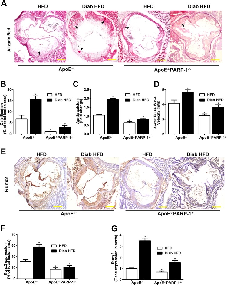
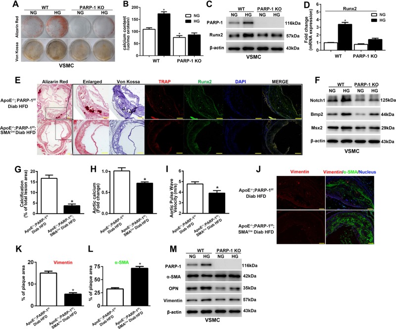
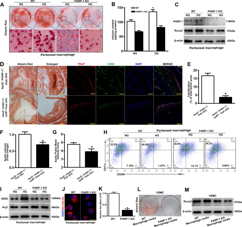
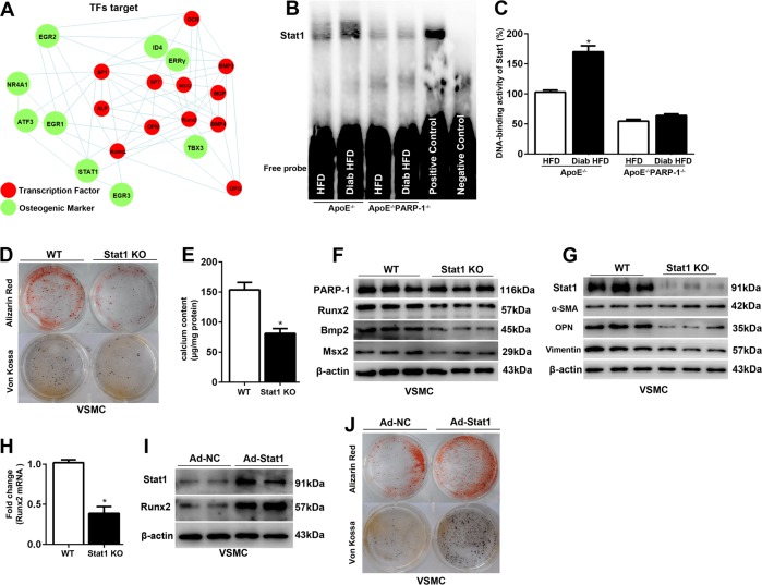
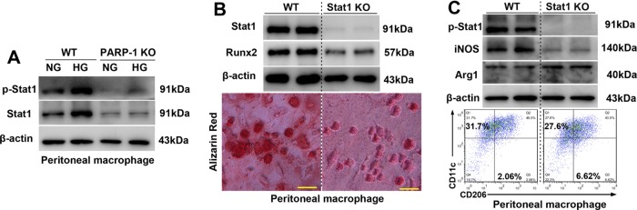
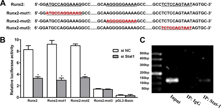
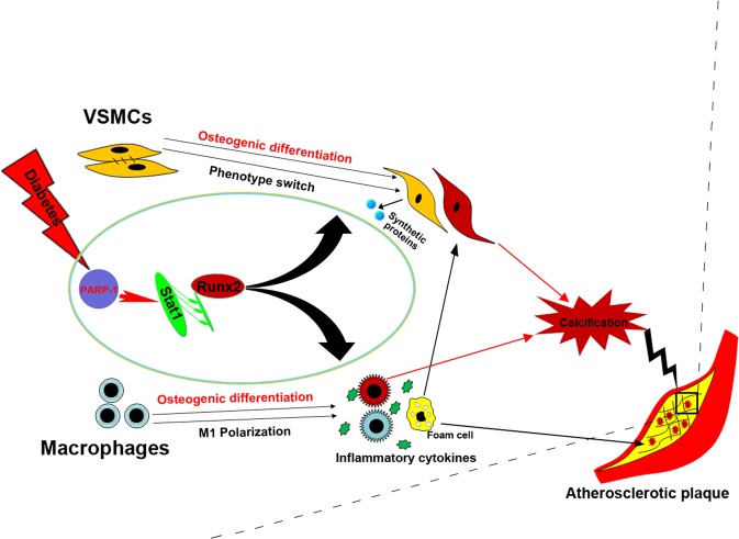
Accelerated atherosclerotic calcification is responsible for plaque burden, especially in diabetes. The regulatory mechanism for atherosclerotic calcification in diabetes is poorly characterized. Here we show that deletion of PARP-1, a main enzyme in diverse metabolic complications, attenuates diabetic atherosclerotic calcification and decreases vessel stiffening in mice through Runx2 suppression. Specifically, PARP-1 deficiency reduces diabetic arteriosclerotic calcification by regulating Stat1-mediated synthetic phenotype switching of vascular smooth muscle cells and macrophage polarization. Meanwhile, both vascular smooth muscle cells and macrophages manifested osteogenic differentiation in osteogenic media, which was attenuated by PARP-1/Stat1 inhibition. Notably, Stat1 acts as a positive transcription factor by directly binding to the promoter of Runx2 and promoting atherosclerotic calcification in diabetes. Our results identify a new function of PARP-1, in which metabolism disturbance-related stimuli activate the Runx2 expression mediated by Stat1 transcription to facilitate diabetic arteriosclerotic calcification. PARP-1 inhibition may therefore represent a useful therapy for this challenging complication.


Conflict of interest statement

The authors declare that they have no conflict of interest.

Figures

Fig. 1
Fig. 1. PARP-1 deletion attenuates diabetic atherosclerotic calcification.
a, b PARP-1 deletion decreased Alizarin Red positively stained atherosclerotic lesion area in diabetic ApoE−/− mice. c, d PARP-1 deletion decreased aortic calcium content and pulse wave velocity. Arrows indicate calcium mineral deposition. e, f Immunohistochemical staining shows increased Runx2-positive areas in diabetic ApoE−/− mice. g Real-time PCR shows increased Runx2 gene expression in the aortas of diabetic ApoE−/− mice. Scale bar = 100 µm. Bar values represent the means ± SD. n = 8 in each group. Asterisks indicate statistically significant differences (*P < 0.01, vs. ApoE−/− HFD mice). The statistical tests are justified as appropriate and meet the assumptions of the tests. The variance between the groups is similar.
Fig. 2
Fig. 2. PARP-1 depletion attenuates hyperglycemia-induced VSMCs calcification and synthetic phenotype switching in vitro and in vivo.
a VSMCs were isolated from WT and PARP-1−/− mice and cultured in osteogenic medium with (HG) or without (NG) high glucose (27.5 mM) for 3 weeks. Calcification was determined by Alizarin red and Von Kossa staining (n = 5). b Total calcium content was quantified using a Calcium Colorimetric Assay Kit. Results shown are normalized to the total protein amount (n = 5). *P < 0.01, vs. WT VSMCs in osteogenic medium without high glucose. c, d Western blot and RT-PCR analyses were performed to determine the effect of PARP-1 deletion on the expression of Runx2 (n = 5). *P < 0.01, vs. WT VSMCs in osteogenic medium without high glucose. e Consecutive sections stained with Alizarin Red or Von Kossa to detect calcification, and immunofluorescence staining of TRAP and Runx2 are shown. VSMC-specific PARP-1 deletion inhibited the expression of Runx2 and formation of TRAP-positive cells. Scale bar = 50 µm. f PARP-1 depletion attenuates HG-induced VSMCs calcification involving Notch1 repression. Western blot analysis of Notch1 and osteogenic genes including Bmp2, Msx2, and OPN in VSMCs isolated from WT and PARP-1−/− mice (n = 5). gi VSMC-specific PARP-1 depletion decreased atherosclerotic calcification lesion area, aortic calcium content, and pulse wave velocity (n = 8). *P < 0.01, vs. diabetic ApoE−/−/PARP-1f/f mice. j Immunofluorescence staining of vimentin (red) and α-SMA (green) in atherosclerotic plaques. VSMC-specific PARP-1 depletion reverses hyperglycemia-induced synthetic phenotype switching of VSMCs in atherosclerotic plaques. k, l Quantification of vimentin and α-SMA coverage. Scale bar = 50 µm (n = 8). *P < 0.01, vs. diabetic ApoE−/−/PARP-1f/f mice. m Western blot analysis of contractile (α-SMA) and synthetic proteins (vimentin and OPN) in VSMCs isolated from WT and PARP-1−/− mice (n = 5). The statistical tests are justified as appropriate and meet the assumptions of the tests. The variance between the groups is similar.
Fig. 3
Fig. 3. PARP-1 plays a critical role in hyperglycemia-induced arteriosclerotic calcification by regulating macrophage calcification and macrophage polarization.
Peritoneal macrophages were isolated from thioglycollate-injected WT and PARP-1−/− mice. a, b Macrophages exhibited evident calcification and increased calcium content after 3-week exposure to osteogenic medium with high glucose (HG) compared with normal glucose (NG), which was reversed by PARP-1 deletion (n = 5). Scale bar = 10 µm. c Western blot analysis was performed to determine the effect of PARP-1 deletion on the expression of Runx2 (n = 5). d Colocalization of TRAP and CD68 revealed that macrophages participated in atherosclerotic calcification. Scale bar = 50 µm. eg Macrophage-specific PARP-1 depletion decreased atherosclerotic calcification lesion area, aortic calcium content, and pulse wave velocity (n = 8). *P < 0.01, vs. diabetic ApoE−/−/PARP-1f/f mice. h, i Flow cytometry and immunoblotting were performed to detect the effect of PARP-1 depletion on macrophage phenotypes switching. j, k Effect on endocytosis of Dil-labeled ox-LDL by PARP-1 depletion. Scale bar = 10 µm. n = 5. Asterisks indicate statistically significant differences (*P < 0.01). l, m VSMCs exhibited evident calcification by stimulation with conditioned medium from WT mouse macrophages for 2 weeks, accompanied by higher expression levels of Runx2. PARP-1 depletion abolished the procalcification effect of macrophages (n = 5). The statistical tests are justified as appropriate and meet the assumptions of the tests. The variance between the groups is similar.
Fig. 4
Fig. 4. PARP-1 deficiency attenuates Stat1-mediated VSMCs calcification and synthetic phenotype switching.
a Protein–protein interaction networks illustrate the osteogenic genes downstream of osteogenic TFs. b, c Stat1 binding activity following PARP-1 deletion in the aortas of diabetic ApoE−/− mice. n = 3 in each group. Asterisks indicate statistically significant differences (*P < 0.01, vs. ApoE−/− mice). d, e Stat1 depletion attenuates VSMC calcification and decreases calcium content after 3-week exposure to osteogenic medium with high glucose (n = 5). *P < 0.01, vs. WT VSMC. f, g Western blot were performed to determine the effect of Stat1 depletion on VSMCs osteogenic differentiation and synthetic phenotype switching (n = 5). h Real-time PCR analysis were performed to determine the effect of Stat1 depletion on the mRNA expression of Runx2 (n = 5). *P < 0.01, vs. WT VSMCs. i, j Effect of Stat1 overexpression on Runx2 expression and VSMC calcification as measured by Alizarin Red and Von Kossa staining after 2-week exposure to osteogenic medium with high glucose (n = 5). The statistical tests are justified as appropriate and meet the assumptions of the tests. The variance between the groups is similar.
Fig. 5
Fig. 5. PARP-1 deficiency attenuates Stat1-mediated macrophage calcification and macrophage polarization.
a Immunoblotting was performed to detect the effect of PARP-1 depletion on total and phosphorylated Stat1 levels (n = 5). b Stat1 depletion attenuates high glucose-promoted macrophage calcification in vitro (n = 5). Scale bar = 10 µm. c Immunoblotting and flow cytometry were performed to detect the effect of Stat1 depletion on macrophage phenotype switching (n = 5).
Fig. 6
Fig. 6. Stat1 directly binds to the Runx2 promoter and contributes to PARP-1-mediated arteriosclerotic calcification.
a Predicted Stat1 binding site (underlined) within the human Runx2 promoter. Mutants with deletion of the predicted binding site (Runx2-mut1, Runx2-mut2, and Runx2-mut3) are shown. b Luciferase activity assay was performed after transfection with the human Runx2 promoter or Runx2 promoter mutants in 293T cells (n = 5).*P< 0.01, vs. transfection with con siRNA. c ChIP assay to verify binding of Stat1 to the Runx2 promoter (n = 5). The statistical tests are justified as appropriate and meet the assumptions of the tests. The variance between the groups is similar.
Fig. 7
Fig. 7. Proposed working schematic of PARP-1 depletion-mediated attenuation of atherosclerotic calcification in diabetes.
Diabetes activates Runx2 expression and induces the osteogenic differentiation of both VSMCs and macrophages. Concurrently, diabetes promotes phenotype switching of VSMCs from the contractile phenotype to a dedifferentiated synthetic phenotype, and of macrophages to a proinflammatory M1 phenotype, which in turn aggravates VSMC calcification. PARP-1 acts on Stat1 transcription, which functions as a regulator of Runx2 expression and osteogenic differentiation. PARP-1 depletion reversed the hyperglycemia-induced synthetic phenotype switching of VSMCs and macrophage polarization by targeting Stat1. As a result, PARP-1 depletion suppresses diabetic atherosclerotic calcification.

Similar articles

Cited by

References

    1. Luscher TF, Creager MA, Beckman JA, Cosentino F. Diabetes and vascular disease: pathophysiology, clinical consequences, and medical therapy: Part II. Circulation. 2003;108:1655–1661. doi: 10.1161/01.CIR.0000089189.70578.E2. - DOI - PubMed
    1. Lanzer P, et al. Medial vascular calcification revisited: review and perspectives. Eur. Heart J. 2014;35:1515–1525. doi: 10.1093/eurheartj/ehu163. - DOI - PMC - PubMed
    1. Ndip A, Wilkinson FL, Jude EB, Boulton AJ, Alexander MY. RANKL-OPG and RAGE modulation in vascular calcification and diabetes: novel targets for therapy. Diabetologia. 2014;57:2251–2260. doi: 10.1007/s00125-014-3348-z. - DOI - PubMed
    1. Mori S, Takemoto M, Yokote K, Asaumi S, Saito Y. Hyperglycemia-induced alteration of vascular smooth muscle phenotype. J. Diabetes Complic. 2002;16:65–68. doi: 10.1016/S1056-8727(01)00189-1. - DOI - PubMed
    1. Tang R, et al. High glucose mediates endothelial-to-chondrocyte transition in human aortic endothelial cells. Cardiovasc. Diabetol. 2012;11:113. doi: 10.1186/1475-2840-11-113. - DOI - PMC - PubMed

Publication types

MeSH terms

Substances