. 2020 Jan 10;11(1):21.
doi: 10.1038/s41419-019-2212-y.

Piperlongumine regulates epigenetic modulation and alleviates psoriasis-like skin inflammation via inhibition of hyperproliferation and inflammation

Affiliations

Piperlongumine regulates epigenetic modulation and alleviates psoriasis-like skin inflammation via inhibition of hyperproliferation and inflammation

Sowjanya Thatikonda et al. Cell Death Dis. .

Abstract

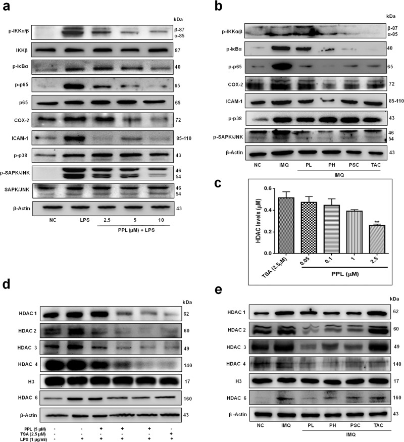
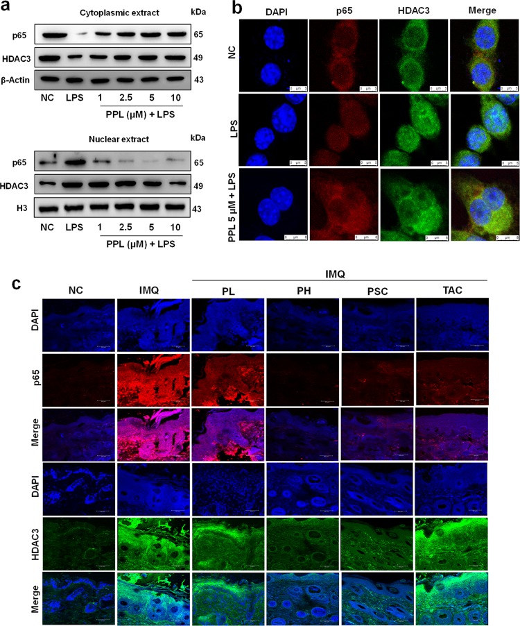
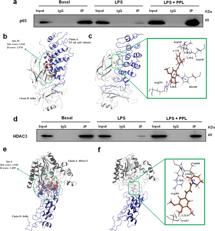
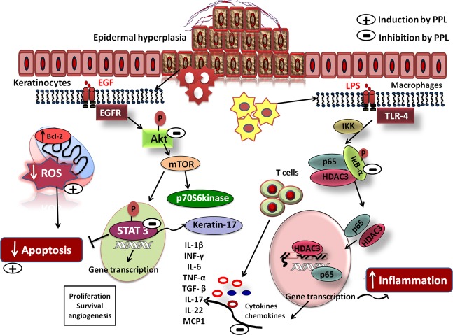
Psoriasis is an autoimmune skin disease, where chronic immune responses due to exaggerated cytokine signaling, abnormal differentiation, and evasion of keratinocytes apoptosis plays a crucial role in mediating abnormal keratinocytes hyperproliferation. From the therapeutic perspective, the molecules with strong anti-proliferative and anti-inflammatory properties could have tremendous relevance. In this study, we demonstrated that piperlongumine (PPL) treatment effectively abrogated the hyperproliferation and differentiation of keratinocytes by inducing ROS-mediated late apoptosis with loss of mitochondrial membrane potential. Besides, the arrest of cell cycle was found at Sub-G1 phase as a result of DNA fragmentation. Molecularly, inhibition of STAT3 and Akt signaling was observed with a decrease in proliferative markers such as PCNA, ki67, and Cyclin D1 along with anti-apoptotic Bcl-2 protein expression. Keratin 17 is a critical regulator of keratinocyte differentiation, and it was found to be downregulated with PPL significantly. Furthermore, prominent anti-inflammatory effects were observed by inhibition of lipopolysaccharide (LPS)/Imiquimod (IMQ)-induced p65 NF-κB signaling cascade and strongly inhibited the production of cytokine storm involved in psoriasis-like skin inflammation, thus led to the restoration of normal epidermal architecture with reduction of epidermal hyperplasia and splenomegaly. In addition, PPL epigenetically inhibited histone-modifying enzymes, which include histone deacetylases (HDACs) of class I (HDAC1-4) and class II (HDAC6) evaluated by immunoblotting and HDAC enzyme assay kit. In addition, our results show that PPL effectively inhibits the nuclear translocation of p65 and a histone modulator HDAC3, thus sequestered in the cytoplasm of macrophages. Furthermore, PPL effectively enhanced the protein-protein interactions of HDAC3 and p65 with IκBα, which was disrupted by LPS stimulation and were evaluated by Co-IP and molecular modeling. Collectively, our findings indicate that piperlongumine may serve as an anti-proliferative and anti-inflammatory agent and could serve as a potential therapeutic option in treating psoriasis.


Conflict of interest statement

The authors declare that they have no conflict of interest.

Figures

Fig. 1
Fig. 1. Piperlongumine (PPL) intervention attenuates Imiquimod (IMQ)-induced psoriasis severity in BALB/c mice.
Mice were received IMQ, PPL, and Tacrolimus (TAC). a After the completion of study, mice were anesthetized and mages of IMQ-induced hyperplasia on mouse back skin with and without PPL treatment were taken by digital camera. b PPL attenuated splenomegaly induced by topical IMQ. PASI scoring was recorded on days 0, 2, 4, and 7 and the cumulative scores of c redness, d scaling, and e thickness were measured. f Skin fold thickness in all groups was measured by Vernier calipers. Representative histological sections of the back skins of mice where g H & E and h DAPI staining show reduced epidermal hyperplasia and parakeratosis induced by IMQ, and i western blotting analysis was performed with the indicated antibodies, where PPL inhibits IMQ-induced Akt pathway-, apoptosis-, and proliferation-mediated protein expression. The data represent mean ± SD (n = 5 mice per group). *P < 0.05 vs. NC group. ^^P < 0.01 and ^^^P < 0.001 are significantly different from the IMQ group. Here PL = PPL 10 mg/kg and PH = PPL 30 mg/kg topically, PSC = PPL 1 mg/kg subcutaneously, and TAC = Tacrolimus 20 mg/kg topically.
Fig. 2
Fig. 2. PPL inhibits keratinocytes hyperproliferation by induction of apoptosis.
HaCaT cells were pretreated with PPL at the indicated concentrations; after 2 h, EGF (50 ng/ml) was stimulated for 48 h and assessed for a morphological changes induced by PPL under phase contrast microscope. b AO/EB dual staining and c DAPI staining were performed to visualize the apoptotic and nuclear changes by using fluorescent microscope at ×200 magnifications. Flow cytometric analysis was performed to determine the d distribution of PI labeling in different phases. The peaks in the histograms correspond to Sub G1, G0/G1, S, and G2/M phases of the cell cycle. e JC-1 staining was performed to analyze the loss of mitochondrial membrane potential (ΔΨm) by PPL. P1 represents the formation of J-aggregates in healthy mitochondria, whereas P2 represents the loss of ΔΨm in cells due to the presence of J-monomers. f Apoptosis in cells was measured by staining with Alexa Flour 488 Annexin V and PI. The percentage of cells positive for Annexin V-Alexa Flour 488 and/or PI in the quadrants were quantified. Cells in the upper left quadrant (Q1-UL; AV−/PI+): necrotic cells; lower left quadrant (Q2-LL; AV−/PI−): live cells; lower right quadrant (Q3-LR; AV+/PI−): early apoptotic cells and upper right quadrant (Q4-UR; AV+/PI+): late apoptotic cells. g Increase in the TUNEL positivity as an indicative of apoptotic DNA fragmentation with PPL was measured by TUNEL staining after 48 h incubation and images were captured at ×400 magnification. h For checking the phosphorylation of Akt signaling, HaCaT cells were pretreated with PPL for 12 h and stimulated the cells with EGF (50 ng/ml) for 30 min, whereas mTOR, Ki67, PCNA, Cyclin D1, and Bcl-2 expression was determined by pretreating the cells with PPL for 2 h and stimulated the cells for 12 h with EGF. Later, whole-cell extract was subjected to western blotting. PPL counteracts EGF-induced proliferation and apoptosis marker protein expression in HaCaT cells. β-Actin or respective totals were used as an endogenous loading controls.
Fig. 3
Fig. 3. PPL suppresses keratin 17 (K17) by inhibiting STAT3 phosphorylation.
a HaCaT cells were pretreated with PPL for 12 h and stimulated with EGF (50 ng/ml) for 1 h. The protein was isolated and the samples were subjected to western blot analysis to determine the expression of p-STAT3, p-ERK 1/2, and K17 in a HaCaT cells and b skin tissue lysates. c Immunofluorescence was performed to analyze the expression of STAT3 and K17 in keratinocytes. d IF analysis shows attenuation of STAT3 and K17 expression in skin tissue sections with PPL treatment. Here PL = PPL 10 mg/kg and PH = PPL 30 mg/kg topically, PSC = PPL 1 mg/kg subcutaneously, and TAC = Tacrolimus 20 mg/kg topically.
Fig. 4
Fig. 4. PPL suppresses inflammatory stimuli-mediated cytokines and chemokine levels.
MTT assay was performed to determine the cell viability of RAW 264.7 cells by stimulation with LPS (1 μg/ml) after pretreatment of PPL for 2 h and incubated for a 24 and b 48 h. c Cytokine and chemokine profiling was performed in murine macrophages, where cells were pretreated with 0.5, 1, and 2.5 μM concentrations of PPL for 2 h and then stimulated with 1 μg/ml of LPS and incubated for another 24 h and the levels of cl cytokines and mo chemokines were determined by multiplex analysis. pv In addition, PPL effectively ameliorates inflammatory cytokine production in skin tissues. The data represent mean ± SD (in vitro, n = 3; in vivo, n = 5 mice per group). *P < 0.05, **P < 0.01, ***P < 0.001, and ****P < 0.0001 vs. NC group. ^P < 0.05, ^^P < 0.01, ^^^P < 0.001, and ^^^^P < 0.0001 are significantly different from the LPS/IMQ group. Here PL = PPL 10 mg/kg and PH = PPL 30 mg/kg topically, PSC = PPL 1 mg/kg subcutaneously, and TAC = Tacrolimus 20 mg/kg topically.
Fig. 5
Fig. 5. PPL inhibits the protein expression of the inflammatory signaling cascade and exhibits potent HDAC inhibitory activity in murine macrophages and skin tissues.
PPL potently inhibits LPS- and IMQ-induced IKK-mediated downstream signaling but no modulation in the MAP kinase pathways. a RAW 264.7 cells were pretreated with PPL for 12 h and stimulated with LPS for 30 min, and western blot analysis was performed with the indicated antibodies. b Immunoblots of p65 and downstream signaling markers with PPL treatment observed in skin tissues. c HDAC inhibitory activity of PPL was analyzed by HDAC fluorometric kit at 0.05, 0.1, 1, and 2.5 µM concentrations of PPL, and HDAC inhibitory activity was compared with trichostatin A (TSA) at 2.5 µM concentrations. d RAW 264.7 cells were pretreated with PPL at the indicated concentrations for 2 h, and later cells were stimulated with LPS (1 µg/ml) and incubated for 12 h. Nuclear lysate was extracted and analyzed for the preferential overexpression of HDAC1–4 with LPS, abrogation in the expression by PPL treatment were analyzed by western blotting and compared with H3 expression, and HDAC6 expression was analyzed in whole-cell extract and compared with β-Actin expression. e Similarly, PPL epigenetic regulation by reducing HDAC1–4 expression were analyzed in psoriatic and PPL-treated skin tissue nuclear extracts; HDAC6 were analyzed in the whole-cell extracts of skin tissue. The data represent mean ± SD (n = 3). **P < 0.01 is significantly different from the TSA. Here PL = PPL 10 mg/kg and PH = PPL 30 mg/kg topically, PSC = PPL 1 mg/kg subcutaneously, and TAC = Tacrolimus 20 mg/kg topically.
Fig. 6
Fig. 6. PPL potently inhibits the nuclear translocation of p65 and HDAC3 induced by LPS.
a RAW 264.7 cells were grown in full medium and then serum starved for 6 h with 0.1% FBS and treated with PPL at different concentrations indicated; after 12 h, cells were stimulated with LPS (1 µg/ml) and further incubated for 30 min. p65 and HDAC3 expression was analyzed in both cytoplasmic and nuclear fractions, where β-Actin and H3 were the loading controls, respectively. b Immunocytochemical analysis was performed to determine the cellular distribution of p65 and HDAC3 in RAW 264.7 cells and stained with p65 antibody (red), HDAC3 antibody (green), and DAPI for nuclear staining (blue) following treatment of the cells with LPS (1 μg/ml) and PPL (5 µM) + LPS (1 μg/ml). Images were captured at ×630 magnification. c IF results showed that PPL downregulated the expression of p65 (Red) and HDAC3 (Green) in the skin tissue sections. Here PL = PPL 10 mg/kg and PH = PPL 30 mg/kg topically, PSC = PPL 1 mg/kg subcutaneously, and TAC = Tacrolimus 20 mg/kg topically.
Fig. 7
Fig. 7. PPL enhances IκBα-HDAC3-p65 complex.
a Co-immunoprecipitation (Co-IP) was performed in the cytoplasmic extract from RAW 264.7 cells to determine the association of IκBα-p65 in basal condition, upon LPS stimulation and with PPL treatment. IgG was used as a control for nonspecific signal. b Site points (white spheres) and SiteMap surface (red and blue color contours) for Site-IV-binding site of p65/IκBα protein complex. c Docking model of PPL in binding site (Site-IV) of p65/IκBα protein complex and its ligand–protein interactions. The dark red dashed lines represent hydrogen bonds. H-bond distances (in Å) between hetero atoms of ligand and amino acid residues are as follows: Arg143 (1.77 and 3.26), Asn145 (2.59), and Arg253 (3.00). The blue line indicates arene–arene interaction with His184 and Arg253. d Co-IP was performed in the cytoplasmic extract from RAW 264.7 cells to determine the association of IκBα-HDAC3 in basal condition, upon LPS stimulation and with PPL treatment. e Site points (white spheres) and SiteMap surface (red and blue color contours) for Site-I-binding site of HDAC3/IκBα protein complex. f Docking model of PPL in binding site (Site-I) of HDAC3/IκBα protein complex and its ligand–protein interactions. The dark red dashed lines represent hydrogen bonds. H-bond distances (in Å) between hetero atoms of ligand and amino acid residues are as follows: Cys167 (2.21), Val300 (2.14), and Arg301 (2.04 and 2.58).
Fig. 8
Fig. 8. PPL ameliorates psoriatic-like skin inflammation by inhibiting hyperproliferation and inflammation.
Schematic illustration of the effect of PPL on Akt/mTOR/STAT3 cascade and apoptosis. PPL inhibits upstream and downstream signaling in keratinocytes resulting in decreased cell proliferation and induction of apoptosis and promotes differentiation by inhibiting keratin17. Simultaneously abrogation of HDAC3 and p65 nuclear translocation was observed in murine macrophages by inducing association of HDAC3 and p65 to IκBα, thus reducing inflammation and cytokine/chemokine levels and also normalizing the tissue architecture.

References

    1. Albanesi. C, Madonna S, Gisondi P, Girolomoni G. The interplay between keratinocytes and immune cells in the pathogenesis of psoriasis. Front. Immunol. 2018;9:1549. doi: 10.3389/fimmu.2018.01549. - DOI - PMC - PubMed
    1. Lowes MA, Bowcock AM, Krueger JG. Pathogenesis and therapy of psoriasis. Nature. 2007;445:866–873. doi: 10.1038/nature05663. - DOI - PubMed
    1. Wang S, Zhang Z, Peng H, Zeng K. Recent advances on the roles of epidermal growth factor receptor in psoriasis. Am. J. Transl. Res. 2019;11:520–528. - PMC - PubMed
    1. Chen W, Wu L, Zhu W, Chen X. The polymorphisms of growth factor genes (VEGFA & EGF) were associated with response to acitretin in psoriasis. Per. Med. 2018;15:181–188. doi: 10.2217/pme-2017-0085. - DOI - PubMed
    1. Li HH, et al. Interleukin-19 upregulates keratinocyte growth factor and is associated with psoriasis. Br. J. Dermatol. 2005;153:591–595. doi: 10.1111/j.1365-2133.2005.06665.x. - DOI - PubMed

Publication types

MeSH terms