. 2020 Jan 10;115(2):14.
doi: 10.1007/s00395-020-0775-5.

TRPV4 deletion protects heart from myocardial infarction-induced adverse remodeling via modulation of cardiac fibroblast differentiation

Affiliations

TRPV4 deletion protects heart from myocardial infarction-induced adverse remodeling via modulation of cardiac fibroblast differentiation

Ravi K Adapala et al. Basic Res Cardiol. .

Abstract

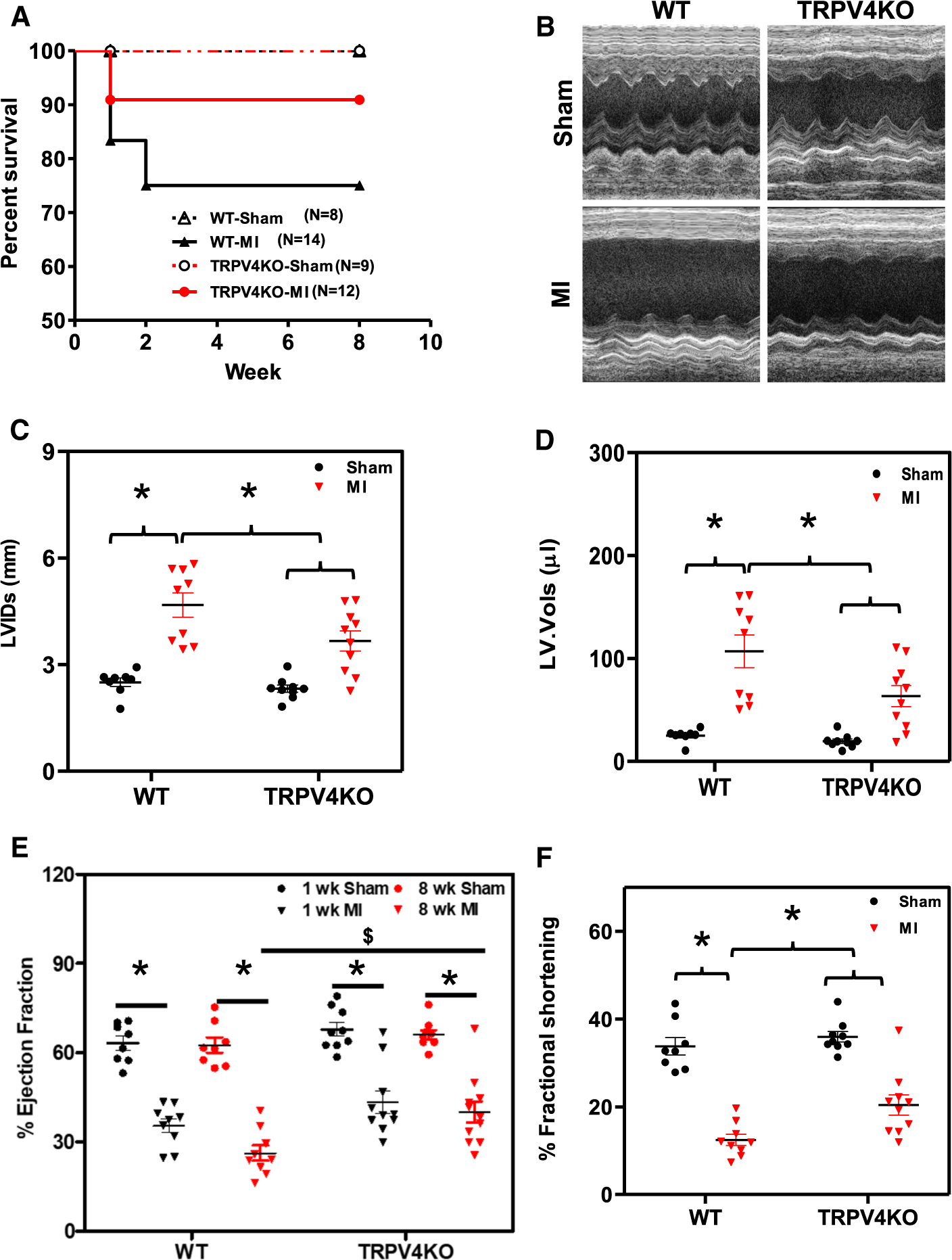
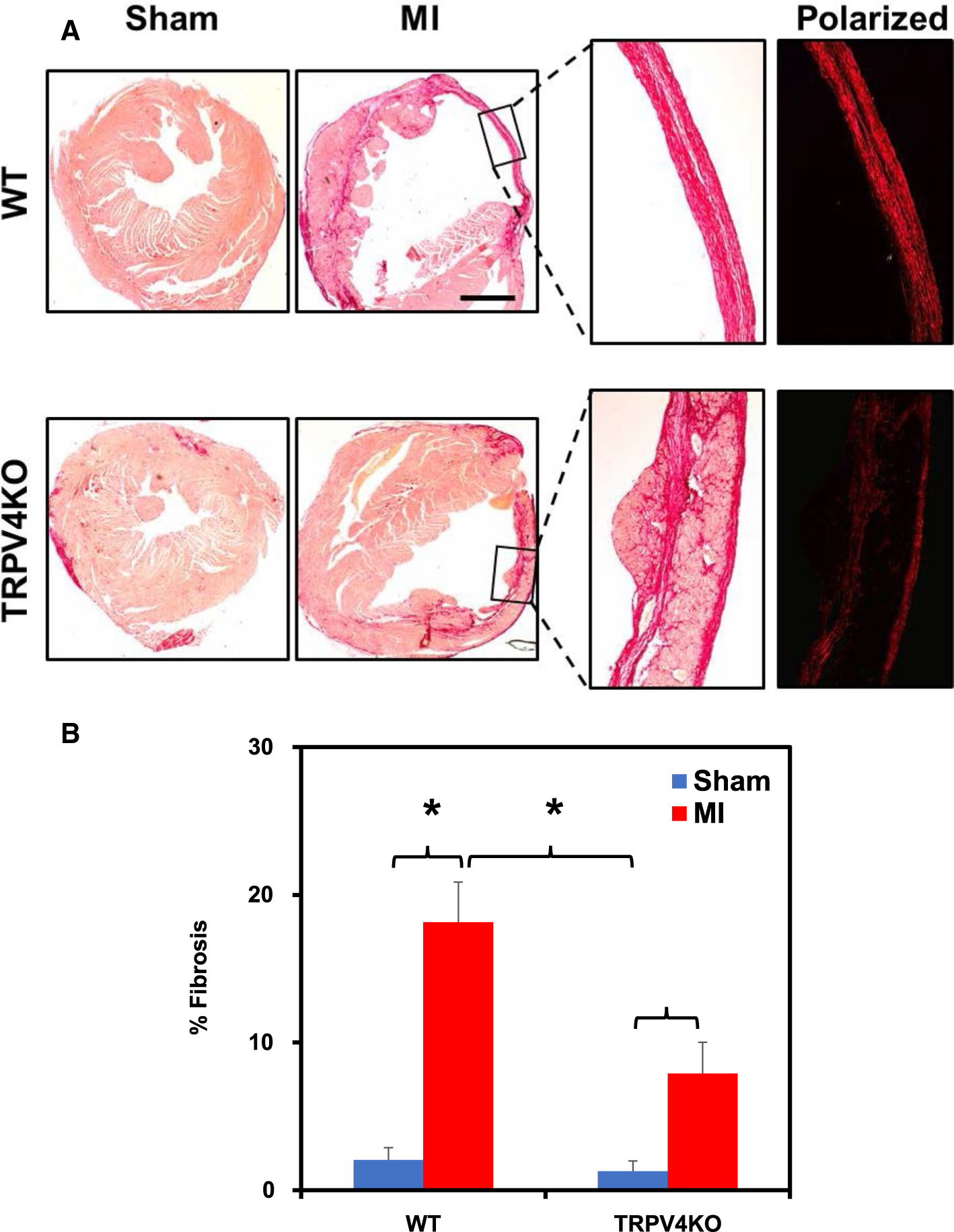
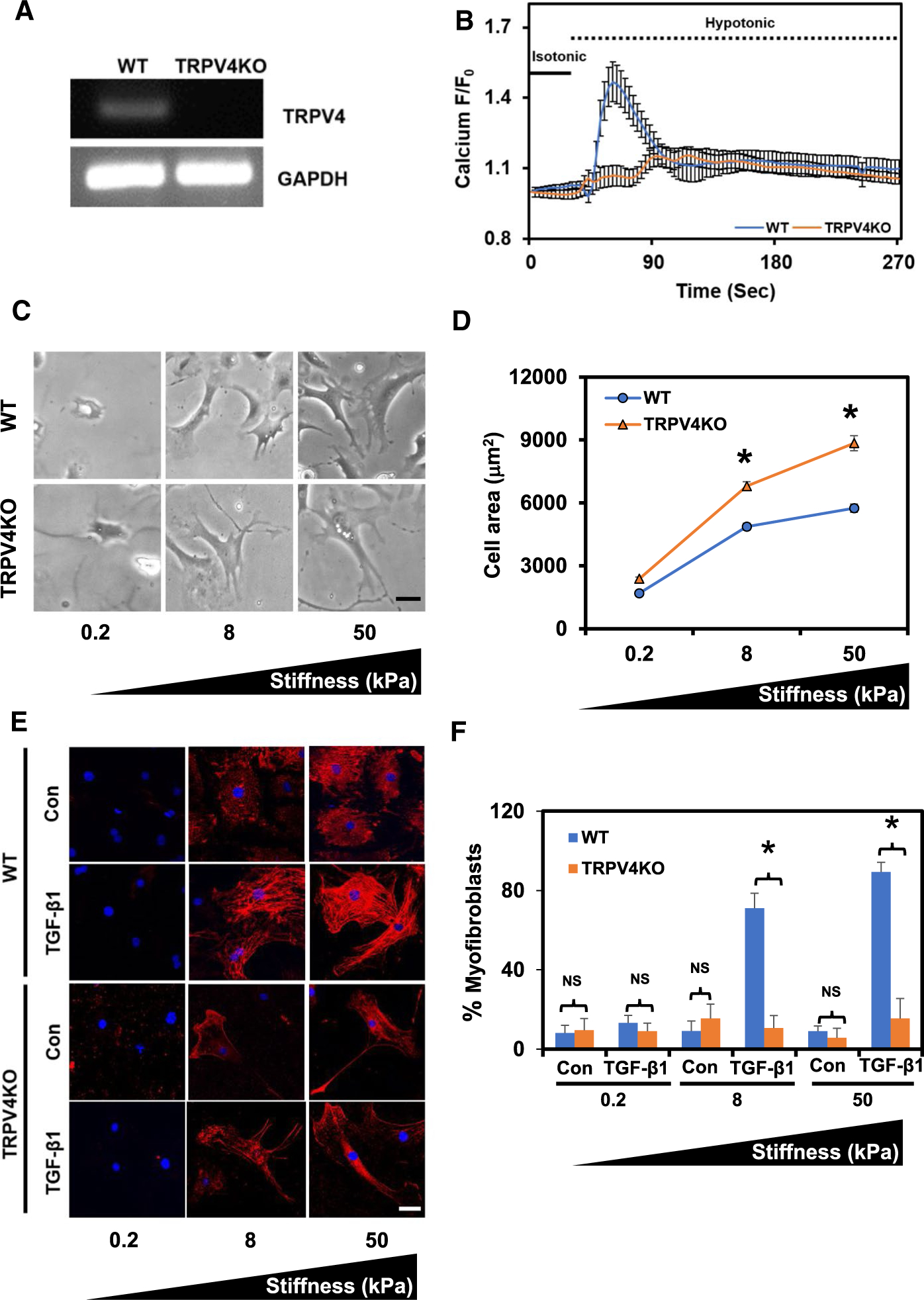
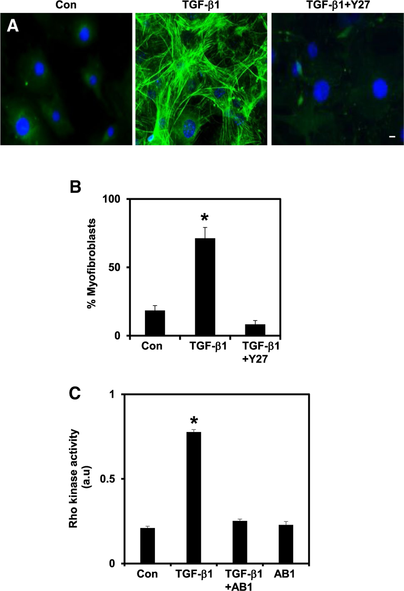
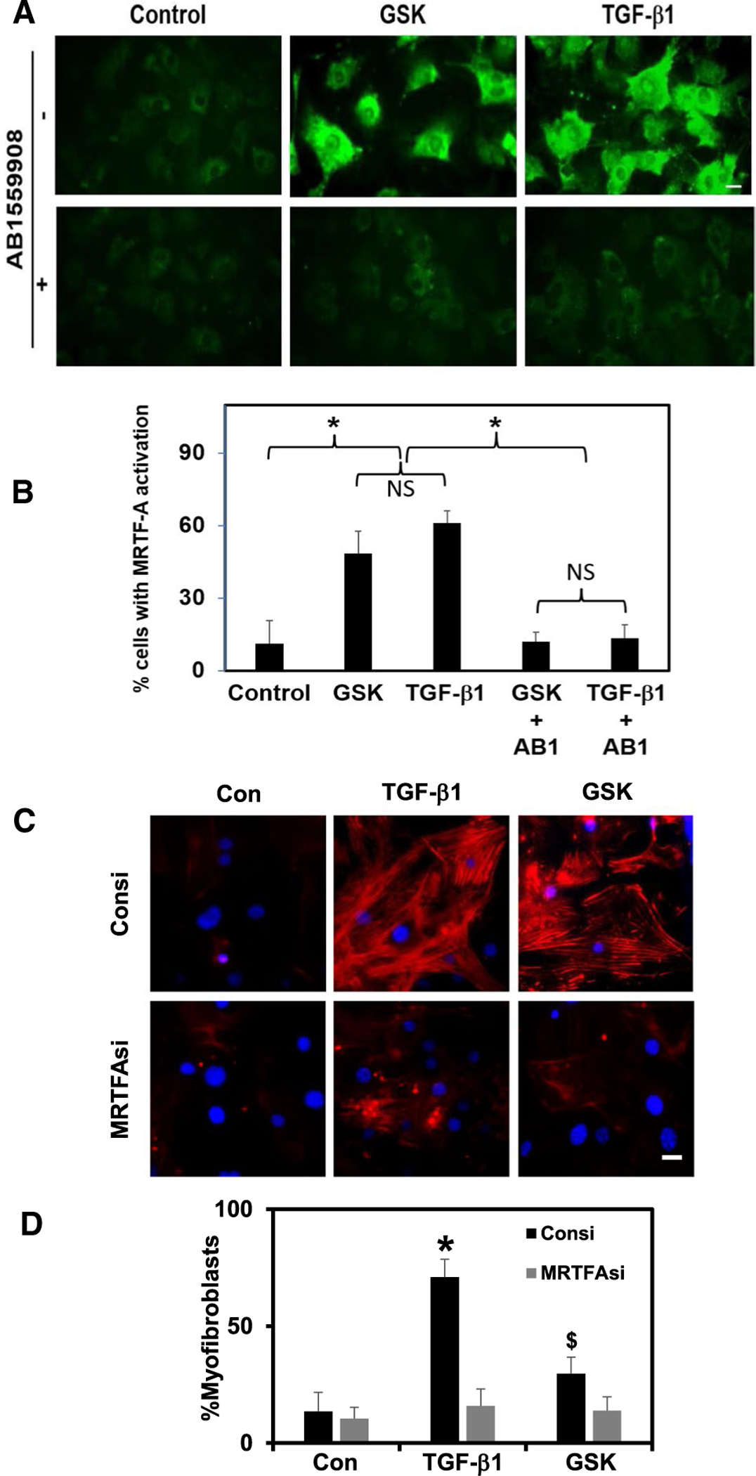
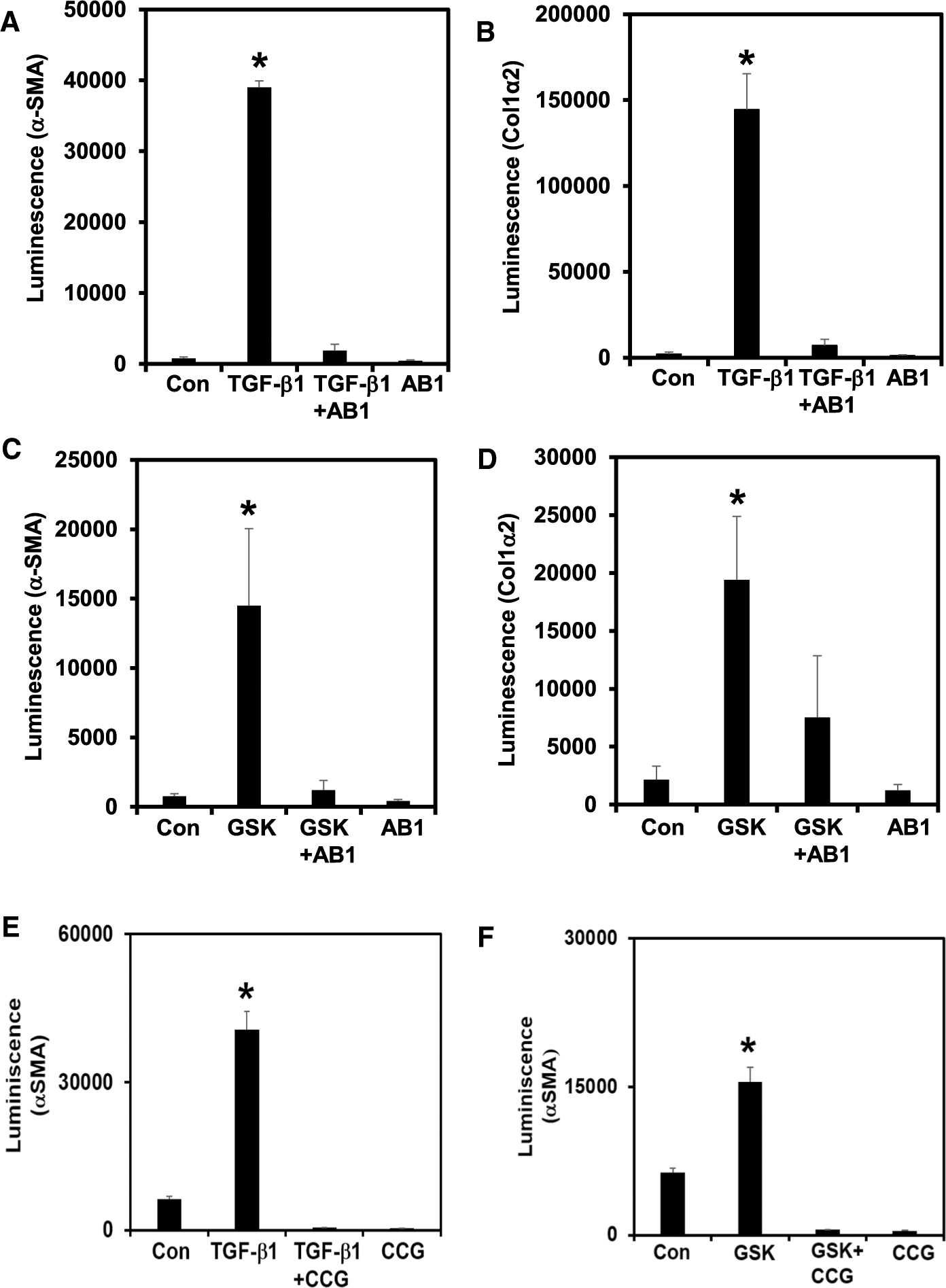
Cardiac fibrosis caused by adverse cardiac remodeling following myocardial infarction can eventually lead to heart failure. Although the role of soluble factors such as TGF-β is well studied in cardiac fibrosis following myocardial injury, the physiological role of mechanotransduction is not fully understood. Here, we investigated the molecular mechanism and functional role of TRPV4 mechanotransduction in cardiac fibrosis. TRPV4KO mice, 8 weeks following myocardial infarction (MI), exhibited preserved cardiac function compared to WT mice. Histological analysis demonstrated reduced cardiac fibrosis in TRPV4KO mice. We found that WT CF exhibited hypotonicity-induced calcium influx and extracellular matrix (ECM)-stiffness-dependent differentiation in response to TGF-β1. In contrast, TRPV4KO CF did not display hypotonicity-induced calcium influx and failed to differentiate on high-stiffness ECM gels even in the presence of saturating amounts of TGF-β1. Mechanistically, TRPV4 mediated cardiac fibrotic gene promoter activity and fibroblast differentiation through the activation of the Rho/Rho kinase pathway and the mechanosensitive transcription factor MRTF-A. Our findings suggest that genetic deletion of TRPV4 channels protects heart from adverse cardiac remodeling following MI by modulating Rho/MRTF-A pathway-mediated cardiac fibroblast differentiation and cardiac fibrosis.

Keywords: Cardiac fibroblast; Cardiac fibrosis; Mechanotransduction; Myocardial infarction; Rho/Rho kinase; TGF-β1; TRPV4.


Conflict of interest statement

Conflict of interest CKT filed a patent application based on some of the findings in the MS. Rest of the authors declare no conflict of interest.

Figures

Fig. 1
Fig. 1
Genetic deletion of TRPV4 preserves cardiac function post-MI. a Kaplan–Meier survival graph showing that survival rate was improved in TRPV4KO mice compared to WT mice following 8 weeks after MI. WT-sham (n = 8); WT-MI (n = 14); TRPV4KO-sham (n = 9); TRPV4KO-MI (n = 11). b Representative 2D-echocardiographic M-mode images showing increased wall integrity in TRPV4KO mice compared to WT mice, post-MI. Quantitative analysis of cardiac function LV internal diamerer (c), LV volume (d); % ejection fraction (e) and % fractional shortening (f) from sham and MI, WT and TRPV4KO mice, 1 and 8 weeks after the surgey (n = 8–10; two-way ANOVA followed by Tukey post hoc analysis; significance was set at p ≤ 0.05). *Sham vs MI; $WT vs TRPV4KO, 8 weeks, post-MI
Fig. 2
Fig. 2
Absence of TRPV4 ameliorates cardiac fibrosis following MI. a Representative images of picrosirius red-stained heart sections from WT and TRPV4KO mice, 8 weeks post-sham or MI surgeries. Polarized microscopy of picrosirius red-stained hearts revealed deposition of matured collagen in WT which was absent in TRPV4KO post-MI hearts. Scale bar 500 μm. b Quantification of percent of collagen deposition (fibrosis) in WT and TRPV4KO hearts, 8 weeks after sham or MI surgeries (n = 3–5; one-way ANOVA followed by Tukey post hoc analysis and significance was set at *p ≤ 0.05)
Fig. 3
Fig. 3
Absence of TRPV4 attenuates TGF-β1-induced mouse CF differentiation. a Representative RT-PCR agarose gel shows TRPV4 expression in WT mCF but not in the TRPV4KO mCF. b Average calcium traces depicting calcium influx in response to the replacement of isotonic solution (~ 300 mOsm) (line) with hypo-tonic solution (~ 140 mOsm) (dashed line) in Fluo-4-loaded WT and TRPV4KO mCF. Note that hypotonic stimulation failed to induce calcium influx in TRPV4KO-mCF. The data presented are mean ± SEM of three separate experiments (n = 60 cells). c Bright-field images showing spreading of WT and TRPV4KO mCF on the ECM gels of varying stiffnesses coated with collagen I (0.2, 8, and 50 kPa). Scale bar 50 μm. d Quantification of cell area shows ECM stiffness-dependent spreading of WT mCF which reached platue at 50 kPa, while TRPV4KO mCF exhibited continuously increased spreading with an increase in stiffness. e Immunofluorescence images (× 40) showing α-SMA expression and incorporation into stress fibers in isolated WT and TRPV4KO mCF on different stiffness gels (0.2, 8 and 50 kPa) in response to TGF-β1 (10 ng/mL) (α-SMA: red and DAPI/nuclei: blue). Scale bar 50 μm. f Quantitative analysis of fibroblast differentiation (α-SMA-positive cells). The data presented are mean ± SEM of three independent experiments. Student’s t test; significance was set at *p ≤ 0.05
Fig. 4
Fig. 4
TRPV4 mediates CF differentiation via Rho/Rho kinase pathway. a Representative immunofluorescence images (× 20) displaying TGF-β1 (10 ng/mL)-induced differentiation of WT mCF in the presence or absence of Rho kinase inhibitor Y27632 (Y27–10 μM) (α-SMA: green and DAPI/nuclei: blue). Scale bar 10 μm. b Quantitative analysis of fibroblast differentiation (α-SMA-positive cells). c Rho kinase activity was measured in response to TGF-β1 in WT mCF either untreated or treated with TRPV4 antagonist AB1 (10 μM). The data presented are mean ± SEM of three separate experiments. Student’s t test; significance was set at *p ≤ 0.05
Fig. 5
Fig. 5
TRPV4 mediates TGF-β1-induced CF differentiation via activation of MRTF-A. a Representative immunofluorescence images (× 20) showing activation of MRTF-A (green) in WT mCF in response to TGF-β1 (10 ng/mL) or GSK (100 nM), in the presence or absence of TRPV4 antagonist, AB1. Scale bar 10 μm. b Quantitative analysis of MRTF-A activation (cytosolic and nuclear translocation). c Immunofluorescence images (× 20) showing TGF-β1/GSK-induced differentiation of CF to MF (α-SMA: red, and nuclei were stained with DAPI, blue) in control siRNA and MRTF-A siRNA-treated mCF. d Quantitative analysis of fibroblast differentiation (α-SMA-positive cells). The data presented are mean ± SEM of three separate experiments. Student’s t test; significance was set at p ≤ 0.05. *Significance between TGF-β1 vs control or TGF-β1 + AB1 or AB1; $significance between GSK vs control, GSK + AB1 and AB1
Fig. 6
Fig. 6
TRPV4 activation regulates cardiac fibrotic gene expression. rCFs (rat cardiac fibroblasts) transfected with either pGL3-α-SMA or pGL3-Col1a2 along with β-galactosidase plasmid were stimulated with TGF-β1 (a, b, e) or GSK (c, d, f) in the presence or absence of TRPV4 antagonist AB1 (10 μM) (ad) or MRTF-A antagonist CCG 1423 (10 μM) (e, f); luciferase activity was measured 24 h after the stimulation. The data presented are mean ± SEM of three independent experiments. Student’s t test; significance was set at *p ≤ 0.05

References

    1. Adapala RK, Talasila PK, Bratz IN, Zhang DX, Suzuki M, Meszaros JG, Thodeti CK (2011) PKCalpha mediates acetylcho-line-induced activation of TRPV4-dependent calcium influx in endothelial cells. Am J Physiol Heart Circ Physiol 301:H757–H765. 10.1152/ajpheart.00142.2011 - DOI - PMC - PubMed
    1. Adapala RK, Thoppil RJ, Ghosh K, Cappelli HC, Dudley AC, Paruchuri S, Keshamouni V, Klagsbrun M, Meszaros JG, Chilian WM, Ingber DE, Thodeti CK (2016) Activation of mechanosensitive ion channel TRPV4 normalizes tumor vasculature and improves cancer therapy. Oncogene 35:314–322. 10.1038/onc.2015.83 - DOI - PMC - PubMed
    1. Adapala RK, Thoppil RJ, Luther DJ, Paruchuri S, Meszaros JG, Chilian WM, Thodeti CK (2013) TRPV4 channels mediate cardiac fibroblast differentiation by integrating mechanical and soluble signals. J Mol Cell Cardiol 54:45–52. 10.1016/j.yjmcc.2012.10.016 - DOI - PMC - PubMed
    1. Al Hattab D, Czubryt MP (2017) A primer on current progress in cardiac fibrosis. Can J Physiol Pharmacol 95:1091–1099. 10.1139/cjpp-2016-0687 - DOI - PubMed
    1. Andrei SR, Ghosh M, Sinharoy P, Dey S, Bratz IN, Damron DS (2017) TRPA1 ion channel stimulation enhances cardiomyocyte contractile function via a CaMKII-dependent pathway. Channels (Austin) 11:587–603. 10.1080/19336950.2017.1365206 - DOI - PMC - PubMed

Publication types

MeSH terms