Review
. 2021 Sep;24(3):317-321.
doi: 10.1007/s40477-019-00422-0. Epub 2020 Jan 10.

Insight into Dunbar syndrome: color-Doppler ultrasound findings and literature review

Affiliations
Review

Insight into Dunbar syndrome: color-Doppler ultrasound findings and literature review

Ciro Acampora et al. J Ultrasound. 2021 Sep.

Abstract

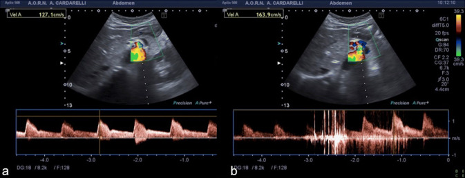
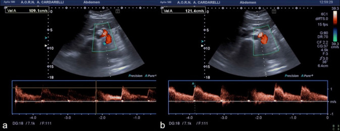
Dunbar syndrome, also known as median arcuate ligament syndrome, is a rare clinical condition due to the external compression of the celiac trunk by the median arcuate ligament causing abdominal angina. We report a case of Dunbar syndrome and its borderline imaging findings focused on the crucial diagnostic role of color-Doppler ultrasound. We also reviewed the current literature, delineating the clinical manifestations and the diagnostic workup of the Dunbar syndrome with the objective to increase the knowledge of this clinical entity as a cause of postprandial abdominal pain and to underline the pivotal role of color-Doppler ultrasound to avoid incorrect or delayed diagnosis.

Keywords: Celiac trunk compression; Color-Doppler ultrasound; Dunbar syndrome; Median arcuate ligament.


Conflict of interest statement

The Authors declare that they have no conflict of interest.

Figures

Fig. 1
Fig. 1
Spectral Doppler US with patient in supine decubitus, during inspiration (a) and expiration (b) at the proximal celiac trunk. Significantly elevated peak systolic velocity (163.9 cm/s) is seen on expiration with aliasing artifact at color-Doppler mode (b)
Fig. 2
Fig. 2
Computed tomography angiography in sagittal (a) and axial (b) planes, showing a slight notch (a marker) along the superior border of the proximal portion of the celiac artery wall and the median arcuate ligament (b marker)
Fig. 3
Fig. 3
Dissection of median arcuate ligament by laparoscopy. The laparoscopic view shows the median arcuate ligament (arrows) at the level of celiac trunk and the crus of diaphragm (double-headed arrow)
Fig. 4
Fig. 4
Postoperative spectral Doppler US with patient in supine decubitus, during inspiration (a) and expiration (b), showing the change in the peak systolic velocity during expiration after surgery, that became normal

References

    1. Torres O, Pereira Gama-Filho O, Torres C, Medeiros R, et al. Laparoscopic treatment of Dunbar syndrome: a case report. Int J Surg Case Rep. 2017;37:230–232. doi: 10.1016/j.ijscr.2017.06.056. - DOI - PMC - PubMed
    1. Rubinkiewicz M, Ramakrishnan PK, Henry BM, Roy J, Budzynski A. Laparoscopic decompression as treatment for median arcuate ligament syndrome. Ann R Coll Surg Engl. 2015;97:e96–e99. doi: 10.1308/rcsann.2015.0025. - DOI - PMC - PubMed
    1. Dunbar JD, Molnar W, Beman FF, Marable SA. Compression of celiac trunkand abdominal angina. Am J Roentgenol Radium Ther Nucl Med. 1965;95:731–744. doi: 10.2214/ajr.95.3.731. - DOI - PubMed
    1. Harjola PT. A rare obstruction of the coeliac artery; report of case. Ann Chir Gynecol Fenn. 1963;52:547–550. - PubMed
    1. Duran M, Simon F, Ertas N, Schelzig H, et al. Open vascular treatment of median arcuate ligament syndrome. BMC Surg. 2017;17(1):95. doi: 10.1186/s12893-017-0289-8. - DOI - PMC - PubMed

MeSH terms

LinkOut - more resources