. 2020 Mar;87(3):442-455.
doi: 10.1002/ana.25675. Epub 2020 Jan 22.

Intravenous Immunomodulatory Nanoparticle Treatment for Traumatic Brain Injury

Affiliations

Intravenous Immunomodulatory Nanoparticle Treatment for Traumatic Brain Injury

Sripadh Sharma et al. Ann Neurol. 2020 Mar.

Abstract

Objective: There are currently no definitive disease-modifying therapies for traumatic brain injury (TBI). In this study, we present a strong therapeutic candidate for TBI, immunomodulatory nanoparticles (IMPs), which ablate a specific subset of hematogenous monocytes (hMos). We hypothesized that prevention of infiltration of these cells into brain acutely after TBI would attenuate secondary damage and preserve anatomic and neurologic function.

Methods: IMPs, composed of US Food and Drug Administration-approved 500nm carboxylated-poly(lactic-co-glycolic) acid, were infused intravenously into wild-type C57BL/6 mice following 2 different models of experimental TBI, controlled cortical impact (CCI), and closed head injury (CHI).

Results: IMP administration resulted in remarkable preservation of both tissue and neurological function in both CCI and CHI TBI models in mice. After acute treatment, there was a reduction in the number of immune cells infiltrating into the brain, mitigation of the inflammatory status of the infiltrating cells, improved electrophysiologic visual function, improved long-term motor behavior, reduced edema formation as assessed by magnetic resonance imaging, and reduced lesion volumes on anatomic examination.

Interpretation: Our findings suggest that IMPs are a clinically translatable acute intervention for TBI with a well-defined mechanism of action and beneficial anatomic and physiologic preservation and recovery. Ann Neurol 2020;87:442-455.


Conflict of interest statement

Potential Conflicts of Interest

Nothing to report.

Figures
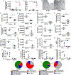
FIGURE 1:
FIGURE 1:
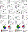
Immunomodulatory nanoparticle (IMP) treatment reduces quantity and quality of infiltrating cells into the controlled cortical impact (CCI) lesion. (A) Absolute number of live cell events detected by flow cytometry from a 3mm3 lesional/perilesional area microdissected 24 or 72 hours after CCI injury. A significant increase in MARCO+ cells was observed in the central nervous system at 72 hours postinjury (hpi). (B, C) MARCO+ cells were identified by immunohistochemistry in the perilesional brain parenchyma at 72 hpi in the CCI model. No MARCO+ cells were noted at 2 hpi, and only rare cells were noted at 24 hpi. Cell counts obtained from 3 images immediately adjacent to the injury cavity on each section, 3 sections per animal; n = 3 animals/group. One-way analysis of variance with Tukey post hoc analysis. Asterisk indicates p < 0.05, double asterisk indicates p < 0.01, and triple asterisk indicates p < 0.01. (D–N) The absolute number of live cell events detected by flow cytometry using cell-specific gating from a 3mm3 lesional/perilesional area that was microdissected at 72 hours after CCI injury. (O–S) Further subanalysis of the previously gated cells examined for the percentage of cells expressing MHC II and CD86. (T, U) Pie charts representing all pertinent live cell recorded events compartmentalized by specific cell types and further subanalysis of the populations of myeloid cells in the lesion area in (T) vehicle- and (U) IMP-treated animals. All statistics shown were by 2-tailed t test with α = 0.05. Asterisk indicates p < 0.05, double asterisk indicates p < 0.01, and triple asterisk indicates p < 0.001. Myeloid cells = CD45hi CD11b+; macrophages (Macro)/monocytes (Mono) = CD45hi CD11b+ Ly6G CD11c; inflammatory monocytes (Inflammatory Mφ) = CD45hi CD11b+ Ly6G CD11c Ly6chi; noninflammatory monocytes (Noninflammatory φ) = CD45hi CD11b+ Ly6G CD11c Ly6clo; dendritic cells (mDCs) = CD45hi CD11b+ Ly6G CD11c+; neutrophils = CD45hi CD11b+ Ly6G+; lymphocytes = CD45hi CD11b; and microglia = CD45int CD11b+ Ly6Clo. Sample size was n = 3 for each group. Each data point (n) is composed of 5 animals’ microdissected lesion areas pooled together.
FIGURE 2:
FIGURE 2:
Immunomodulatory nanoparticle (IMP) treatment reduces the lesion volume in controlled cortical impact (CCI) injury. (A) Whole brains of vehicle-treated (top row) and IMP-treated (bottom row) animals 10 weeks after visual cortex CCI injury. (B) Representative cryostat brain sections of vehicle- and IMP-treated animals taken through the center of the lesion cavity. The coronal sections correspond to the top middle (vehicle) and bottom middle (IMP) in (A). (C) Representative perilesional confocal images stained for GFAP+ astrocytes and 4, 6-diamidino-2-phenylindole (DAPI) at 10 weeks postinjury to showcase the attenuated glial scar in the IMP-treated animals. (D) Quantified lesion volumes of the brains in (A) and (B) via cryosectioning and extrapolating to 3 dimensions. (E) Quantification of the GFAP intensity in perilesional areas in 10 weeks postinjury vehicle- and IMP-treated animals. All statistics shown in this figure were by 2-tailed t test with α = 0.05. Asterisk indicates p < 0.05, double asterisk indicates p < 0.01, and triple asterisk indicates p < 0.001. Sample size was n = 7 for lesion volumes and n = 4 for immunohistochemistry.
FIGURE 3:
FIGURE 3:
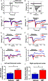
Immunomodulatory nanoparticle (IMP) treatment preserves electrophysiologic function in spared visual cortex after parieto-occipital controlled cortical impact injury. (A) Representative trace of a single visual evoked potential (VEP) in an uninjured mouse. (B) A time dilated trace of the VEP to showcase the P1 and N1 waveforms and the bounded area for area under the curve (AUC) measurements. (C, D) Superimposed grand average VEP waveforms of uninjured, vehicle, and IMP groups from the left eye/uninjured right visual cortex (C) and from the right eye/injured left visual cortex (D). (E, F) Time dilated VEPs from (C) and (D), respectively. (G, H) Transformation with the voltage in the uninjured group becoming the x-axis, and the vehicle and IMP waveforms from (E) and (F) expressed in relationship to the uninjured voltage for the left eye (uninjured right visual cortex; G) and the right eye (injured left visual cortex; H). (I) Voltage/potential summated in the time between the P1 response and the end of the stimulus for AUC for the N1 waveform. (J) Voltage/potential summated after the end of the stimulus for AUC for the subsequent minor waveforms. Statistics were by 2-sided t test with α = 0.05. Asterisk indicates p < 0.05 and double asterisk indicates p < 0.01. Sample size was n = 7 in each group.
FIGURE 4:
FIGURE 4:
Immunomodulatory nanoparticle (IMP) treatment after unilateral motor cortex controlled cortical impact (CCI) injury decreases lesion volume and attenuates motor deficits. (A) A 16-week postinjury magnetic resonance imaging T2-weighted sequence with serial cuts from the frontal lobe to the caudal aspect of the cerebral cortex to showcase the lesion volume and ventricle size differences among uninjured, vehicle, and IMP groups. (B) Lesion volume quantification of the vehicle and IMP groups. (C) Three-dimensional reconstructions of the brains shown in (A; top) and the ventricles isolated in red (bottom; see Supplementary Videos 1–3). (D) Quantification of the cerebrospinal fluid/ventricular volumes of the different groups. (E) Quantification of ladder rung behavior done at various time points after the CCI measuring percentage of right forelimb (RF) errors. (F) Correlation of lesion volume versus the number of RF strides. (G) Correlation of lesion volume versus the number of RF strides by Digigait. For statistical analysis, (B) uses a 2-sided t test, (D) and (E) use one-way analysis of variance with post hoc Tukey all comparisons, and (F) and (G) use a Pearson correlation. All statistics in this figure used α = 0.05. Asterisk indicates p < 0.05, double asterisk indicates p < 0.01, triple asterisk indicates p < 0.001, and quadruple asterisk indicates p < 0.0001. Sample size in (A) and (D) was n = 4. Sample size in (E) and (F) was n = 5–10 in each group. Blue asterisks indicate comparing uninjured and vehicle groups, red asterisks indicate comparing uninjured and IMP groups, and pink asterisks indicate comparing vehicle and IMP groups; ns = not significant.
FIGURE 5:
FIGURE 5:
Mechanism of action of immunomodulatory nanoparticle (IMP) treatment in closed head injury (CHI)/traumatic brain injury (TBI) parallels controlled cortical impact (CCI)/TBI. (A) Absolute number of live cell events detected by flow cytometry from whole brain at 24 and 72 hours postinjury (hpi). After CHI, a significant increase in MARCO+ cells was observed in the central nervous system at 72 hpi. (B–O) Total number of live cell events by cell-specific flow sorting from whole brain tissue 72 hpi. (P–T) Subanalysis of the flow sorted cells showing the percentage of MCH II and CD86 expression. (U, V) Pie charts representing all pertinent live cell recorded events compartmentalized by specific cell types and further subanalysis of the populations of myeloid cells in the lesion area in (U) vehicle- and (V) IMP-treated animals. All statistics shown were by 2-sided t test with α = 0.05. Asterisk indicates p < 0.05, double asterisk indicates p < 0.01, triple asterisk indicates p < 0.001. Myeloid cells = CD45hi CD11b+; macrophages (Macro)/monocytes (Mono) = CD45hi CD11b+ Ly6G CD11c; inflammatory monocytes (Inflammatory Mφ): CD45hi CD11b+ Ly6G CD11c Ly6chi; noninflammatory monocytes (noninflammatory φ) = CD45hi CD11b+ Ly6G CD11c Ly6clo; dendritic cells (mDCs) = CD45hi CD11b+ Ly6G CD11c+; neutrophils = CD45hi CD11b+ Ly6G+; lymphocytes = CD45hi CD11b; microglia = CD45int CD11b+ Ly6Clo. Sample size was n = 3 for each group. Each data point (n) is composed of 5 animals’ microdissected lesion areas pooled together; a total of 15 animals were used for each group. All statistics in this figure used α = 0.05. Asterisk indicates p < 0.05, double asterisk indicates p < 0.01, and triple asterisk indicates p < 0.001.
FIGURE 6:
FIGURE 6:
Immunomodulatory nanoparticle (IMP) treatment acutely attenuates edema and increase in intracranial pressure and preserves motor behavior. (A) Representative slices of quantitative magnetic resonance imaging R2* sequences of vehicle- and IMP-treated animals at 24 hours after closed head injury. (B) Quantification of the total amount of R2* hyperintense volume. (C) Representative 3-dimensional reconstruction of the ventricles of the vehicle- and IMP-treated animals at 24 hours postinjury showing a decreased ventricle size in the vehicle-treated group. (D) Quantification of the ventricular volumes of the vehicle- and IMP-treated group. (E) Pearson correlation of amount of R2* hyperintensity volume versus the ventricular volume. (F) Quantification of the percentage of errors in all limbs at 24 hours postinjury on ladder-rung assay. For statistical analysis, (B) and (D) use 2-sided t tests, (E) uses Pearson correlation, and (F) uses a one-way analysis of variance with post hoc Tukey all comparisons. All statistics in this figure used α = 0.05. Asterisk indicates p < 0.05, double asterisk indicates p < 0.01, and triple asterisk indicates p < 0.001. NS = not significant; n = 5 animals per group.

References

    1. Centers for Disease Control and Prevention. Report to Congress on traumatic brain injury in the United States: epidemiology and rehabilitation. Atlanta, GA: National Center for Injury Prevention and Control; Division of Unintentional Injury Prevention, 2015.
    1. Thurman DJ, Alverson C, Dunn KA, et al. Traumatic brain injury in the United States: a public health perspective. J Head Trauma Rehabil 1999;14:602–615. - PubMed
    1. de Souza RS, Pinheiro PP, de Lima Silva JMF, et al. Traumatic brain injury (TBI): morbidity, mortality and economic implications. Int Arch Med 2015. Available at: http://imed.pub/ojs/index.php/iam/article/view/1109. Last accessed on June 6, 2019.
    1. Bramlett HM, Dietrich WD. Long-term consequences of traumatic brain injury: current status of potential mechanisms of injury and neurological outcomes. J Neurotrauma 2015;32:1834–1848. - PMC - PubMed
    1. Gruenbaum SE, Zlotnik A, Gruenbaum BF, et al. Pharmacologic neuroprotection for functional outcomes after traumatic brain injury: a systematic review of the clinical literature. CNS Drugs 2016;30: 791–806. - PMC - PubMed

Publication types

MeSH terms

Substances