. 2020 Apr;153(1):103-119.
doi: 10.1111/jnc.14955. Epub 2020 Feb 25.

A simple, versatile and robust centrifugation-based filtration protocol for the isolation and quantification of α-synuclein monomers, oligomers and fibrils: Towards improving experimental reproducibility in α-synuclein research

Affiliations

A simple, versatile and robust centrifugation-based filtration protocol for the isolation and quantification of α-synuclein monomers, oligomers and fibrils: Towards improving experimental reproducibility in α-synuclein research

Senthil T Kumar et al. J Neurochem. 2020 Apr.

Abstract

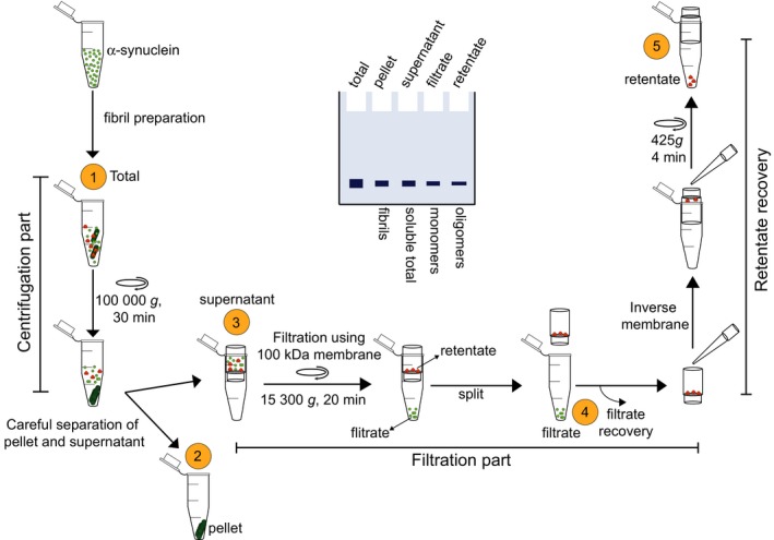
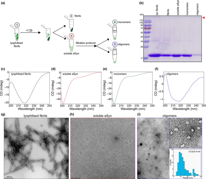
Increasing evidence suggests that the process of alpha-synuclein (α-syn) aggregation from monomers into amyloid fibrils and Lewy bodies, via oligomeric intermediates plays an essential role in the pathogenesis of different synucleinopathies, including Parkinson's disease (PD), multiple system atrophy and dementia with Lewy bodies (DLB). However, the nature of the toxic species and the mechanisms by which they contribute to neurotoxicity and disease progression remain elusive. Over the past two decades, significant efforts and resources have been invested in studies aimed at identifying and targeting toxic species along the pathway of α-syn fibrillization. Although this approach has helped to advance the field and provide insights into the biological properties and toxicity of different α-syn species, many of the fundamental questions regarding the role of α-syn aggregation in PD remain unanswered, and no therapeutic compounds targeting α-syn aggregates have passed clinical trials. Several factors have contributed to this slow progress, including the complexity of the aggregation pathways and the heterogeneity and dynamic nature of α-syn aggregates. In the majority of experiment, the α-syn samples used contain mixtures of α-syn species that exist in equilibrium and their ratio changes upon modifying experimental conditions. The failure to quantitatively account for the distribution of different α-syn species in different studies has contributed not only to experimental irreproducibility but also to misinterpretation of results and misdirection of valuable resources. Towards addressing these challenges and improving experimental reproducibility in Parkinson's research, we describe here a simple centrifugation-based filtration protocol for the isolation, quantification and assessment of the distribution of α-syn monomers, oligomers and fibrils, in heterogeneous α-syn samples of increasing complexity. The protocol is simple, does not require any special instrumentation and can be performed rapidly on multiple samples using small volumes. Here, we present and discuss several examples that illustrate the applications of this protocol and how it could contribute to improving the reproducibility of experiments aimed at elucidating the structural basis of α-syn aggregation, seeding activity, toxicity and pathology spreading. This protocol is applicable, with slight modifications, to other amyloid-forming proteins.

Keywords: Parkinson's disease; alpha-synuclein; amyloid fibrils; oligomers and monomers.


Figures

Figure 1
Figure 1
Schematic depiction of the centrifugation‐based filtration protocol. Briefly, the protocol includes a centrifugation step for the isolation of the fibril (pellet) from soluble α‐syn (monomers and oligomers), a filtration step (through 100 kDa MWCO) for the separation of the monomers from the oligomers, and a final step for the recovery of the oligomers from the spin filters. A depiction of the sodium dodecyl sulphate–polyacrylamide gel electrophoresis (SDS‐PAGE) analysis of the samples at each step is shown in the middle. Quantification of band intensity should allow for a quick assessment of the distribution of coexisting α‐syn species
Figure 2
Figure 2
Schematic illustrations of (a) the separation of α‐syn fibrils from a crude mixture of total α‐syn fibrils following fibrillization and (b) following sonication (20% amplitude, one‐second ON/OFF pulse cycle for 20 s), to assess the amounts of monomers/oligomers present in the supernatant fraction. (c) sodium dodecyl sulphate–polyacrylamide gel electrophoresis (SDS‐PAGE) analysis of the different batches of α‐syn fibrils during fibrillization; mono: α‐syn monomers prior to initiating fibrillization; p. fibrils: pelleted fibrils after resuspension; and sup: supernatant α‐syn species following centrifugation at the end of fibrillization. (d) Bar chart displaying the percentage of α‐syn monomers converted to fibrils in different batches of fibril preparation. Bullet points represent the different batches of fibril preparation (n = 15). (e) EM images of total α‐syn fibrils at the end of fibrillization showing three representative images from three random batches of fibril preparations. (f) EM images of sonicated α‐syn fibrils showing three representative images from three random batches. (g) Differences in the lengths of the fibrils before and after sonication. (h) SDS‐PAGE analysis of different batches of α‐syn fibrils following sonication; pf. seeds: the fibril seeds sedimented as pellets after sonication; sup: the soluble supernatant α‐syn species following centrifugation. * in (c) and (h) denotes the increase in the amount of soluble α‐syn species in the supernatant fractions in some non‐reproducible fibril batches
Figure 3
Figure 3
Size‐based separation of α‐syn oligomers using Superose 6 column and filtration analysis. (a) Superose 6 based purification of α‐syn oligomers. Oligomeric fractions (highlighted in red box) eluted from 8 ml to 14 ml and monomeric fractions from 16 ml to 19 ml. (b) Highlighted oligomeric peak from (a) showing the elution of different fractions (fraction 1, fraction 2, fraction 3 and fraction 4 in varying colours) of oligomers collected for filtration analysis. (c) EM images based size distribution analysis of different fractions of oligomeric populations. (d, e, f and g) EM analysis of total (left) and filtrate (right) of oligomeric fraction 1 (d), fraction 2 (e), fraction 3 (f) and fraction 4 (g). Scale bars of total and filtrate samples are 50 nm. Zoomed EM images (scale bars: 25 nm) on the top right of each fraction shows the length (ln) or diameter (d) of differently sized oligomers measured in (c). Coomassie‐stained sodium dodecyl sulphate–polyacrylamide gel electrophoresis (SDS‐PAGE) gel in the middle of each oligomeric fraction shows the total (tot) oligomeric fraction used for filtration analysis using 100 kDa MWCO membrane, filtrate (fil) and retentate (ret) samples following the filtration protocol
Figure 4
Figure 4
(a) A scheme illustrating the experimental setup for assessing efficiency to separate α‐syn species from a prepared mixture containing known concentration of α‐syn monomers, oligomers and fibril. (b) sodium dodecyl sulphate–polyacrylamide gel electrophoresis (SDS‐PAGE) analysis of the samples collected at each step of the protocol. Red arrow head points the band of SDS‐resistant oligomers. (c) Comparison of protein concentration estimation (μM; based on BCA assay and AAA) of α‐syn samples (fibrils, oligomers and monomers) from the initial concentrations, that is before the preparation of α‐syn mixture versus the obtained concentrations of α‐syn fractions isolated from the α‐syn mixture during the steps of the protocol. (d–h) CD spectra of the total α‐syn mixture (d), fibrils (e), monomers and oligomers (mono + oligo) in the soluble α‐syn supernatant (f), monomers (mono) in the filtrate (g) and oligomers (oligo) in the retentate (h) samples. (i–m) Representative electron micrograph images of the α‐syn mixture (i), fibrils (j), monomers and oligomers (mono + oligo) in the soluble α‐syn supernatant (k), monomers (mono) in the filtrate (l) and oligomers (oligo) in the retentate (m) samples isolated from the different steps of the protocol
Figure 5
Figure 5
(a) Schematic illustration showing the experimental setup of the centrifugation‐based filtration analysis of lyophilized and resuspended α‐syn fibrils (lyophilized fibrils). (b) sodium dodecyl sulphate–polyacrylamide gel electrophoresis (SDS‐PAGE) analysis of α‐syn samples isolated from the different steps of the protocol. Red arrow head points the band of SDS‐resistant oligomers. (c–f) CD spectra of lyophilized fibrils (c), soluble α‐syn (d), monomers (filtrate, e) and oligomers (retentate, f) in the samples. (g–i) Representative EM images of lyophilized fibrils (g), soluble α‐syn (h), and oligomers (retentate, i) in the samples from the different steps of the protocol. The top insert on (i) is the zoom of the EM images of the oligomer sample. The bottom insert of I shows the distribution of the diameter of oligomeric particles based on the quantification from EM images

References

    1. Abdelmotilib, H. , Maltbie, T. , Delic, V. , Liu, Z., Hu, X. , Fraser, K. B. … West, A. (2017). Alpha‐Synuclein fibril‐induced inclusion spread in rats and mice correlates with dopaminergic Neurodegeneration. Neurobiology of Diseases, 105, 84–98. - PMC - PubMed
    1. Alam, P. , Bousset, L. , Melki, R. , & Otzen, D. E. (2019). Alpha‐synuclein oligomers and fibrils: A spectrum of species, a spectrum of toxicities. Journal of Neurochemistry, 150, 522–534. - PubMed
    1. Baba, M. , Nakajo, S. , Tu, P. H. , Tomita, T. , Nakaya, K. , Lee, V. M. , … Iwatsubo, T. (1998). Aggregation of alpha‐synuclein in Lewy bodies of sporadic Parkinson's disease and dementia with Lewy bodies. American Journal of Pathology, 152, 879–884. - PMC - PubMed
    1. Bieschke, J. , Russ, J. , Friedrich, R. P. , Ehrnhoefer, D. E. , Wobst, H. , Neugebauer, K. , & Wanker, E. E. (2010). EGCG remodels mature alpha‐synuclein and amyloid‐beta fibrils and reduces cellular toxicity. Proceedings of the National Academy of Sciences of the United States of America, 107, 7710–7715. - PMC - PubMed
    1. Braak, H. , Del Tredici, K. , Rub, U. , de Vos, R. A. , Jansen Steur, E. N. , & Braak, E. (2003). Staging of brain pathology related to sporadic Parkinson's disease. Neurobiology of Aging, 24, 197–211. 10.1016/S0197-4580(02)00065-9 - DOI - PubMed

Publication types