. 2020 Feb;32(2):e12829.
doi: 10.1111/jne.12829. Epub 2020 Jan 29.

Neural connectivity between the hypothalamic supramammillary nucleus and appetite- and motivation-related regions of the rat brain

Affiliations

Neural connectivity between the hypothalamic supramammillary nucleus and appetite- and motivation-related regions of the rat brain

Fabrice Plaisier et al. J Neuroendocrinol. 2020 Feb.

Abstract

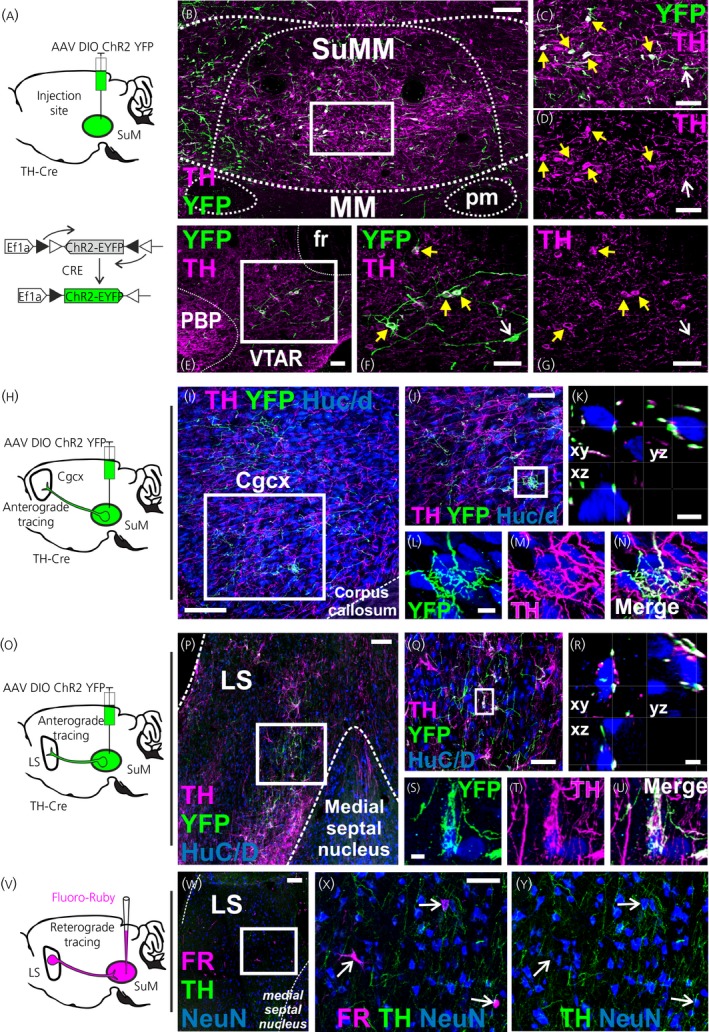
The supramammillary nucleus (SuM) has an emerging role in appetite control. We have shown that the rat SuM is activated during hunger or food anticipation, or by ghrelin administration. In the present study, we characterised the connectivity between the SuM and key appetite- and motivation-related nuclei in the rat. In adult wild-type rats, or rats expressing Cre recombinase under the control of the tyrosine hydroxylase (TH) promoter (TH-Cre rats), we used c-Fos immunohistochemistry to visualise and correlate the activation of medial SuM (SuMM) with activation in the lateral hypothalamic area (LH), the dorsomedial hypothalamus (DMH) or the ventral tegmental area (VTA) after voluntary consumption of a high-sugar, high-fat food. To determine neuroanatomical connectivity, we used retrograde and anterograde tracing methods to specifically investigate the neuronal inputs and outputs of the SuMM. After consumption of the food there were positive correlations between c-Fos expression in the SuMM and the LH, DMH and VTA (P = 0.0001, 0.01 and 0.004). Using Fluoro-Ruby as a retrograde tracer, we demonstrate the existence of inputs from the LH, DMH, VTA and ventromedial hypothalamus (VMH) to the SuMM. The SuMM showed reciprocal inputs to the LH and DMH, and we identified a TH-positive output from SuMM to DMH. We co-labelled retrogradely-labelled sections for TH in the VMH, or for TH, orexin and melanin-concentrating hormone in the LH and DMH. However, we did not observe any colocalisation of immunoreactivity with any retrogradely-labelled cells. Viral mapping in TH-Cre rats confirms the existence of a reciprocal SuMM-DMH connection and shows that TH-positive cells project from the SuMM and VTA to the lateral septal area and cingulate cortex, respectively. These data provide evidence for the connectivity of the SuMM to brain regions involved in appetite control, and form the foundation for functional and behavioural studies aiming to further characterise the brain circuitry controlling eating behaviours.

Keywords: food; motivation; neuroanatomy; supramammillary nucleus; tracing.


Figures

Figure 1
Figure 1
Expression of Fos immunoreactivity in the medial supramammillary nucleus (SuMM) and appetite‐reward related areas in response to the voluntary consumption of sweetened condensed milk (SCM). Data are the mean ± SEM. **P < 0.01, ***P < 0.001. A‐D, Representative images of Fos (arrowheads) and tyrosine hydroxylase (TH) (arrows) immunohistochemistry within SuMM in control (A, B) and SCM‐fed rats (C, D) in coronal sections at bregma −4.5 mm. E, Number of cells expressing Fos per brain section in SuMM for control (n = 8) and SCM (n = 6) groups (P = 0.0007, Mann‐Whitney test). F, Percentage of SuMM TH‐immunoreactive cells expressing Fos‐positive nuclei per brain section for control (n = 8) and SCM (n = 6) groups (P = 0.0007, Mann‐Whitney test). G and H, Representative images of Fos expression in the lateral hypothalamic area (LH) (bregma −1.72 mm), dorsomedial nucleus of the hypothalamus (DMH) (K, L; bregma −3 mm) and ventral tegmental area (VTA) (O, P; bregma −5.04 mm) in control and SCM‐fed rats. I, M, and Q, Number of cells expressing Fos per brain section for control and SCM groups in the LHA (I; control n = 7, SCM n = 6; P = 0.0012, Mann‐Whitney test), DMH (M; control n = 8, SCM n = 6; P = 0.0013, Mann‐Whitney test) and VTA (Q; control n = 8, SCM n = 4; P = 0.0081, Mann‐Whitney test). J, N, and R, Linear regression plots and coefficients (with significance indicated as *P ≤ 0.05, **P ≤ 0.01 or ***P ≤ 0.001) showing the correlation between Fos expression in the SuMM and Fos expression in the LHA (J; r 2 = 0.7562, P = 0.0001), DMH (N; r 2 = 0.434, P = 0.0104) and VTA (R; r 2 = 0.5827, P = 0.0039). Scale bars represent 200 µm (G, H, K, L, O, P), 50 µm (A, C) and 25 µm (B, D). 3V, third ventricle; AH, anterior hypothalamic nucleus; EA, sublenticular extended amygdala; f, fornix; fr, fasciculus retroflexus; LH, lateral hypothalamic area; ml, medial lemniscus; OC, optic tract; PB, parabrachial pigmented nucleus; pm, principal mammillary tract; PVN, paraventricular nucleus; SNR, substantia nigra; SuM, supramammillary nucleus; VMH, ventromedial nucleus
Figure 2
Figure 2
Retrograde labelling from the medial supramammillary nucleus (SuMM) in appetite and reward related areas. A, Schematic of the experimental procedure. Injection of Fluoro‐Ruby (FR) into the SuMM results in a fluorescent signal in regions that project to the SuMM. B, Representative image showing the iontophoretic injection site of FR confined to the SuMM at bregma −4.5 mm. (C‐O) Representative images of FR+ retrogradely labelled neurones (NeuN+) in the rostral part of the ventral tegmental area (VTAR) (C‐F; bregma −5.04 mm; yellow arrowheads point to neurones triple‐labelled for FR, tyrosine hydroxylase (TH) and a neurone‐specific marker (NeuN) and white arrows point to TH‐negative FR+ neurones), the ventromedial nucleus (VMH) (G‐I; bregma −2.52 mm; white arrows point to FR‐labelled neurones that were TH‐negative and black arrows point to nonspecific FR labelling), the lateral hypothalamic area (LHA) (J‐L; bregma −1.56 mm [J] and −2.52 mm [K, L]; white arrows point to FR‐labelled neurones that were TH‐negative, ORX‐negative or MCH‐negative) and the dorsomedial nucleus of the hypothalamus (DMH) (M‐O; bregma −2.76 mm [M] and −3.12 mm [N, O]; white arrows point to FR‐labelled neurones that were TH‐negative, ORX‐negative or MCH‐negative). Scale bars represent 200 µm (B), 100 µm (C, D), 50 µm (E‐I) and 25 µm (J‐O). ARC, arcuate nucleus; fr, fasciculus retroflexus; MM, medial mammillary nucleus; PBP, parabrachial pigmented nucleus; pm, principal mammillary tract; SUM, supramammillary nucleus; VTA, ventral tegmental area
Figure 3
Figure 3
Retrograde labelling from the lateral hypothalamic area (LHA) and dorsomedial nucleus of the hypothalamus (DMH) in the medial supramammillary nucleus (SuMM). A, Scheme of the experimental procedure. Injection of Fluoro‐Ruby (FR) into the LHA results in a fluorescent signal in SuMM cells that project to the LHA. B, Representative image showing the iontophoretic injection site FR in the LHA at 2.4 mm posterior to bregma. Double‐labelling immunofluorescence for a neurone‐specific marker (NeuN) and tyrosine hydroxylase (TH) was performed to count and determine the chemical nature of the retrogradely labelled (FR+) neurones by confocal microscopy. C, Distribution of retrogradely labelled TH‐negative SuMM neurones (n = 2). D‐H, Representative confocal image of retrogradely labelled neurones in the SuMM taken at 4.5 mm posterior to bregma. The region in the white box in (D) is shown at higher magnification in (E‐H). To identify the labelled cells, TH, FR and NeuN‐labelling of the same cells are shown in separate images (E‐H). The white arrows indicate examples of FR‐labelled neurones that were TH‐negative. The black arrow indicates nonspecific labelling that can be observed following injection of FR. I, Scheme of the experimental procedure. Injection of FR into the DMH results in a fluorescent signal in SuMM cells that project to the DMH. J, Representative image showing the iontophoretic injection site of FR in the DMH at 3 mm posterior to bregma. K, Distribution of retrogradely labelled TH+ and TH‐negative SuMM neurones (n = 1). L‐R, Representative confocal images of retrogradely labelled SuMM neurones at 4.5 mm posterior to bregma. Regions in the white boxes 1 and 2 in (L) are shown at higher magnification in (M‐O) and (P‐R), respectively. (M‐R) To facilitate identification of the labelled cells, TH, FR and NeuN‐labelling of the same cells is shown in separate images. The white arrows indicate one example of FR‐labelled neurone that was TH‐negative (M‐O), the yellow arrowhead indicate one example of FR‐labelled neurone that were TH+ (P‐R). Scale bars represent 100 µm in (B, D, J, L) and 25 µm in (E‐H, M‐R). 3V, third ventricle; f, fornix; pm, principal mammillary tract; MM, medial mammillary nucleus; sor, supraoptic nucleus retrochiasmatic part; sox, supraoptic decussation; SUM, supramammillary nucleus; VMH, ventromedial nucleus
Figure 4
Figure 4
Connections between the medial supramammillary nucleus (SuMM) and the ventral tegmental area (VTA), lateral septal nucleus and cingulate cortex. A, Scheme of the experimental procedure. Injection of a DIO‐ChR2‐yellow fluorescent protein (YFP) adenoassociated virus (AAV) results in YFP expression in the SuMM. B‐D, Representative confocal images showing the injection site of DIO‐ChR2‐YFP AAV in the SuMM of rats expressing Cre recombinase under the tyrosine hydroxylase (TH) promoter (TH‐Cre) rats (4.5 mm posterior to bregma). E‐G, YFP expression in cell bodies of neurones in the rostral VTA (5.04 mm posterior to bregma). Regions in the white boxes in (B) and (E) are shown at higher magnification in (C, D) and (F, G), respectively. Yellow arrowheads indicate examples of YFP expression in cell bodies of TH+ neurones. White arrows indicate example of YFP expression in cell bodies of TH‐negative neurones. H and O, Schemes of the experimental procedure. Injection of DIO‐ChR2‐YFP AAV in the SuMM results in YFP‐labelled fibres in the cingulate cortex and lateral septal nucleus. Representative confocal images showing the AAV‐mediated anterograde labelling observed in the cingulate cortex (I‐K) and lateral septal nucleus (P‐R) at 0.48 mm anterior to bregma following the injection in the SuMM. Confocal photomicrographs (flattened stack of 30 optical sections at intervals of 1 μmol L‐1) illustrate neurones contacted by YFP+/TH+ labelled fibres in the cingulate cortex (L‐N) and lateral septal nucleus (S‐U). K and R, Contacts were confirmed in 3D with single optical sections in the xy‐, xz‐ and yz‐plane. V, Scheme of the experimental procedure. Injection of Fluoro‐Ruby (FR) into the SuMM resulted in a fluorescent signal in the lateral septal nucleus. W, Representative confocal images showing FR retrogradely labelled neurones in the lateral septal nucleus (0.48 mm anterior to bregma). The region in the white box in (W) is shown in higher magnification in (X, Y). The white arrows indicate three examples of FR+ retrogradely labelled neurones (NeuN+) that were TH‐negative. Scale bars represent 100 µm (B, I, P, W), 50 µm (C‐G, J, Q, X, Y) and 5 µm (K, L‐N, R, S‐U). Cgcx, cingulate cortex; LS, lateral septal nucleus; MM, medial mammillary nucleus; SUM, supramammillary nucleus; VTAR, ventral tegmental area (rostral part)
Figure 5
Figure 5
Reciprocal connections between the medial supramammillary nucleus (SuMM) and the dorsomedial nucleus of the hypothalamus (DMH). A, Scheme of the experimental procedure. Injection of DIO‐ChR2‐yellow fluorescent protein (YFP) adenoassociated virus (AAV) in the SuMM results in YFP‐labelled fibres in the DMH. B and C, Representative confocal images showing the AAV‐mediated anterograde labelling in the DMH (3 mm posterior to bregma). YFP‐labelled fibres in the white box in (B) are shown at higher magnification in (C). Regions in the white boxes 1 and 2 in (C) are shown at higher magnification in (D‐F) and (H‐J), respectively. D‐K, Confocal photomicrograph (D, H; flattened stack of 30 optical sections at intervals of 1 μmol L‐1) and 3D reconstructions (E‐G and I‐K) illustrate two examples of DMH neurones contacted by YFP+/TH+ (D‐G) and YFP+/TH‐negative labelled fibres (H‐K). G, K, All close appositions are confirmed in 3D with single optical sections in the xy‐, xz‐ and yz‐plane. L, Scheme of the experimental procedure. Injection of DIO‐ChR2‐YFP AAV in the DMH results in YFP‐labelled fibres in the SuMM. M, Representative confocal images showing the injection site of DIO‐ChR2‐YFP AAV in the DMH (3 mm posterior to bregma). N, YFP labelling was observed in the SuMM (4.5 mm posterior to bregma). Regions in the white boxes 1 and 2 in (N) are shown at higher magnification in (O‐Q) and (S‐U), respectively. (O‐V) Confocal photomicrographs (O, S; flattened stack of 30 optical sections at intervals of 1 μmol L‐1) and 3D reconstructions (P‐R and T‐V) illustrate examples of SUMM TH+ (O‐R) and SUMM TH‐negative neurones (S‐V) observed with YFP+‐TH‐negative appositions (arrows). R, V, All close appositions are confirmed in 3D with single optical sections in the xy‐, xz‐ and yz‐plane. Scale bars represent 100 µm (B, M), 50 µm (C) and 5 µm in all other images. MM, medial mammillary nucleus; pm, principal mammillary tract; SUM, supramammillary nucleus; VMH, ventromedial nucleus; VTA, ventral tegmental area
Figure 6
Figure 6
Depiction of tyrosine hydroxylase (TH)‐positive and TH‐negative connections between the medial supramammillary nucleus (SuMM) and selected appetite/reward areas. Regions in blue show increased levels of Fos expression after consumption of sweetened condensed milk (SCM), the region shown in yellow show no change after consumption of SCM. DMH, dorsomedial nucleus of the hypothalamus; LHA, lateral hypothalamic area; SuM, supramammillary nucleus; VMH, ventromedial nucleus; VTA, ventral tegmental area

Similar articles

Cited by

References

    1. Le May MV, Hume C, Sabatier N, et al. Activation of the rat hypothalamic supramammillary nucleus by food anticipation, food restriction or ghrelin administration. J Neuroendocrinol. 2018;31:e12676. - PubMed
    1. Vogel H, Wolf S, Rabasa C, et al. GLP‐1 and estrogen conjugate acts in the supramammillary nucleus to reduce food‐reward and body weight. Neuropharmacology. 2016;110:396‐406. - PubMed
    1. Lopez‐Ferreras L, Eerola K, Mishra D, et al. GLP‐1 modulates the supramammillary nucleus‐lateral hypothalamic neurocircuit to control ingestive and motivated behavior in a sex divergent manner. Mol Metab. 2019;20:178‐193. - PMC - PubMed
    1. Ikemoto S. The supramammillary nucleus mediates primary reinforcement via GABA(A) receptors. Neuropsychopharmacology. 2005;30:1088‐1095. - PMC - PubMed
    1. Shin R, Ikemoto S. Administration of the GABAA receptor antagonist picrotoxin into rat supramammillary nucleus induces c‐Fos in reward‐related brain structures. Supramammillary picrotoxin and c‐Fos expression. BMC Neurosci. 2010;11:101. - PMC - PubMed

Publication types

MeSH terms

Substances