

Interstitial chromosomal deletion of the tuberous sclerosis complex 2 locus is a signature for radiation-associated renal tumors in Eker rats
- PMID: 31925975
- PMCID: PMC7060461
- DOI: 10.1111/cas.14307
Interstitial chromosomal deletion of the tuberous sclerosis complex 2 locus is a signature for radiation-associated renal tumors in Eker rats
Abstract
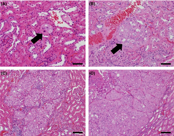
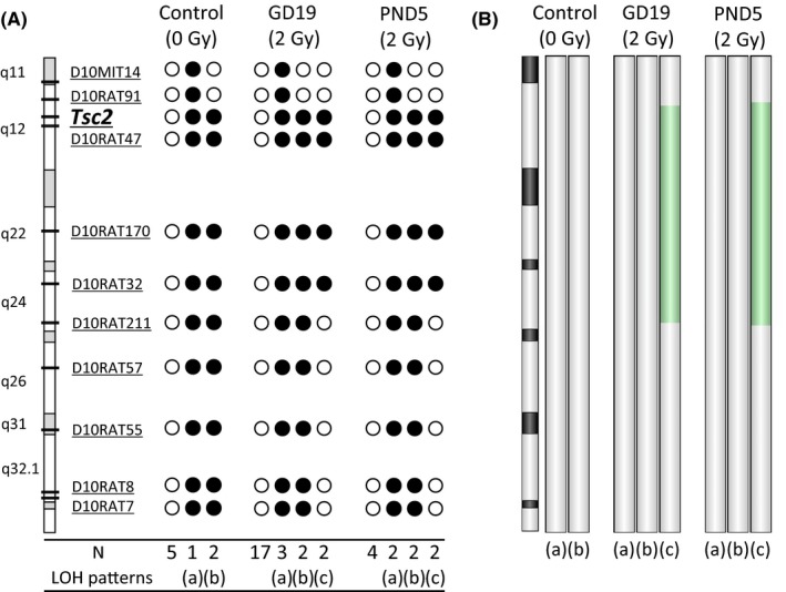
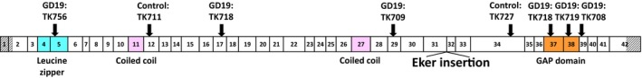
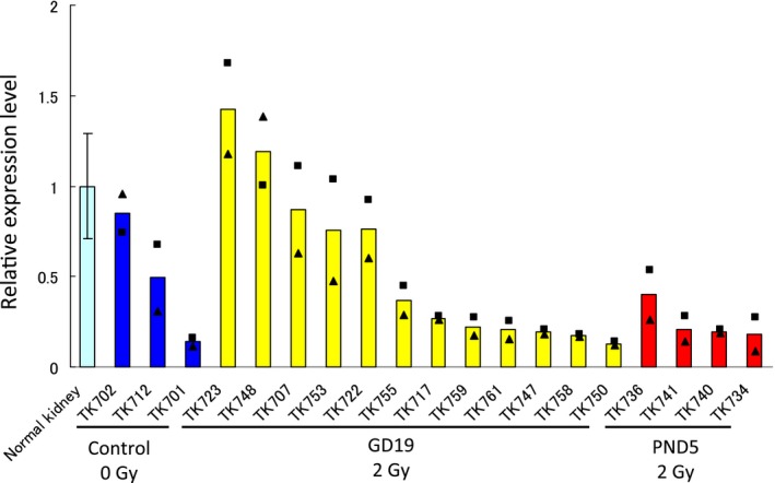
Ionizing radiation can damage DNA and, therefore, is a risk factor for cancer. Eker rats, which carry a heterozygous germline mutation in the tumor-suppressor gene tuberous sclerosis complex 2 (Tsc2), are susceptible to radiation-induced renal carcinogenesis. However, the molecular mechanisms involved in Tsc2 inactivation are unclear. We subjected Fischer 344 × Eker (Long Evans Tsc2+/- ) F1 hybrid rats to gamma-irradiation (2 Gy) at gestational day 19 (GD19) or postnatal day 5 (PND5) and investigated the patterns of genomic alterations in the Tsc2 allele of renal tumors that developed at 1 year after irradiation (N = 24 tumors for GD19, N = 10 for PND5), in comparison with spontaneously developed tumors (N = 8 tumors). Gamma-irradiation significantly increased the multiplicity of renal tumors. The frequency of LOH at the chromosome 10q12 region, including the Tsc2 locus, was 38%, 29% and 60% in renal carcinomas developed from the nonirradiated, GD19 and PND5 groups, respectively. Array comparative genomic hybridization analysis revealed that the LOH patterns on chromosome 10 in renal carcinomas were classified into chromosomal missegregation, mitotic recombination and chromosomal deletion types. LOH of the interstitial chromosomal deletion type was observed only in radiation-associated carcinomas. Sequence analysis for the wild-type Tsc2 allele in the LOH-negative carcinomas identified deletions (nonirradiated: 26%; GD19: 21%) and base-substitution mutations (GD19: 4%). Reduced expression of Tsc2 was also observed in the majority of the LOH-negative carcinomas. Our results suggest that interstitial chromosomal deletion is a characteristic mutagenic event caused by ionizing radiation, and it may contribute to the assessment of radiation-induced cancer risk.
Keywords: Tsc2; Eker rat; genomic signature; ionizing radiation; renal carcinoma.
© 2020 The Authors. Cancer Science published by John Wiley & Sons Australia, Ltd on behalf of Japanese Cancer Association.
Conflict of interest statement
The authors declare that there are no conflicts of interest.
Figures
Similar articles
-
Allelic loss at the tuberous sclerosis 2 locus in spontaneous tumors in the Eker rat.Mol Carcinog. 1995 Sep;14(1):28-36. doi: 10.1002/mc.2940140107. Mol Carcinog. 1995. PMID: 7546222
-
Age dependence of radiation-induced renal cell carcinomas in an Eker rat model.Cancer Sci. 2010 Mar;101(3):616-23. doi: 10.1111/j.1349-7006.2009.01456.x. Epub 2009 Dec 4. Cancer Sci. 2010. PMID: 20132221 Free PMC article.
-
Allelic loss at the tuberous sclerosis (Tsc2) gene locus in spontaneous uterine leiomyosarcomas and pituitary adenomas in the Eker rat model.Jpn J Cancer Res. 1995 Sep;86(9):828-32. doi: 10.1111/j.1349-7006.1995.tb03092.x. Jpn J Cancer Res. 1995. PMID: 7591959 Free PMC article.
-
Tuberous sclerosis complex and DNA repair.Adv Exp Med Biol. 2010;685:84-94. doi: 10.1007/978-1-4419-6448-9_8. Adv Exp Med Biol. 2010. PMID: 20687497 Review.
-
TSC2 gene mutant (Eker) rat model of a Mendelian dominantly inherited cancer.Prog Exp Tumor Res. 1999;35:95-108. doi: 10.1159/000062006. Prog Exp Tumor Res. 1999. PMID: 10377754 Review. No abstract available.
Cited by
-
Individual response of humans to ionising radiation: governing factors and importance for radiological protection.Radiat Environ Biophys. 2020 May;59(2):185-209. doi: 10.1007/s00411-020-00837-y. Epub 2020 Mar 7. Radiat Environ Biophys. 2020. PMID: 32146555 Review.
-
Establishment and activity of the planning and acting network for low dose radiation research in Japan (PLANET): 2016-2023.J Radiat Res. 2024 Sep 24;65(5):561-574. doi: 10.1093/jrr/rrae049. J Radiat Res. 2024. PMID: 39007844 Free PMC article.
-
Calorie restriction alters the mechanisms of radiation-induced mouse thymic lymphomagenesis.PLoS One. 2023 Jan 20;18(1):e0280560. doi: 10.1371/journal.pone.0280560. eCollection 2023. PLoS One. 2023. PMID: 36662808 Free PMC article.
-
Morphology dynamics in intestinal crypt during postnatal development affect age-dependent susceptibility to radiation-induced intestinal tumorigenesis in ApcMin/+ mice: possible mechanisms of radiation tumorigenesis.Carcinogenesis. 2023 May 15;44(1):105-118. doi: 10.1093/carcin/bgac100. Carcinogenesis. 2023. PMID: 36546734 Free PMC article.
-
Newly discovered genomic mutation patterns in radiation-induced small intestinal tumors of ApcMin/+ mice.PLoS One. 2023 Oct 12;18(10):e0292643. doi: 10.1371/journal.pone.0292643. eCollection 2023. PLoS One. 2023. PMID: 37824459 Free PMC article.
References
MeSH terms
Substances
Supplementary concepts
Associated data
- Actions
Grants and funding
LinkOut - more resources
Full Text Sources
Medical
Molecular Biology Databases
