Case Reports
. 2020 Jan 11;15(1):4.
doi: 10.1186/s13000-019-0913-2.

Uterine intravenous leiomyomatosis with an isolated large metastasis to the right atrium: a case report

Affiliations
Case Reports

Uterine intravenous leiomyomatosis with an isolated large metastasis to the right atrium: a case report

Mitsutake Yano et al. Diagn Pathol. .

Abstract

Background: An intravenous leiomyomatosis is a special type of uterine leiomyoma characterized by the formation of benign leiomyomatous tissue within the vascular wall. Although histologically benign, intracardiac metastasis can lead to circulatory failure, and death, if untreated. Herein, we report on a case of a uterine intravenous leiomyomatosis with an isolated large adherent metastasis in the right atrium of the heart.

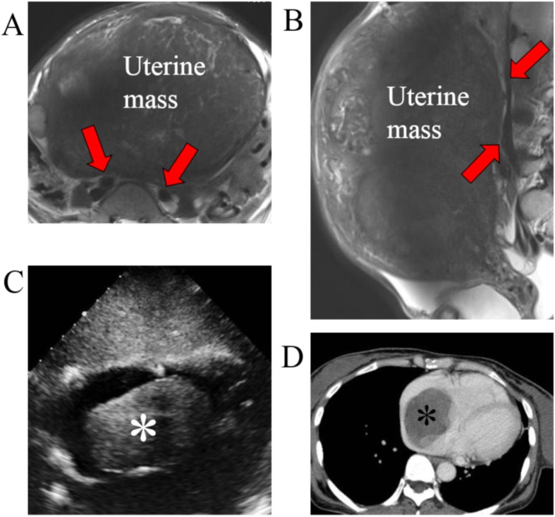
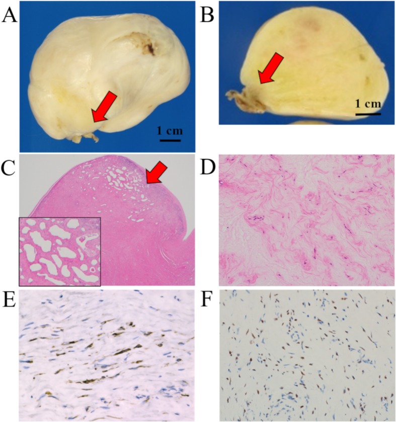
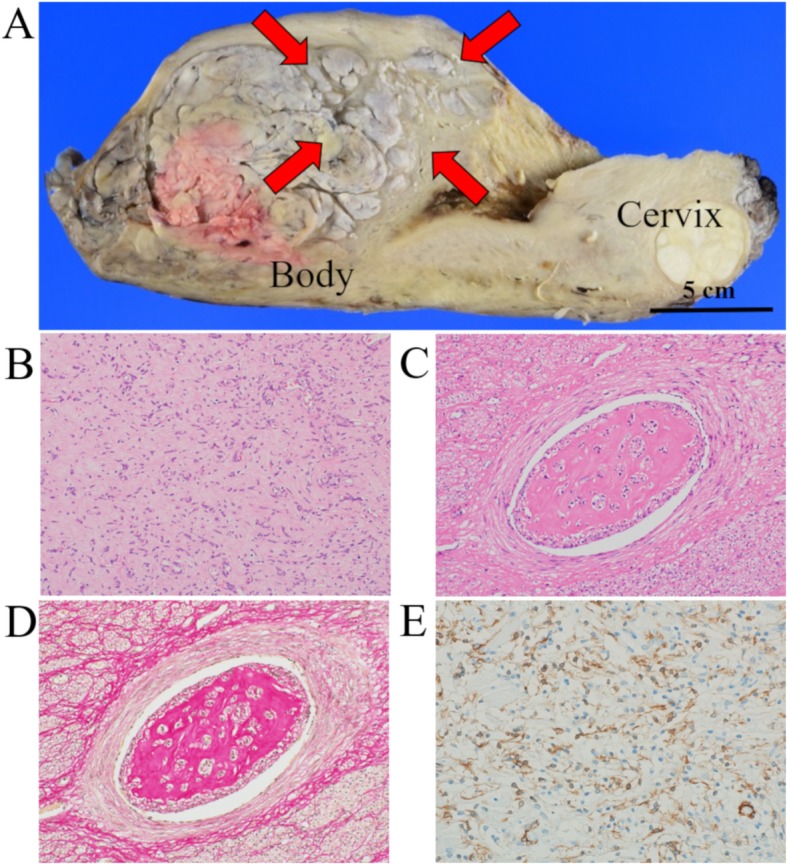
Case presentation: A 52-year-old Japanese woman sought medical attention at our hospital for lower abdominal pain. A 27-cm uterine mass was detected on clinical imaging, with a 78 × 47-mm mass in the right atrium detected on preoperative echocardiography. Intracardiac mass resection and tricuspid annuloplasty were performed as the first-stage surgery. The pedicle of the tumor was adherent to the wall of the atrium. On histological examination, the tumor was found to consist of spindle-shaped cells with eosinophilic cytoplasm, without atypia, but with a myxoid change, and rich microvascularization of the pedicle. Total abdominal hysterectomy was performed as the second-stage surgery, with confirmation of the diagnosis as uterine intravenous leiomyomatosis with an isolated metastasic lesion to the right atrium. There has been no evidence of tumor recurrence in the 15 months since surgery.

Conclusion: We report a unique case in which a large right atrial leiomyoma was identified following a uterine intravenous leiomyomatosis. Our case exemplifies that intravenous leiomyomatosis metastatic tumors have the potential to grow via their vascularization.

Keywords: Cardiac metastasis; Case report; Intravenous leiomyomatosis; Right atrium; Uterus.


Conflict of interest statement

The authors declare that they have no competing interests.

Figures

Fig. 1
Fig. 1
Clinical imaging findings: a and b T2-weighted MRI revealed a 27-cm mass in the uterus. No tumor was detected in the iliac vein (A, red arrows) or in the inferior vena cava (B, red arrows). c Echocardiography imaging revealed a 78 × 47-mm mass (asterisk) in the right atrium. d Enhanced computed tomography imaging confirmed a 75-mm mass (asterisk) in the right atrium
Fig. 2
Fig. 2
The macroscopic and microscopic findings and immunohistochemical staining of the intracardiac tumor: a and b Macroscopically, the tumor was a 78 × 58 × 55-mm mass, with a pedicle (red arrows) clinging to the right atrium. b The cut surface showed a gray-white colored solid mass. c and d Microscopically, the rich microvasculature of the pedicle was shown (red arrows); d The tumor consisted of spindle-shaped cells, with eosinophilic cytoplasm, lacked atypia, and was accompanied by a myxoid change (high power view). e and f Immunohistochemically, the tumor cells were positive for (e) desmin and (f) ER
Fig. 3
Fig. 3
The macroscopic and microscopic findings and immunohistochemical staining of the uterine mass: a Macroscopically, the cut surface presented as a gray-white colored mass, with a “worm-like” appearance (red arrows). b Microscopically, the uterine large myoma presented spindle-like smooth muscle cells without atypia and mitosis. c and d The tumor showed a growth of benign leiomyomatous tissues within vascular vessels (C, H&E; D, Elastica van Gieson). e Immunohistochemically, the tumor was positive for α-SMA

References

    1. Du J, Zhao X, Guo D, Li H, Sun B. Intravenous leiomyomatosis of the uterus: a clinicopathologic study of 18 cases, with emphasis on early diagnosis and appropriate treatment strategies. Hum Pathol. 2011;42:1240–1246. doi: 10.1016/j.humpath.2010.10.015. - DOI - PubMed
    1. Chiang CS, Chen PL, Kuo TT, Chen IM, Wu NY, Chang HH. One-stage surgery for removal of intravascular leiomyomatosis extending to right ventricle. Medicine. 2018;97:e0051. doi: 10.1097/MD.0000000000010051. - DOI - PMC - PubMed
    1. Burke M, Opeskin K. Death due to intravenous leiomyomatosis extending to the right pulmonary artery. Pathology. 2004;36:202–203. doi: 10.1080/00313020410001672075. - DOI - PubMed
    1. Wang H, Nie P, Chen B, Hou F, Dong C, He F, et al. Contrast-enhanced CT findings of intravenous leiomyomatosis. Clin Radiol. 2018;73:503.e1–503.e6. doi: 10.1016/j.crad.2017.12.016. - DOI - PubMed
    1. Thukkani N, Ravichandran PS, Das A, Slater MS, et al. Leiomyomatosis metastatic to the tricuspid valve complicated by pelvic hemorrhage. Ann Thorac Surg. 2005;79:707–709. doi: 10.1016/j.athoracsur.2003.08.038. - DOI - PubMed

Publication types