. 2020 Feb 1;106(2):440-448.
doi: 10.1016/j.ijrobp.2019.10.049.

Design, Implementation, and in Vivo Validation of a Novel Proton FLASH Radiation Therapy System

Affiliations

Design, Implementation, and in Vivo Validation of a Novel Proton FLASH Radiation Therapy System

Eric S Diffenderfer et al. Int J Radiat Oncol Biol Phys. .

Abstract

Purpose: Recent studies suggest that ultrahigh-dose-rate, "FLASH," electron radiation therapy (RT) decreases normal tissue damage while maintaining tumor response compared with conventional dose rate RT. Here, we describe a novel RT apparatus that delivers FLASH proton RT (PRT) using double scattered protons with computed tomography guidance and provide the first report of proton FLASH RT-mediated normal tissue radioprotection.

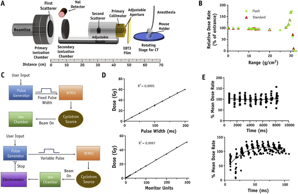
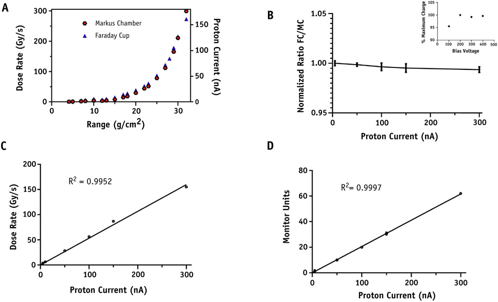
Methods and materials: Absolute dose was measured at multiple depths in solid water and validated against an absolute integral charge measurement using a Faraday cup. Real-time dose rate was obtained using a NaI detector to measure prompt gamma rays. The effect of FLASH versus standard dose rate PRT on tumors and normal tissues was measured using pancreatic flank tumors (MH641905) derived from the KPC autochthonous PanCa model in syngeneic C57BL/6J mice with analysis of fibrosis and stem cell repopulation in small intestine after abdominal irradiation.

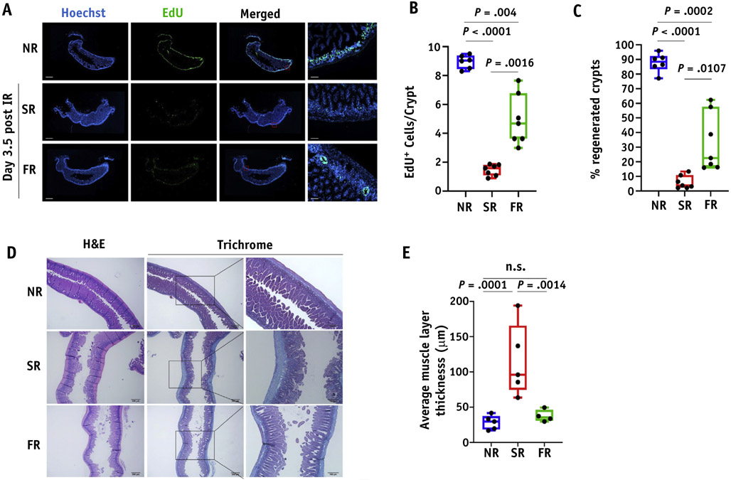
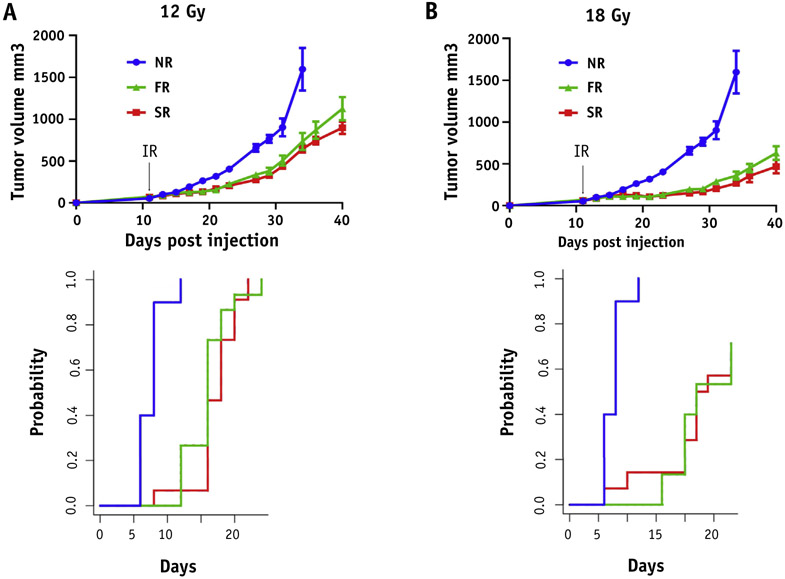
Results: The double scattering and collimation apparatus was dosimetrically validated with dose rates of 78 ± 9 Gy per second and 0.9 ± 0.08 Gy per second for the FLASH and standard PRT. Whole abdominal FLASH PRT at 15 Gy significantly reduced the loss of proliferating cells in intestinal crypts compared with standard PRT. Studies with local intestinal irradiation at 18 Gy revealed a reduction to near baseline levels of intestinal fibrosis for FLASH-PRT compared with standard PRT. Despite this difference, FLASH-PRT did not demonstrate tumor radioprotection in MH641905 pancreatic cancer flank tumors after 12 or 18 Gy irradiation.

Conclusions: We have designed and dosimetrically validated a FLASH-PRT system with accurate control of beam flux on a millisecond time scale and online monitoring of the integral and dose delivery time structure. Using this system, we found that FLASH-PRT decreases acute cell loss and late fibrosis after whole-abdomen and focal intestinal RT, whereas tumor growth inhibition is preserved between the 2 modalities.


Figures

Fig. 1.
Fig. 1.
Proton radiation therapy set up. Schematic diagram of FLASH proton radiation therapy set up (A). Beam line vacuum ends at the primary ionization chamber (IC) with dual channel readout electrometers interlocked to the primary dose counter and the cyclotron rf chain for a positive failsafe shutoff. After the primary IC is a 2 mm lead first scatterer and secondary IC providing online dose verification. Adjacent to the primary IC exit window, a NaI-gamma detector was introduced for time structure measurements of proton flux. Downstream from the secondary IC is the secondary scatterer consisting of a 5 mm diameter lead shot embedded in a plastic torus which itself is inserted into an acrylic column providing adjustment of distance between scattering centers for optimal beam profile parameters. Custom brass collimators of proton stopping thickness can be inserted into the acrylic column for variation of beam size. (B) Comparison of proton depth dose distribution using FLASH versus standard dose rate. (C) Schematics of proton flux modulation by fixed versus variable pules sent to the beam current regulation unit of the IBA system and (D) linearity of dose versus pulse width or monitor unit count for these designs. (E) Representative dose rate time structure delivered throughout each pulse for standard (18 beam pulses, upper panel) and FLASH (20 beam pulses, lower panel) dose rates.
Fig. 2.
Fig. 2.
Parameters and dose rate verification of FLASH proton radiation therapy set up. (A) Comparison of dose rate measured by Markus Chamber and Proton current measured by Faraday cup versus proton range at target isocenter. (B) Normalized ratio of Faraday cup to Marcus Chamber (MC) readings for proton currents up to 300 nA at a range of 32 g/cm2 which was used for animal irradiations. MC saturation curve (inset). (C) Dose rate measured using an MC. (D) The secondary ionization chamber MU versus proton beam current for a fixed pulse width of 100 ms.
Fig. 3.
Fig. 3.
Dose distribution for murine experiments. Dose distribution versus depth in solid water was measured with EBT3 film using the double scattered total body set up depicted in Figure 1 for a 1 x 2 cm collimator in either 0- or 90-degree orientation to allow irradiation of whole versus upper abdomen of mice. These data are presented as x-y line profiles (middle panels) and dose color wash using computed tomography aligned beam geometries (upper and lower panels). (A color version of this figure is available at https://doi.org/10.1016/j.ijrobp.2019.10.049.)
Fig. 4.
Fig. 4.
FLASH dose radiation preserves better the proliferation of intestinal crypts and inhibits fibrosis formation as a long-term effect post-IR. (A) Representative images of EdU staining in optimal cutting temperature-frozen jejunum sections at 3.5 days post 15 Gy of whole abdominal irradiation (scan of whole tissue, scale bar 1 mm; 10x, scale bar 100 μm). (B) Quantification of EdU + cells per crypt. (C) Quantification of the % regenerated crypts. Data expressed as mean ± SEM. (D) Representative Masson’s trichrome stain images of formalin fixed jejunum sections irradiated with 18 Gy at 8 weeks post-IR. (E) Quantification of fibrosis formation by calculating the average muscle layer thickness. Data expressed as mean ± standard error of the mean. Abbreviation: n.s. = not significant.
Fig. 5.
Fig. 5.
FLASH dose rate has no additional impact on tumor growth control. Mice injected with MH641905 cells on their flanks and received 12 (A) or 18 Gy (B) focal irradiation (including tumor and upper intestine) with protons at standard (SR, red) versus FLASH (FR, green) dose rates and followed for tumor growth. Unirradiated mice were served as a control group (NR, blue). Black arrow indicates the time of irradiation. Data expressed as mean ± SEM. Cumulative incidence of tumor regrowth to 4x the starting tumor volume was calculated for each dose (bottom panel) and demonstrated that tumor regrowth was decreased by proton radiation therapy (P < .000001), but not different between FLASH and standard proton radiation therapy (12 Gy P = .68; 18 Gy P = .84). (A color version of this figure is available at https://doi.org/10.1016/j.ijrobp.2019.10.049.)

References

    1. Favaudon V, Caplier L, Monceau V, et al. Ultrahigh dose-rate FLASH irradiation increases the differential response between normal and tumor tissue in mice. Sci Transl Med 2014;6:245ra93. - PubMed
    1. Durante M, Brauer-Krisch E, Hill M. Faster and safer? FLASH ultra-high dose rate in radiotherapy. Br J Radiol 2018;91:20170628. - PMC - PubMed
    1. Harrington KJ. Ultrahigh dose-rate radiotherapy: Next steps for FLASH-RT. Clin Cancer Res 2019;25:3–5. - PubMed
    1. Vozenin MC, De Fornel P, Petersson K, et al. The advantage of FLASH radiotherapy confirmed in mini-pig and cat-cancer patients. Clin Cancer Res 2019;25:35–42. - PubMed
    1. Schmid TE, Dollinger G, Hauptner A, et al. No evidence for a different rbe between pulsed and continuous 20 mev protons. Radiat Res 2009;172:567–574. - PubMed

Publication types

MeSH terms