Review
. 2019:118:273-288.
doi: 10.1016/bs.apcsb.2019.09.005. Epub 2019 Oct 21.

LOXL1 folding in exfoliation glaucoma

Affiliations
Review

LOXL1 folding in exfoliation glaucoma

Audrey M Bernstein et al. Adv Protein Chem Struct Biol. 2019.

Abstract

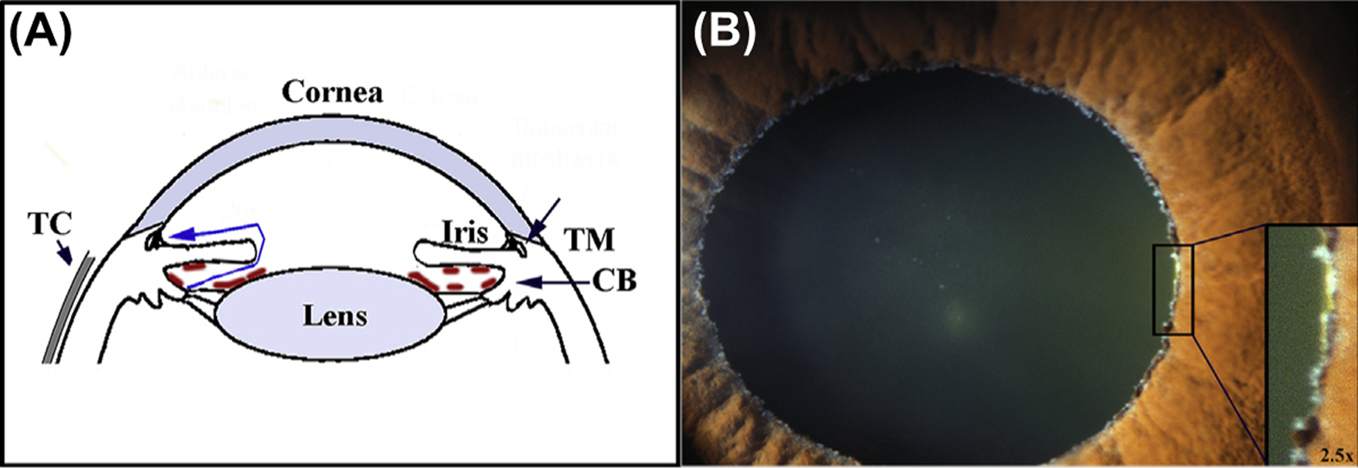
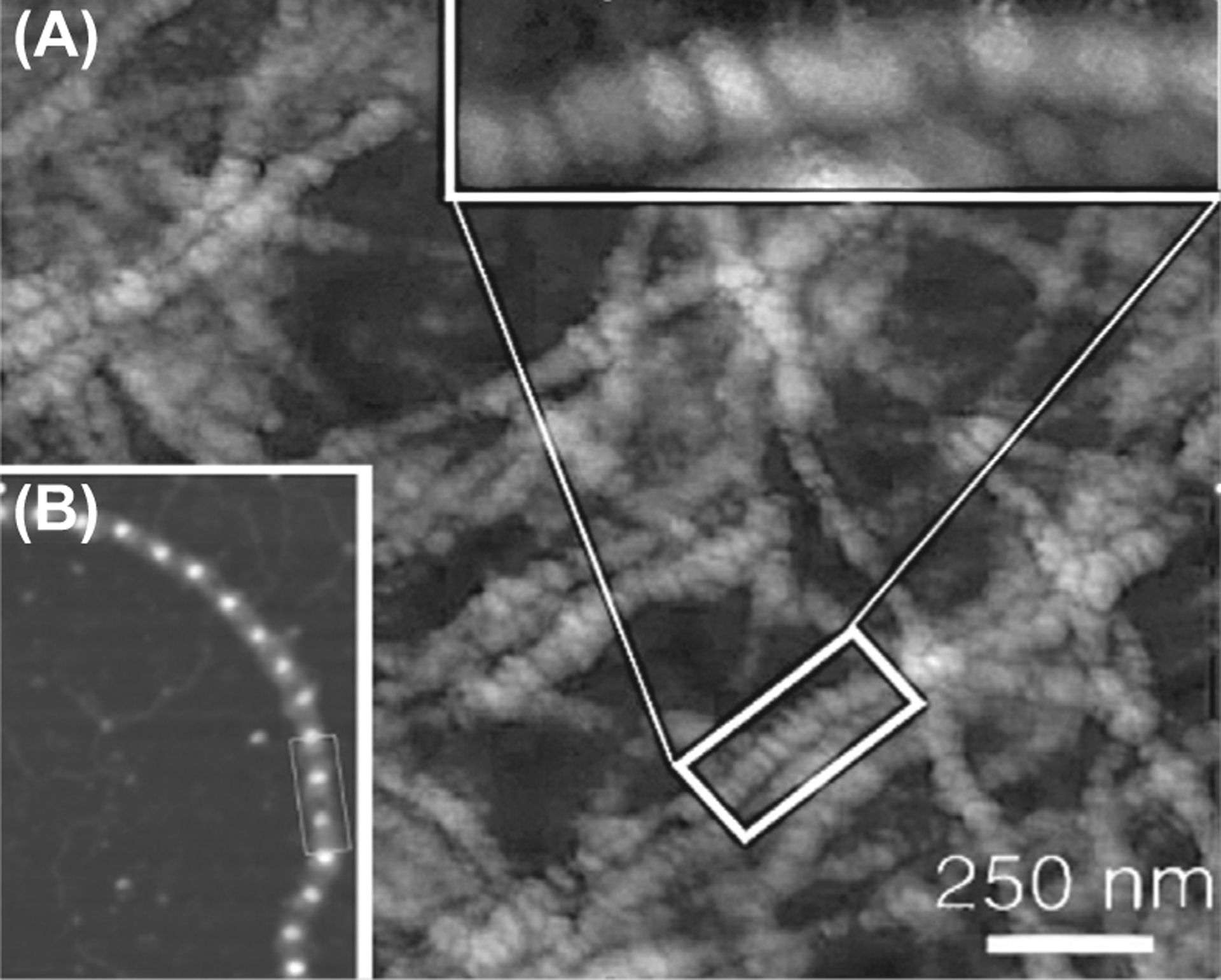
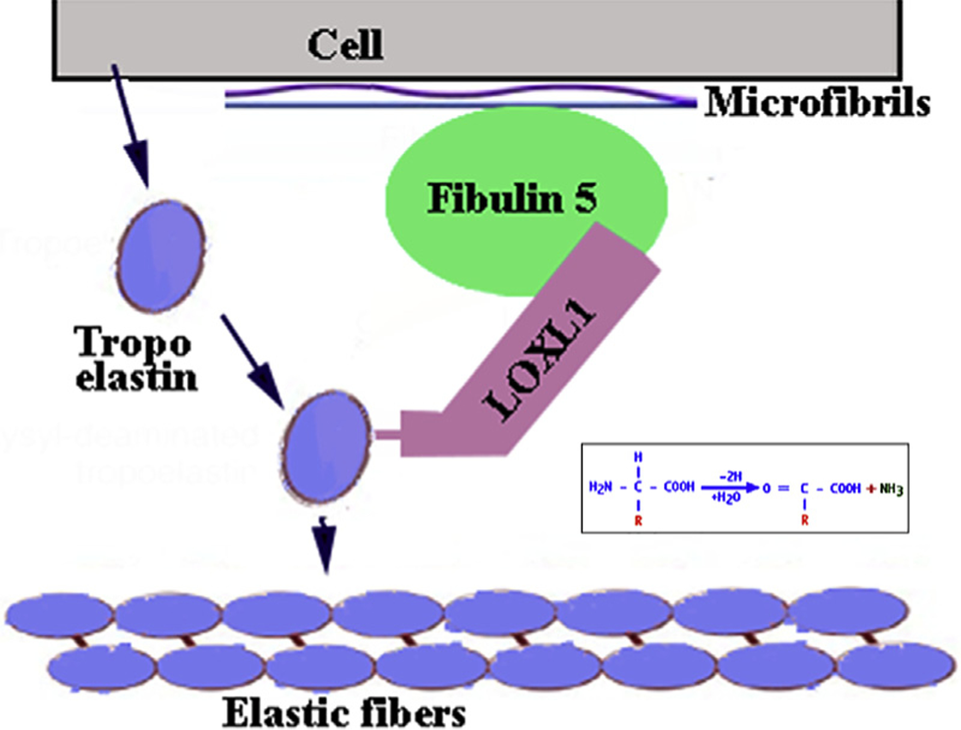
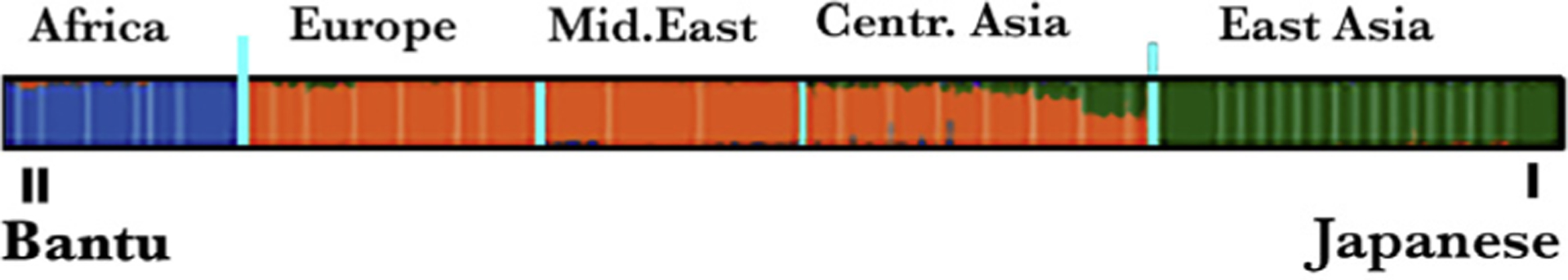
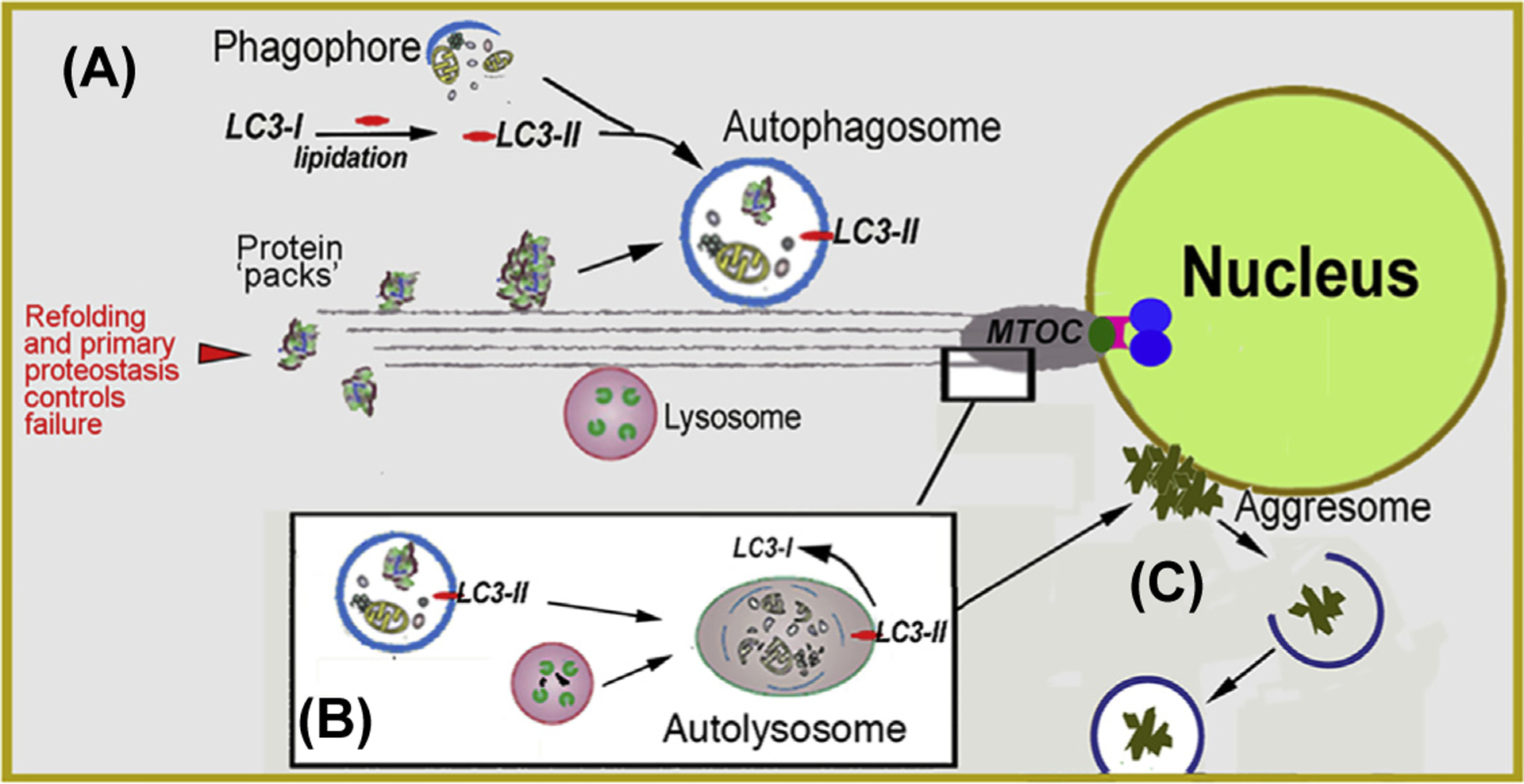
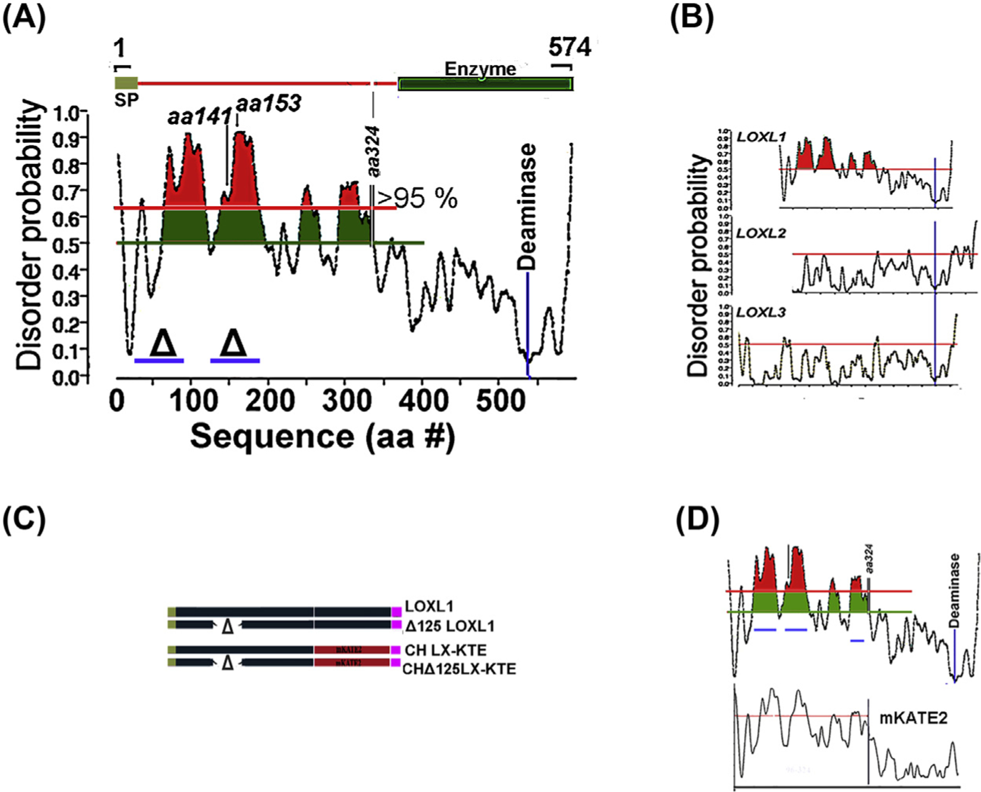
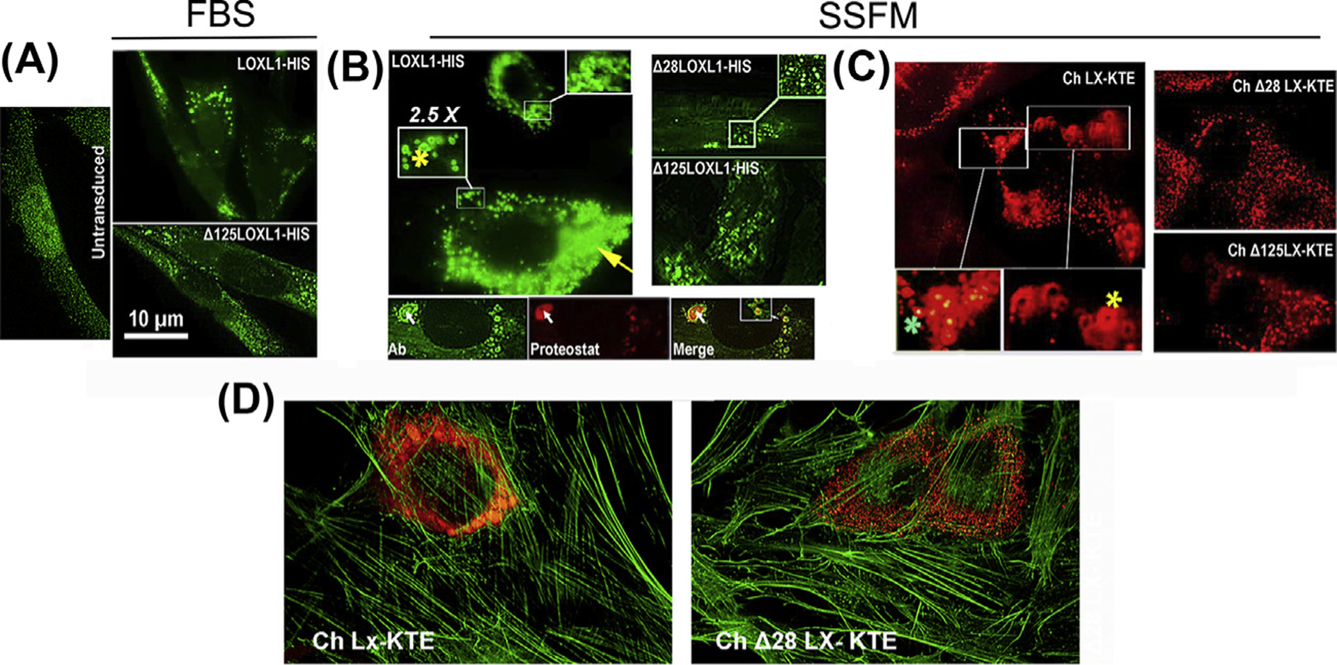
Exfoliation syndrome (XFS) is an age-related disease defined by the deposition of aggregated fibrous material (XFM) in the peri-cellular space. Principal morbidity occurs in the eye, where XFM accumulates on the anterior ocular tissues. GWAS have found that certain genetic variants of lysyl oxidase-like 1 (LOXL1), a matrix cross-linking enzyme that is required for elastic fiber formation confer risk for the development of XFS, but are not a single causative factor as many genetically affected individuals do not develop XFS or subsequent glaucoma (XFG). We have found that XFG cells display defects in lysosomes, microtubules, autophagy, and mitochondria resembling defects found in cells from age-related syndromes, such as the main neurodegenerative diseases. In the majority of these diseases, the determining cellular factor is a protein containing intrinsically disordered regions (IDRs) and displaying a high propensity for aggregation. We have found that in XFG patient-derived cells, LOXL1 protein is actively subjected to autophagic clearance, suggesting that LOXL1 is undergoing aggregation. In silico analysis demonstrates that LOXL1's first 369 aa constitute an IDR with the highest disorder probability peak centering around the known risk positions. Experimentally, we have found over-expression of either unmodified LOXL1 or fluorescent chimeras preserving the well-structured N-terminus cause copious intracellular aggregation and that aggregation wanes when the high IDR peaks are deleted. Overall, our work suggests that XFS/G results from the aggregation of the LOXL1 protein coupled with a reduction of cellular proteostasis capabilities in aging, resulting in a chronic build-up of LOXL1-containing protein aggregates.

Keywords: Aggregopathy; Autophagy; Glaucoma; Intrinsically disordered regions; LOXL1.


Figures

Fig. 1
Fig. 1. (A) Schematic description of the cellular components of the anterior segments of the eye in relation to aqueous flow.
Fluid (blue line) is generated at the inner surface of the epithelium of the ciliary body CB) in the posterior aqueous chamber. From there it flows through the space between the iris and lens into the anterior aqueous chamber to exit the eye through the trabecular meshwork (TM) also referred to as the outflow facility. Increased, abnormal flow resistance of the facility causes elevation of the intraocular pressure (IOP). The increased pressure lead to the development of glaucoma, loss of retinal visual function due to retinal neural death. In individuals affected by exfoliation syndrome (XSF), proteinaceous material (shown tan marks in sketch) forms or deposits on the surface of the epithelial facing the posterior aqueous chamber, including the equator of the lens. The deposition is frequently associated with a type of glaucoma, exfoliation glaucoma, characterized by drug-unresponsive IOP increases and rapid progression of glaucoma. The Tenon’s capsule (TC) is a thin, elastic collagenous lining covering the sclera. (B) Flaky white deposits in the equator of the lens. The pupil has been dilated.
Fig. 2
Fig. 2
(A) XFM structure. (B) Fibrillin microfibrils.
Fig. 3
Fig. 3. Schematic description of elastic fiber development.
At the peri-cellular space the adapter protein Fibulin 5 anchoring at Fibrillin 1, binds the deaminase LOXL1 setting it in the proper spatial configuration to cause deamination of secreted Tropoelastin monomers. The reactive aldehyde groups allow homologous polymerization of Tropoelastin to form elastic fibers.
Fig. 4
Fig. 4. Human genetic clustering.
The South African Bantu and Japanese sit at the opposite sides of the spectrum of the differences in expression of genetic variants. One representative gene cluster, of several studied, is shown.
Fig. 5
Fig. 5. Macroautophagy.
(A) Biogenesis of autophagosomes from precursor phagophores within the cytosol is orchestrated by the lipidated form of LC3 (LC3-II). Autophagosomes engulf ‘macroscopic’ cellular detritus, naturally decaying organelles, in particular, mitochondria (i.e., mitophagy) and misfolded or denatured proteins that have resisted the primary protein folding quality control mechanisms. (B) Autophagosomes fuse with lysosomes at the microtubule organizing center (MTOC). (C) Detritus that resist the combined proteostatic processes may accumulate in body inclusions, (e.g., Lewy body in senile dementia) categorized alternatively as aggresomes. If as demonstrated for tau protein propagation in Alzheimer’s and a-synuclein protein in Parkinson’s, then the aggregates may propagate to adjacent cells by particulate phagocytosis or endocytosis.
Fig. 6
Fig. 6. IDR in LOXL proteins.
(A) Order/disorder probability prediction in the LOXL1 sequence. The zones where prediction confidence is > 95% is highlighted in red and the location for the enzymatic activity is indicated. (B) Comparison of order/disorder in LOXL family. (C) Comparison of order/disorder pattern in parent LOXL1 sequence and its mKATE2 chimera. (D) Examples of transgene LOXL1 constructs and their equivalent chimeras used to test aggregation propensity in overexpression assays.
Fig. 7
Fig. 7. Aggregation in overexpressed LOXL1 and homolog mKATE2 sequences.
Representative images of BJ5a cells transduced with the indicated constructs. LOXL1 constructs were stained with an N-terminus anti-LOXL1 monoclonal Ab (H11 clone). Chimeras are red fluorescent. (A) Cultures in FBS-containing media. Left. Untransduced cells. Top. Cells transduced with LOXL1-HIS. Enlarged vesicles accumulated near the nuclei are the most common feature of cells expressing high levels of the transgene. Bottom. Typical stain profile for the Δ125–185 LOXL1 version. Vesicles are smaller and distribute throughout the cytosol. (B) Left top. The same transduction with LOXL1-HIS cDNA in supplemented-serum free media (SSFM). In addition to engorged vesicles (yellow arrow), very large particles and ring-shaped profiles (yellow asterisks) are frequent. Bottom 3-frame composite. Anti-LOXL1- Proteostat dual stain. Whereas the LOXL1 Ab stains the periphery of particles generating ring-shaped fluorescence, the aggregated-protein dye (Proteostat) labels the center of the same particles (white arrow). Right. LOXL1 with deleted domains lack this grossly vesicular phenotype. (C) The fluorescence patterns of the mKATE2 chimeras. Left. Ch LX-KTE fluorescence. Large vesicles and ring-like profiles (yellow asterisk) and engorged vesicles. There are also vesicles where the fluorescence intensity is higher at the center, rather than absent or lower (green asterisk). Right. The fluorescence of Δ versions. There are no aggregation profiles. (D) Micrographs of cells transduced with whole sequence (1–324) mKATE2 chimera and a chimera with a 28–95 aa deletion (Δ28) and counterstained with FITC-phalloidin.

References

    1. Aboobakar IF, Johnson WM, Stamer WD, Hauser MA, & Allingham RR (2016). Major review: Exfoliation syndrome; advances in disease genetics, molecular biology, and epidemiology. Experimental Eye Research, 154, 88–103. 10.1016/j.exer.2016.11.011. - DOI - PubMed
    1. Abu-Amero KK, Osman EA, Dewedar AS, Schmidt S, Allingham RR, & Al-Obeidan SA (2010). Analysis of LOXL1 polymorphisms in a Saudi Arabian population with pseudoexfoliation glaucoma. Molecular Vision, 16, 2805–2810. Retrieved from https://www.ncbi.nlm.nih.gov/pubmed/21197115. - PMC - PubMed
    1. Anastasopoulos E, Founti P, & Topouzis F (2015). Update on pseudoexfoliation syndrome pathogenesis and associations with intraocular pressure, glaucoma and systemic diseases. Current Opinion in Ophthalmology, 26(2), 82–89. 10.1097/ICU.0000000000000132. - DOI - PubMed
    1. Atkins JD, Boateng SY, Sorensen T, & McGuffin LJ (2015). Disorder prediction methods, their applicability to different protein targets and their usefulness for guiding experimental studies. International Journal of Molecular Sciences, 16(8), 19040–19054. 10.3390/ijms160819040. - DOI - PMC - PubMed
    1. Bernstein AM, Ritch R, & Wolosin JM (2018). Exfoliation syndrome: A disease of autophagy and LOXL1 proteopathy. Journal of Glaucoma. 10.1097/IJG.0000000000000919. - DOI - PMC - PubMed

Publication types

Substances

LinkOut - more resources