. 2020 Mar;73(3):460-468.
doi: 10.1016/j.bjps.2019.11.048. Epub 2019 Dec 11.

Revascularization patterns of nerve allografts in a rat sciatic nerve defect model

Affiliations

Revascularization patterns of nerve allografts in a rat sciatic nerve defect model

Tiam M Saffari et al. J Plast Reconstr Aesthet Surg. 2020 Mar.

Abstract

Introduction: The specific patterns of revascularization of allograft nerves after the addition of vascularization remain unknown. The aim of this study was to determine the revascularization patterns of optimized processed allografts (OPA) after surgically induced angiogenesis to the wound bed in a rat sciatic nerve model.

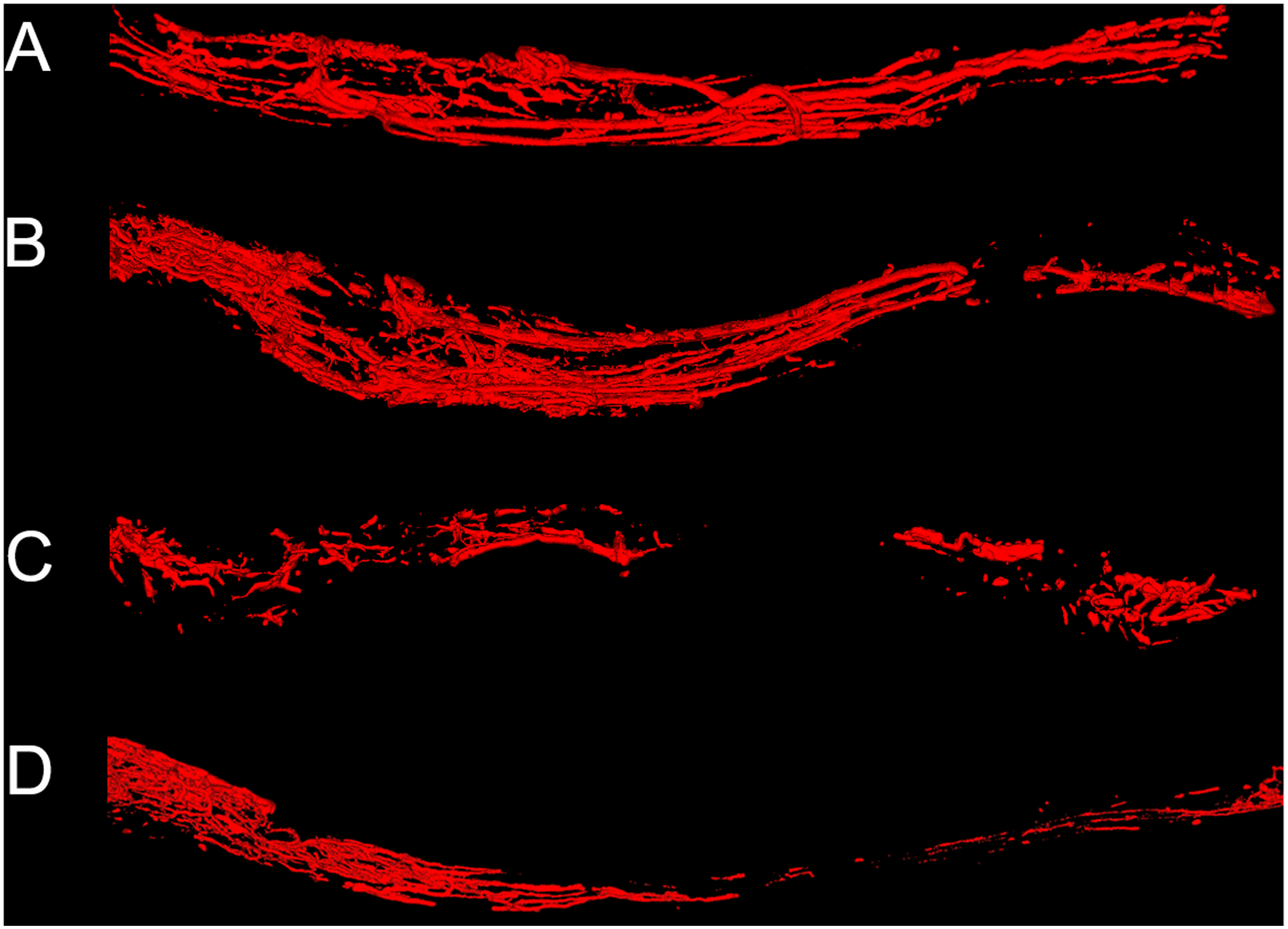
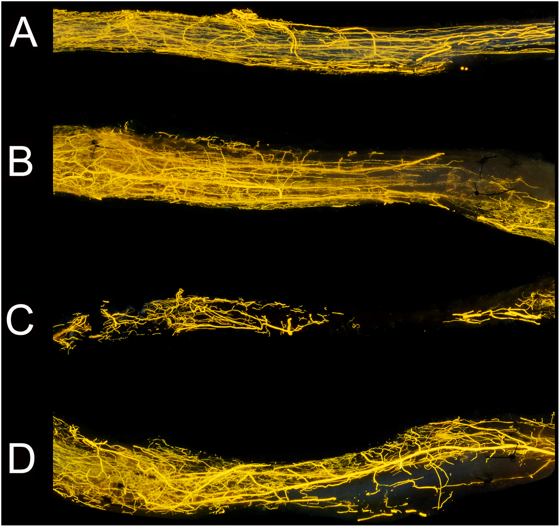
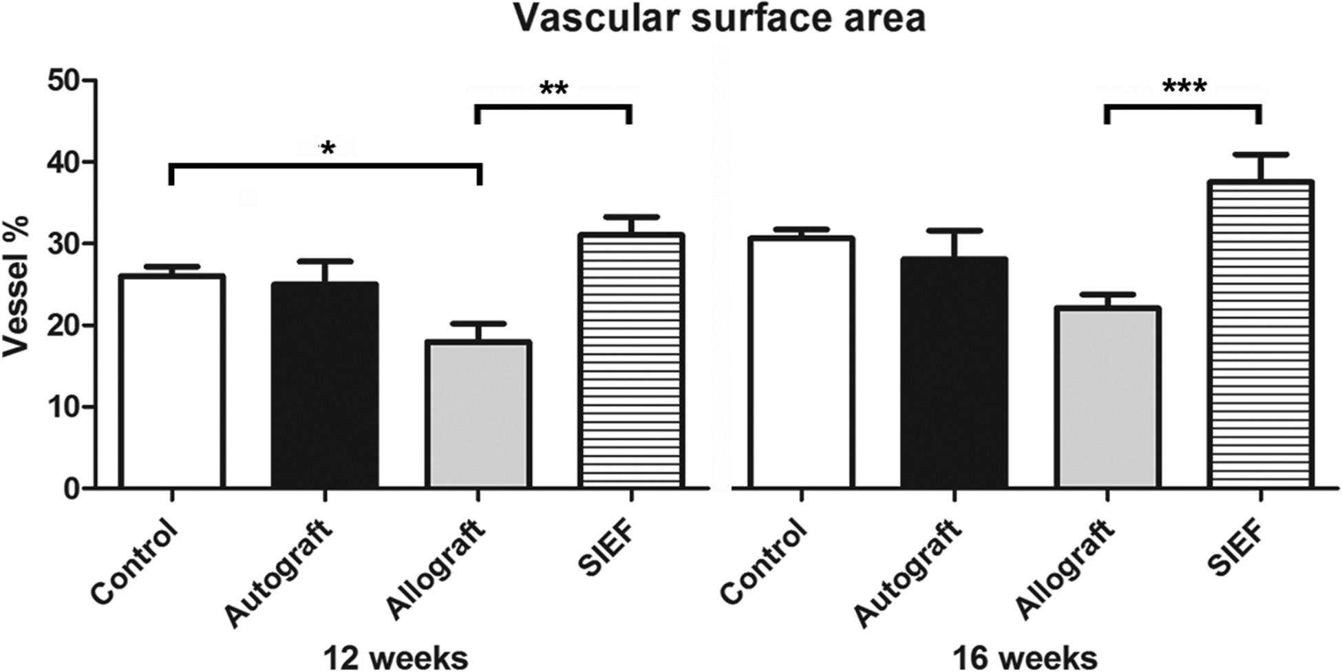
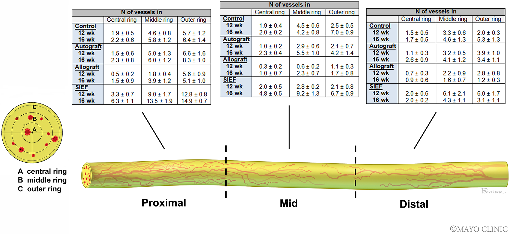
Materials and methods: In 51 Lewis rats, sciatic nerve gaps were repaired with (i) autografts, (ii) OPA and (iii) OPA wrapped in a pedicled superficial inferior epigastric artery fascia flap (SIEF) to provide vascularization to the wound bed. At 2, 12, and 16 weeks, the vascular volume and vascular surface area in nerve samples were measured using micro CT and photography. Cross-sectional images were obtained and the number of vessels was quantified in the proximal, mid, and distal sections of the nerve samples.

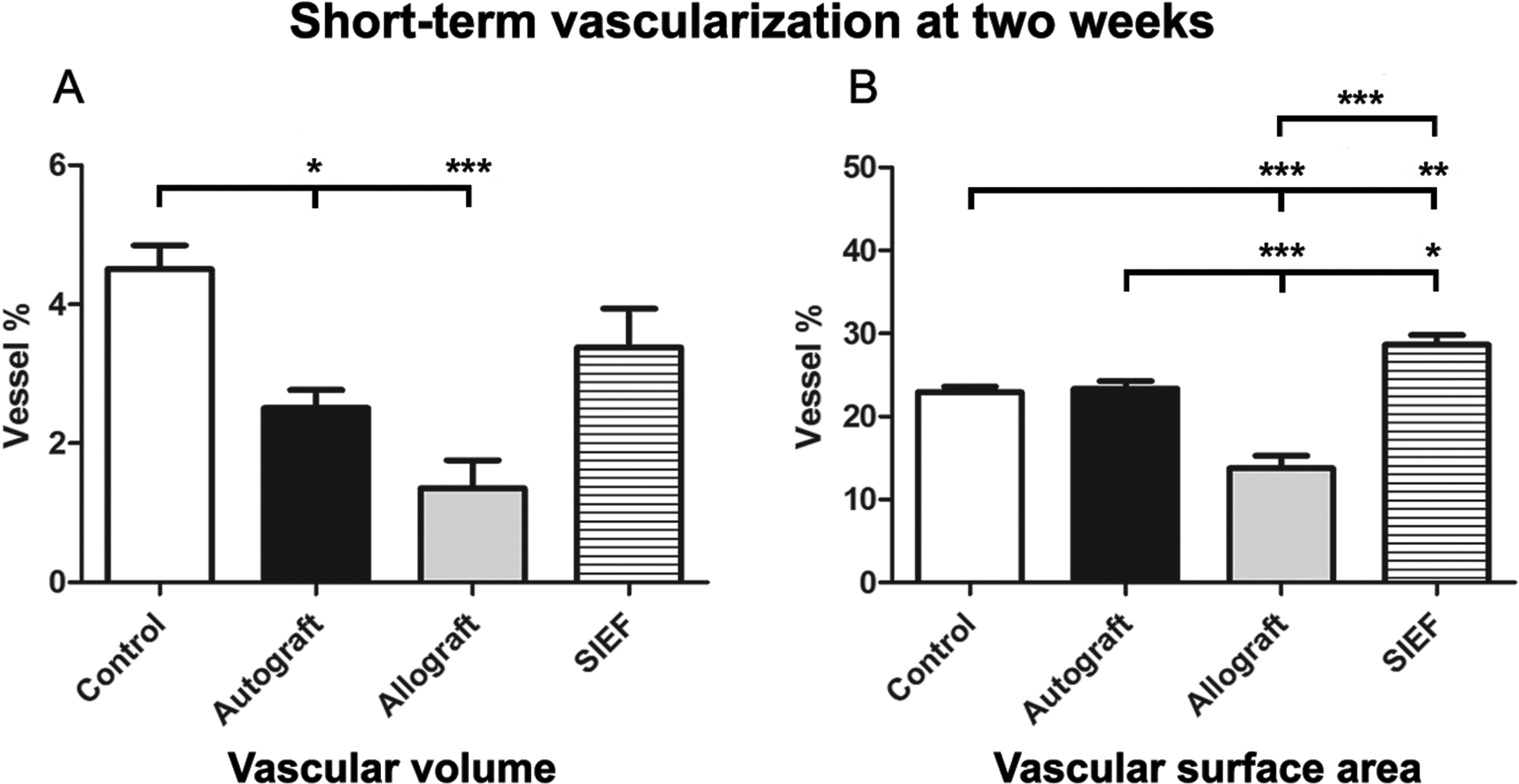
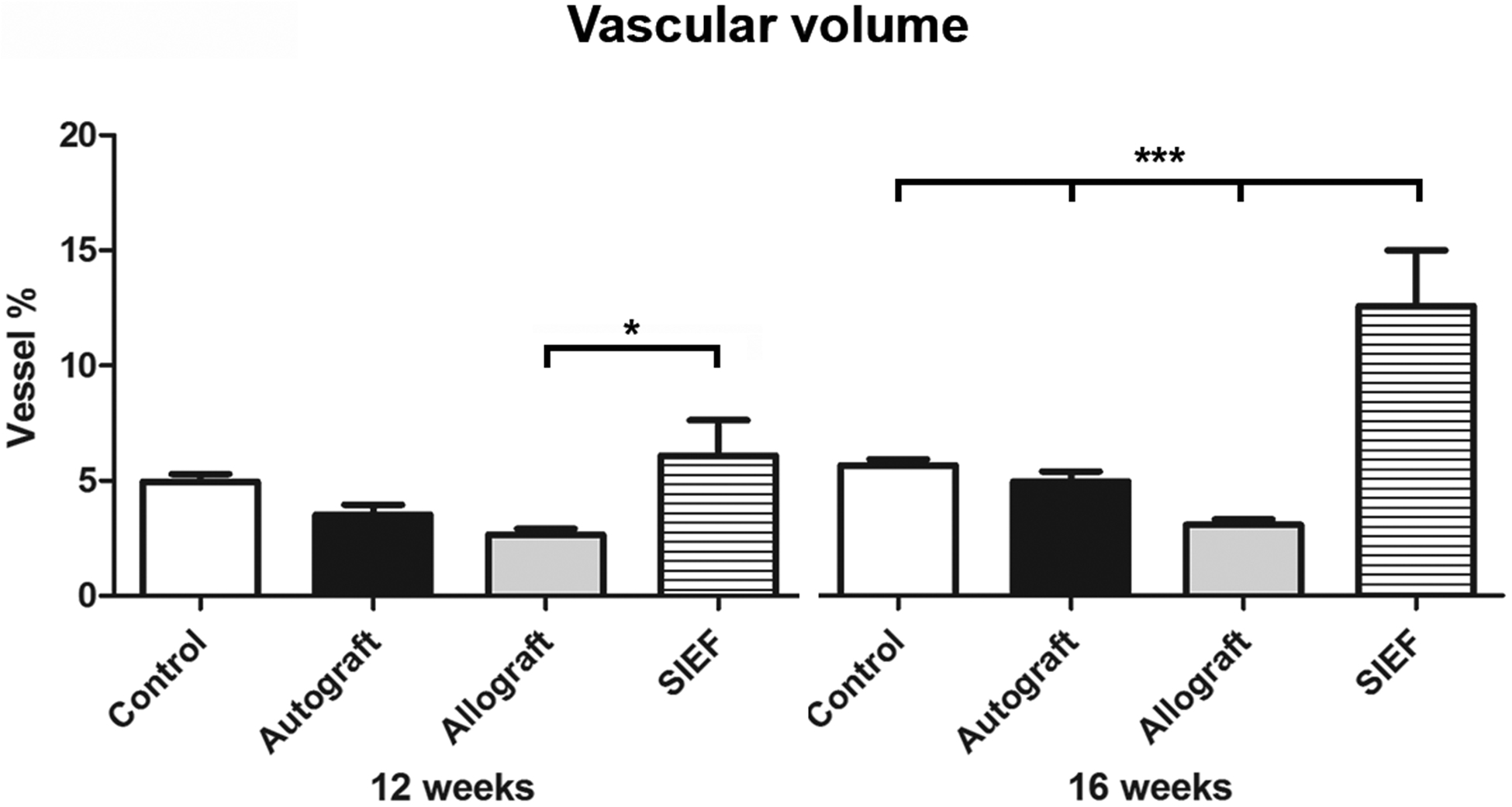
Results: At 2 weeks, the vascular volume of SIEF nerves was comparable to control (P = 0.1). The vascular surface area in SIEF nerves was superior to other groups (P<0.05). At 12 weeks, vascularity in SIEF nerves was significantly higher than allografts (P<0.05) and superior compared to all other groups (P<0.0001) at 16 weeks. SIEF nerves had a significantly increased number of vessels compared to allografts alone in the proximal (P<0.05) and mid-section of the graft (P<0.05).

Conclusions: Addition of surgical angiogenesis to the wound bed greatly improves revascularization. It was demonstrated that revascularization occurs primarily from proximal to distal (proximal inosculation) and not from both ends as previously believed and confirms the theory of centripetal revascularization.

Keywords: Angiogenesis; Nerve regeneration; Peripheral nerve repair; Processed nerve allograft; Revascularization patterns; Vascularized nerve allograft.


Conflict of interest statement

Declaration of Competing Interest None declared.

Figures

Figure 1.
Figure 1.. Micro computed tomography (micro CT) images of nerve samples.
Micro CT images of control nerve (A), autograft (B), allograft (C) and allograft wrapped in a pedicled superficial inferior epigastric fascial (SIEF) flap (D). Images were obtained at 16 weeks. Nerve samples were positioned from proximal to distal (left to right respectively). Scale bar is set at 1 millimeter. -In the supplementary data videos of these nerve samples were provided to give a 3D representation of the micro CT.
Figure 2.
Figure 2.. Macroscopic images of nerve samples obtained with conventional digital photography.
Images of the same samples visualized in Figure 2. Microvessels were clearly seen in the control nerve (A), autograft (B), allograft (C) allograft wrapped in a pedicled superficial inferior epigastric fascial (SIEF) flap (D). These photographs depicted nerve groups at 16 weeks. Sutures that were used to repair the graft were visible in nerve graft groups (B,D) and depicted the border of the analyzed frame. Nerve samples were positioned from proximal to distal (left to right respectively). Scale bar is set at 1 millimeter.
Figure 3.
Figure 3.. Short-term vascularization at two weeks measured by vascular volume (micro CT, 3A) and vascular surface area (conventional digital photography, 3B).
Results of control, autograft, allograft and allograft wrapped in a pedicled superficial inferior epigastric fascial (SIEF) flap expressed as percentage (vessel %) of the total nerve area and were given as the mean ± SEM. Please note that the range of the Y-axes is different. * Indicates significance at P<0.05, ** P<0.01, *** P<0.0001. SEM = Standard error of the mean
Figure 4.
Figure 4.. Vascular volume of nerve groups at 12- and 16 weeks using micro CT.
Results of control, autograft, allograft and allograft wrapped in a pedicled superficial inferior epigastric fascial (SIEF) flap were expressed as a percentage (vessel %) of the total nerve area and were given as the mean ± SEM. *Indicates significance at P<0.05, *** P<0.0001. SEM = Standard error of the mean
Figure 5.
Figure 5.. Vascular surface area of nerve groups at 12- and 16 weeks using digital photography.
Results of control, autograft, allograft and allograft wrapped in a pedicled superficial inferior epigastric fascial (SIEF) flap were expressed as a percentage (vessel %) of the total nerve area and were given as the mean ± SEM. *Indicates significance at P<0.05, *** P<0.0001. SEM = Standard error of the mean
Figure 6.
Figure 6.. Nerve vascularization patterns of nerve groups at 12- and 16 weeks.
Micro CT cross-sectional images throughout the length of the nerve grafts were obtained. The length of the nerve was divided into three sections: (I) proximal, (II) mid and (III) distal. The cross-sectional images were divided into three rings: (A) central ring, (B) middle ring, (C) outer ring to count the number of vessels. Nerve tissue was depicted in yellow and the vessels were depicted in red. Tables describe the number of vessels in each of the rings per nerve section (proximal, mid and distal) for control, autograft, allograft and allograft wrapped in a superficial inferior epigastric artery fascia (SIEF) flap. The first row denotes the number of vessels (mean ± SEM) at 12 weeks and the second row at 16 weeks. SEM = Standard error of the mean

References

    1. Ferretti A, Boschi E, Stefani A, et al. Angiogenesis and nerve regeneration in a model of human skin equivalent transplant. Life Sci 2003;73):1985–1994. - PubMed
    1. Gu XH, Terenghi G, Kangesu T, et al. Regeneration pattern of blood vessels and nerves in cultured keratinocyte grafts assessed by confocal laser scanning microscopy. Br J Dermatol 1995;132:376–383. - PubMed
    1. Manek S, Terenghi G, Shurey C, Nishikawa H, Green CJ, Polak JM. Neovascularisation precedes neural changes in the rat groin skin flap following denervation: an immunohistochemical study. Br J Plast Surg 1993;46:48–55. - PubMed
    1. Kangesu T, Manek S, Terenghi G, et al. Nerve and blood vessel growth in response to grafted dermis and cultured keratinocytes. Plast Reconstr Surg 1998;101:1029–1038. - PubMed
    1. Hobson MI, Brown R, Green CJ, Terenghi G. Inter-relationships between angiogenesis and nerve regeneration: a histochemical study. Br J Plast Surg 1997;50:125–131. - PubMed