. 2020 Jan 12;12(1):844-865.
doi: 10.18632/aging.102659. Epub 2020 Jan 12.

Surgery/Anesthesia disturbs mitochondrial fission/fusion dynamics in the brain of aged mice with postoperative delirium

Affiliations

Surgery/Anesthesia disturbs mitochondrial fission/fusion dynamics in the brain of aged mice with postoperative delirium

Yayuan Lu et al. Aging (Albany NY). .

Abstract

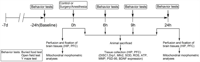
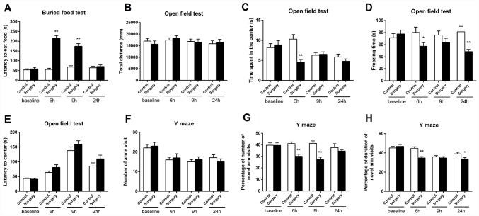
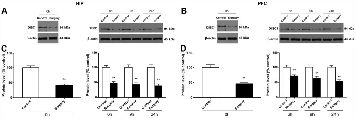
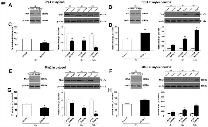
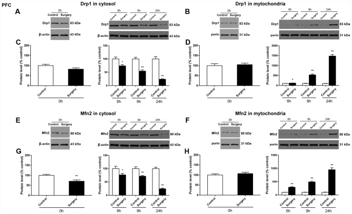
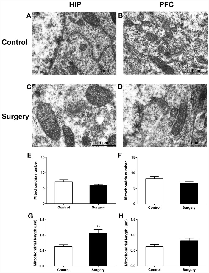
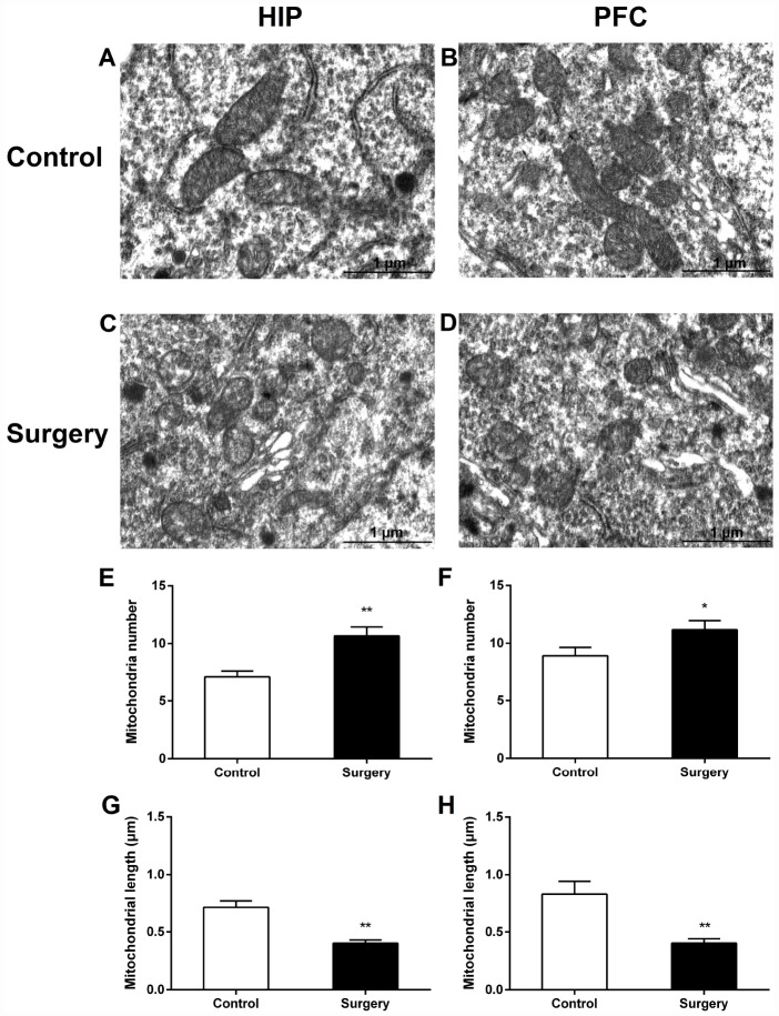
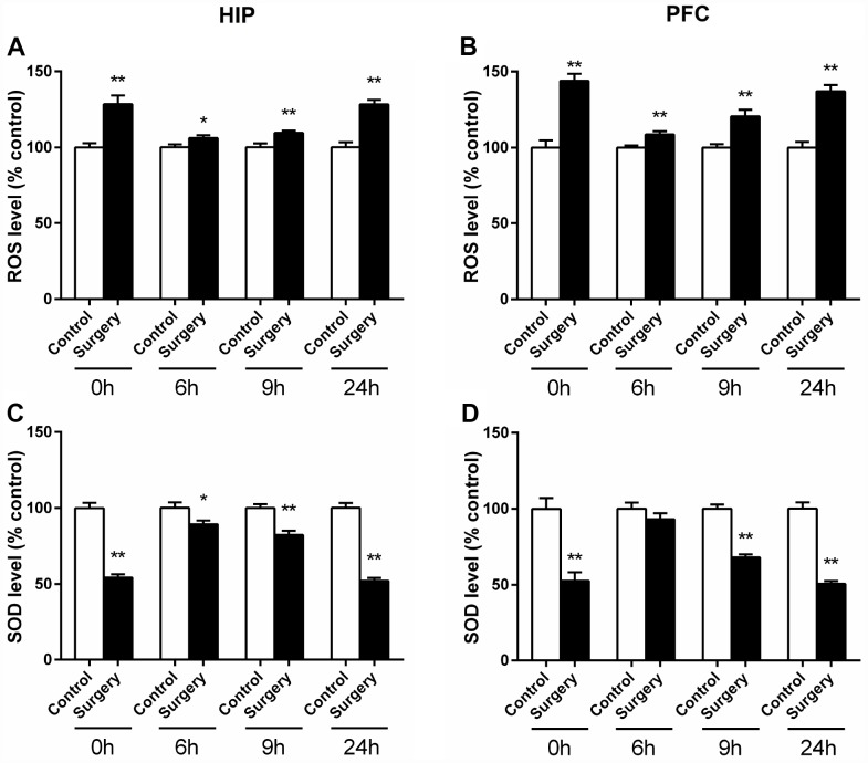
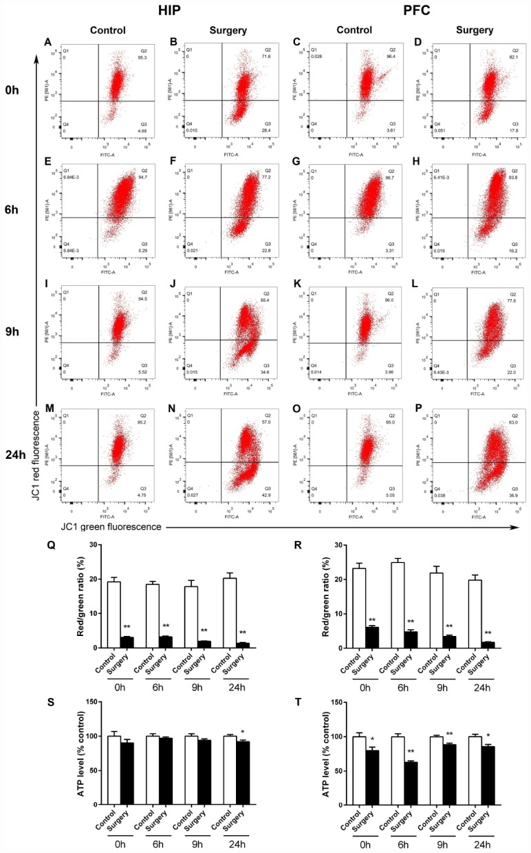
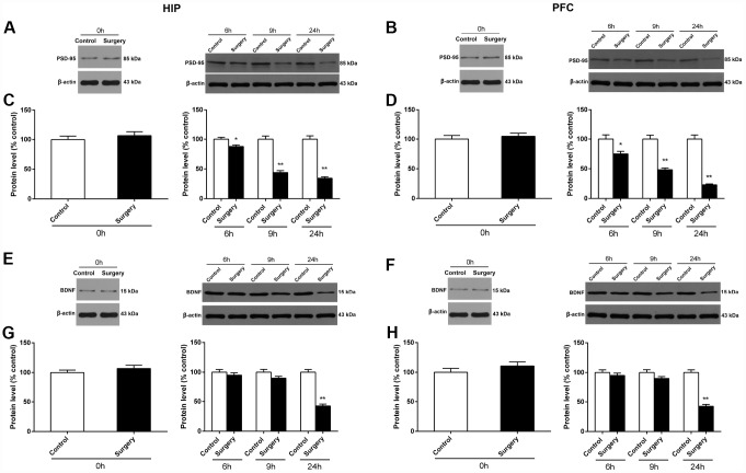
Postoperative delirium (POD) is a common complication following surgery and anesthesia (Surgery/Anesthesia). Mitochondrial dysfunction, which is demonstrated by energy deficits and excessively activated oxidative stress, has been reported to contribute to POD. The dynamic balance between mitochondrial fusion and fission processes is critical in regulating mitochondrial function. However, the impact of Surgery/Anesthesia on mitochondrial fusion/fission dynamics remains unclear. Here, we evaluate the effects of laparotomy under 1.4% isoflurane anesthesia for 2 hours on mitochondrial fission/fusion dynamics in the brain of aged mice. Mice in Surgery/Anesthesia group showed unbalanced fission/fusion dynamics, with decreased DISC1 expression and increased expression of Drp1 and Mfn2 in the mitochondrial fraction, leading to excessive mitochondrial fission and disturbed mitochondrial morphogenesis in the hippocampus and prefrontal cortex. In addition, surgical mice presented mitochondrial dysfunction, demonstrated by abnormally activated oxidative stress (increased ROS level, decreased SOD level) and energy deficits (decreased levels of ATP and MMP). Surgery/Anesthesia also decreased the expression of neuronal/synaptic plasticity-related proteins such as PSD-95 and BDNF. Furthermore, Surgery/Anesthesia induced delirium-like behavior in aged mice. In conclusion, Surgery/Anesthesia disturbed mitochondrial fission/fusion dynamics and then impaired mitochondrial function in the brain of aged mice; these effects may be involved in the underlying mechanism of POD.

Keywords: hippocampus; mitochondrial dynamics; mitochondrial function; postoperative delirium; prefrontal cortex.


Conflict of interest statement

CONFLICTS OF INTEREST: The authors declare no conflicts of interest in this study.

Figures

Figure 1
Figure 1
Diagram of the experimental design. The mice underwent behavioral tests at 24 hours before (baseline) and at 6, 9, and 24 hours after the Surgery/Anesthesia. Within each group, separate cohorts were subjected to assessments at each time point (n = 9 per cohort). Mice were sacrificed immediately after the Surgery/Anesthesia and at 6, 9, and 24 hours postoperatively. The hippocampal and prefrontal cortex tissues were harvested for analysis of DISC1, Drp1, Mfn2, SOD, ROS, ATP, MMP, BDNF, and PSD-95 levels (n = 6 per cohort). Mice were anesthetized and transcardially perfused with ice-cold phosphate-buffered saline (PBS) followed by paraformaldehyde and glutaraldehyde; then, hippocampal and prefrontal cortex tissues were collected and stored in the same fixative for electron microscopy analysis immediately after the Surgery/Anesthesia and at 24 hours postoperatively (n = 3 per cohort). DISC1, disrupted in schizophrenia 1. Drp1, dynamin-related protein 1. Mfn2, mitofusin 2. SOD, superoxide dismutase. ROS, reactive oxygen species. ATP, adenosine triphosphate. MMP, mitochondrial membrane potential. PSD-95, postsynaptic density protein 95. BDNF, brain-derived neurotrophic factor.
Figure 2
Figure 2
Surgery/Anesthesia impaired the behavior of aged mice at 6, 9, and 24 hours postoperatively. (A) Surgery/Anesthesia increased the latency of mice to eat the food as compared to the control condition in the buried food test at 6 and 9 hours postoperatively. Surgery/Anesthesia did not significantly alter the latency of mice to eat food as compared to the control condition at 24 hours postoperatively. (B) Surgery/Anesthesia did not significantly change the total distance travelled by mice in the open field test as compared to the control condition at 6, 9, and 24 hours postoperatively. (C) Surgery/Anesthesia significantly decreased the time spent in the center of the open field as compared to the control condition at 6 but neither 9 nor 24 hours postoperatively. (D) Surgery/Anesthesia significantly decreased the freezing time in the open field test as compared to the control condition at 6 and 24 but not 9 hours postoperatively. (E) Surgery/Anesthesia did not significantly change the time to reach the center (latency to the center) in the open field test as compared to the control condition at 6, 9, and 24 hours postoperatively. (F) Surgery/Anesthesia did not significantly change the number of arm visits in the Y maze test as compared to the control condition at 6, 9, and 24 hours postoperatively. (G) Surgery/Anesthesia significantly decreased the number of entries in the novel arm in the Y maze test as compared to the control condition at 6 and 9 but not 24 hours postoperatively. (H) Surgery/Anesthesia significantly decreased the duration in the novel arm in the Y maze test as compared to the control condition at 6 and 24 but not 9 hours postoperatively. The data are plotted as the mean ± standard error of the mean for each group (n = 9). *p < 0.05 and **p < 0.01, compared to control.
Figure 3
Figure 3
Surgery/Anesthesia decreased the DISC1 expression in the hippocampus and prefrontal cortex of aged mice at postoperative 0, 6, 9, and 24 hours. The expression of DISC1 was estimated using western blotting analysis of fresh homogenates from hippocampal and prefrontal cortex tissues of mice at 0, 6, 9, and 24 hours after Surgery/Anesthesia or control treatment. (A and C) The level of DISC1 in the hippocampus of mice in the Surgery/Anesthesia group decreased significantly compared to that in control mice at postoperative 0, 6, 9, and 24 hours. (B and D) The level of DISC1 in the prefrontal cortex tissue of mice in the Surgery/Anesthesia group decreased significantly compared to that in control mice at postoperative 0, 6, 9, and 24 hours. The data are plotted as the mean ± standard error of the mean for each group (n = 6). *p < 0.05 and **p < 0.01, compared to control.
Figure 4
Figure 4
Surgery/Anesthesia changed the expression of Drp1 and Mfn2 in the cytosolic fraction and in the mitochondrial fraction of the hippocampus in aged mice at 0, 6, 9, and 24 hours postoperatively. The expression of Drp1 and Mfn2 was estimated using western blotting analysis of fresh cytosolic and mitochondrial fractions of the hippocampus obtained from mice at 0, 6, 9, and 24 hours after Surgery/Anesthesia or control treatment. As compared to control condition in mice, Surgery/Anesthesia decreased Drp1 expression in the cytosolic fraction (A and C) and increased Drp1 expression in the mitochondrial fraction (B and D) of homogenates of hippocampal tissue. When compared to the control condition in mice, Surgery/Anesthesia significantly decreased the expression of Mfn2 in the cytosolic fraction (E and G) and increased Mfn2 level in the mitochondrial fraction (F and H) of homogenates of hippocampus tissue at 0, 6, 9, and 24 hours postoperatively. The data are plotted as the mean ± standard error of the mean for each group (n = 6). *p < 0.05 and **p < 0.01, compared to control.
Figure 5
Figure 5
Surgery/Anesthesia changed the expression of Drp1 and Mfn2 in the cytosolic fraction and in the mitochondrial fraction of the prefrontal cortex tissues in aged mice at postoperative 0, 6, 9, and 24 hours. Immediately after Surgery/Anesthesia, there was no significant change in the expression of Drp1 both in the cytosolic fraction (A and C) and the mitochondrial fraction (B and D) as compared to that in the control condition. At 6, 9, and 24 hours postoperatively, Surgery/Anesthesia significantly decreased the expression of Drp1 in the cytosolic fraction (A and C) and increased Drp1 protein level in the mitochondrial fraction (B and D) from the prefrontal cortex tissues in mice when compared to the control condition. Additionally, Surgery/Anesthesia decreased the expression of Mfn2 in the cytosolic fraction (E and G) at 0, 6, 9, and 24 hours postoperatively. When compared to the control condition, Surgery/Anesthesia did not significantly change the expression of Mfn2 in the mitochondrial fraction (F and H) at 0 hour postoperatively. As compared to the control condition, Surgery/Anesthesia significantly increased Mfn2 expression in the mitochondrial fraction (F and H) from the prefrontal cortex tissue in mice at 6, 9, and 24 hours postoperatively. The data are plotted as the mean ± standard error of the mean for each group (n = 6). *p < 0.05 and **p < 0.01, compared to control.
Figure 6
Figure 6
Surgery/Anesthesia caused acute ultrastructural changes in the mitochondria of hippocampal but not prefrontal cortex neurons in aged mice immediately after Surgery/Anesthesia. Mitochondria in the cytoplasm of hippocampal (A) and prefrontal cortex (B) neurons from the control mice resemble long tubules with intact outer and inner membranes and numerous cristae tightly packed in healthy looking matrix. Compared with those in the control group, mitochondria in the cytoplasm of hippocampal neurons (C) from mice in the Surgery/Anesthesia group became swollen, while the ultrastructure of mitochondria in the prefrontal cortex neurons (D) was normal immediately after Surgery/Anesthesia. The number and length of mitochondria were measured in the hippocampus (E, G) and prefrontal cortex (F, H) in 6 different fields of view per animal. (G) Surgery/Anesthesia increased mitochondrial length in the hippocampus compared to the control condition at 0 hour postoperatively. Scale bar: 1 μm. The data are plotted as the mean ± standard error of the mean for each group (n = 3). **p < 0.01, compared to control.
Figure 7
Figure 7
Surgery/Anesthesia caused ultrastructural changes in the mitochondria of hippocampal and prefrontal cortex neurons in aged mice at 24 hours postoperatively. Mitochondria in the cytoplasm of hippocampal (A) and prefrontal cortex (B) neurons from the control mice resemble long tubules with intact outer and inner membranes and numerous cristae tightly packed in healthy looking matrix. The number of mitochondria in the cytoplasm of hippocampal (C and E) and prefrontal cortex (D and F) neurons from mice in the Surgery/Anesthesia group were increased significantly. Compared to the control condition, Surgery/ Anesthesia decreased the mitochondrial length in the hippocampus (G) and prefrontal cortex (H) at 24 hours postoperatively. The mitochondria in the Surgery/Anesthesia group were small, round, and displayed globular morphology. Although the outer and inner membranes appeared somewhat intact, the cristae seemed distorted and difficult to discern. Scale bar: 1 μm. The data are plotted as the mean ± standard error of the mean for each group (n = 3). *p < 0.05 and **p < 0.01, compared to control.
Figure 8
Figure 8
Surgery/Anesthesia altered the activity level of SOD and ROS in the hippocampus and prefrontal cortex of aged mice at 0, 6, 9, and 24 hours postoperatively. SOD and ROS levels were measured in fresh homogenates from hippocampal and prefrontal cortex tissues of mice at 0, 6, 9, and 24 hours after Surgery/Anesthesia or control treatment. The level of ROS in the hippocampus (A) and prefrontal cortex (B) of mice in the Surgery/Anesthesia group increased significantly compared to that in the control mice at all the postoperative timepoints. Conversely, the SOD level in the hippocampus (C) of mice in the Surgery/Anesthesia group decreased significantly compared to that in control mice at 0, 6, 9, and 24 hours postoperatively. (D) The level of SOD in the prefrontal cortex tissues of mice in the Surgery/Anesthesia group decreased significantly as compared to that in control mice at 0, 9, and 24 hours but not 6 hours postoperatively. The data are plotted as the mean ± standard error of the mean for each group (n = 6). *p < 0.05 and **p < 0.01, compared to control.
Figure 9
Figure 9
Surgery/Anesthesia altered the levels of MMP and ATP in the hippocampus and prefrontal cortex of aged mice at 0, 6, 9, and 24 hours postoperatively. Changes in MMP were measured using flow cytometry and JC-1. Representative graphs of flow cytometric analysis of the altered MMP level in the hippocampus (A, B, E, F, I, J, M, N) and prefrontal cortex (C, D, G, H, K, L, O, P) of mice after incubation with JC-1. Statistical bar graphs show the changes of MMP detected using flow cytometry. The changes of MMP in the hippocampal (Q) and prefrontal cortex tissues (R) were defined as the ratio of red/green fluorescence intensity. Surgery/Anesthesia reduced the MMP level in the hippocampus and prefrontal cortex as compared to the control condition in mice immediately after Surgery/ Anesthesia. (S) Surgery/Anesthesia decreased the ATP level in the hippocampus at 24 hours but not at 0, 6, or 9 hours postoperatively. (T) In the prefrontal cortex, Surgery/Anesthesia significantly decreased the level of ATP as compared to control condition in mice at all the postoperative timepoints. The data are plotted as the mean ± standard error of the mean for each group (n = 6). *p < 0.05 and **p < 0.01, compared to control.
Figure 10
Figure 10
Surgery/Anesthesia decreased PSD-95 and BDNF expression in the hippocampus and prefrontal cortex of aged mice at postoperative 0, 6, 9, and 24 hours. The expression of PSD-95 and BDNF was estimated using western blotting analysis of fresh homogenates of hippocampal and prefrontal cortex tissues obtained at 0, 6, 9, and 24 hours after Surgery/Anesthesia or control treatment. Immediately after Surgery/Anesthesia, there were no significant differences in the PSD-95 expression in the hippocampus (A and C) and prefrontal cortex (B and D) between the mice in the Surgery/Anesthesia group and control group. However, the level of PSD-95 in the hippocampus (A and C) and prefrontal cortex (B and D) of mice in the Surgery/Anesthesia group decreased significantly compared to that in control mice at postoperative 6, 9, and 24 hours. Additionally, Surgery/Anesthesia decreased the BDNF expression in the hippocampus (E and G) and the prefrontal cortex tissues (F and H) at 24 but not 0, 6, or 9 hours postoperatively when compared to the control condition in mice. The data are plotted as the mean ± standard error of the mean for each group (n = 6). *p < 0.05 and **p < 0.01, compared to control.

Similar articles

Cited by

References

    1. Martins S, Fernandes L. Delirium in elderly people: a review. Front Neurol. 2012; 3:101. 10.3389/fneur.2012.00101 - DOI - PMC - PubMed
    1. Whitlock EL, Vannucci A, Avidan MS. Postoperative delirium. Minerva Anestesiol. 2011; 77:448–56. - PMC - PubMed
    1. Robinson TN, Eiseman B. Postoperative delirium in the elderly: diagnosis and management. Clin Interv Aging. 2008; 3:351–55. 10.2147/CIA.S2759 - DOI - PMC - PubMed
    1. Aldecoa C, Bettelli G, Bilotta F, Sanders RD, Audisio R, Borozdina A, Cherubini A, Jones C, Kehlet H, MacLullich A, Radtke F, Riese F, Slooter AJ, et al.. European Society of Anaesthesiology evidence-based and consensus-based guideline on postoperative delirium. Eur J Anaesthesiol. 2017; 34:192–214. 10.1097/EJA.0000000000000594 - DOI - PubMed
    1. Inouye SK. Delirium in older persons. N Engl J Med. 2006; 354:1157–65. 10.1056/NEJMra052321 - DOI - PubMed

Publication types