

Crystal structure of an oxidized mutant of human mitochondrial branched-chain aminotransferase
- PMID: 31929181
- PMCID: PMC6957111
- DOI: 10.1107/S2053230X19016480
Crystal structure of an oxidized mutant of human mitochondrial branched-chain aminotransferase
Abstract
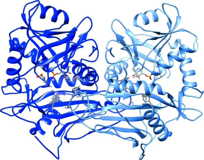
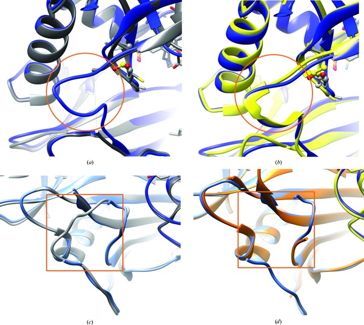
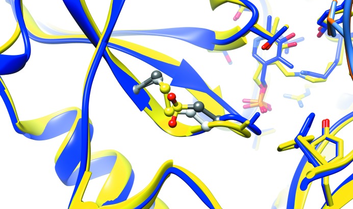
This study presents the crystal structure of a thiol variant of the human mitochondrial branched-chain aminotransferase protein. Human branched-chain aminotransferase (hBCAT) catalyzes the transamination of the branched-chain amino acids leucine, valine and isoleucine and α-ketoglutarate to their respective α-keto acids and glutamate. hBCAT activity is regulated by a CXXC center located approximately 10 Å from the active site. This redox-active center facilitates recycling between the reduced and oxidized states, representing hBCAT in its active and inactive forms, respectively. Site-directed mutagenesis of the redox sensor (Cys315) results in a significant loss of activity, with no loss of activity reported on the mutation of the resolving cysteine (Cys318), which allows the reversible formation of a disulfide bond between Cys315 and Cys318. The crystal structure of the oxidized form of the C318A variant was used to better understand the contributions of the individual cysteines and their oxidation states. The structure reveals the modified CXXC center in a conformation similar to that in the oxidized wild type, supporting the notion that its regulatory mechanism depends on switching the Cys315 side chain between active and inactive conformations. Moreover, the structure reveals conformational differences in the N-terminal and inter-domain region that may correlate with the inactivated state of the CXXC center.
Keywords: CXXC center; N-terminal loop; human mitochondrial branched-chain aminotransferase; interdomain loop; redox regulation; transaminases.
open access.
Figures
Similar articles
-
Roles for cysteine residues in the regulatory CXXC motif of human mitochondrial branched chain aminotransferase enzyme.Biochemistry. 2004 Jun 15;43(23):7356-64. doi: 10.1021/bi0498050. Biochemistry. 2004. PMID: 15182179
-
Identification of a peroxide-sensitive redox switch at the CXXC motif in the human mitochondrial branched chain aminotransferase.Biochemistry. 2002 Jul 23;41(29):9070-8. doi: 10.1021/bi020200i. Biochemistry. 2002. PMID: 12119021
-
Structural determinants for branched-chain aminotransferase isozyme-specific inhibition by the anticonvulsant drug gabapentin.J Biol Chem. 2005 Nov 4;280(44):37246-56. doi: 10.1074/jbc.M506486200. Epub 2005 Sep 1. J Biol Chem. 2005. PMID: 16141215
-
Human mitochondrial branched chain aminotransferase: structural basis for substrate specificity and role of redox active cysteines.Biochim Biophys Acta. 2003 Apr 11;1647(1-2):61-5. doi: 10.1016/s1570-9639(03)00051-7. Biochim Biophys Acta. 2003. PMID: 12686109 Review.
-
An overview of branched-chain amino acid aminotransferases: functional differences between mitochondrial and cytosolic isozymes in yeast and human.Appl Microbiol Biotechnol. 2021 Nov;105(21-22):8059-8072. doi: 10.1007/s00253-021-11612-4. Epub 2021 Oct 8. Appl Microbiol Biotechnol. 2021. PMID: 34622336 Review.
Cited by
-
Crystal structure of CmnB involved in the biosynthesis of the nonproteinogenic amino acid L-2,3-diaminopropionic acid.Acta Crystallogr F Struct Biol Commun. 2023 Jul 1;79(Pt 7):193-199. doi: 10.1107/S2053230X23005769. Epub 2023 Jul 5. Acta Crystallogr F Struct Biol Commun. 2023. PMID: 37405487 Free PMC article.
-
Branched-chain amino acid transferase type 2 (BCAT2) deficiency: Report of an eighth case and literature review.Mol Genet Metab Rep. 2025 Apr 9;43:101213. doi: 10.1016/j.ymgmr.2025.101213. eCollection 2025 Jun. Mol Genet Metab Rep. 2025. PMID: 40248769 Free PMC article.
-
An Extended C-Terminus, the Possible Culprit for Differential Regulation of 5-Aminolevulinate Synthase Isoforms.Front Mol Biosci. 2022 Jul 14;9:920668. doi: 10.3389/fmolb.2022.920668. eCollection 2022. Front Mol Biosci. 2022. PMID: 35911972 Free PMC article. Review.
References
-
- Birolo, L., Sandmeier, E., Christen, P. & John, R. A. (1995). Eur. J. Biochem. 232, 859–864. - PubMed
-
- Brosnan, J. T. & Brosnan, M. E. (2006). J. Nutr. 136, 207S–211S. - PubMed
-
- Christen, P., Mehta, P. K. & Sandmeier, E. (1994). Biochemistry of Vitamin B6 and PQQ, edited by G. Marino, G. Sannia & F. Bossa, pp. 9–13. Basel: Birkhäuser.
-
- Coles, S. J., Easton, P., Sharrod, H., Hutson, S. M., Hancock, J., Patel, V. B. & Conway, M. E. (2009). Biochemistry, 48, 645–656. - PubMed
MeSH terms
Substances
Grants and funding
LinkOut - more resources
Full Text Sources
Other Literature Sources
