

Iatrogenic aortic regurgitation following primary closure of ventricular septal defect: Role of transesophageal echocardiography
- PMID: 31929261
- PMCID: PMC7034207
- DOI: 10.4103/aca.ACA_238_18
Iatrogenic aortic regurgitation following primary closure of ventricular septal defect: Role of transesophageal echocardiography
Abstract
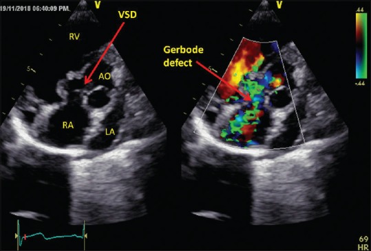
Iatrogenic valvular regurgitation following cardiac surgery has been reported as a result of leaflet perforation or entrapment. Due to its central location, the aortic valve is one of the most vulnerable structures for iatrogenic injuries. Proper assessment of the aortic valve by transesophageal echocardiography (TEE) should be done after a cardiac surgery in the periaortic area. We hereby report a case of iatrogenic aortic regurgitation which was developed after primary closure of perimembranous ventricular septal defect. It was timely diagnosed by TEE after termination of cardiopulmonary bypass and helped in further management.
Keywords: Iatrogenic aortic regurgitation; transesophageal echocardiography; ventricular septal defect.
Conflict of interest statement
None
Figures


References
-
- Hill AC, Bansal RC, Razzouk AJ, Liu M, Bailey LL, Gundry SR. Echocardiographic recognition of iatrogenic aortic valve leaflet perforation. Ann Thorac Surg. 1997;64:684–9. - PubMed
-
- Ducharme A, Courval JF, Dore A, Leclerc Y, Tardif JC. Severe aortic regurgitation immediately after mitral valve annuloplasty. Ann Thorac Surg. 1999;67:1487–9. - PubMed
-
- Aboelnasr M, Rohn V. Aortic valve leaflet perforation after mitral valve repair. Prague Med Rep. 2013;114:172–6. - PubMed
-
- Dogan M, Acikel S, Arslantas U, Cimen T, Yeter E. Inadvertent complication of prosthetic valve surgery: Leaflet perforation. Acta Medica (Hradec Kralove) 2013;56:167–9. - PubMed
Publication types
MeSH terms
LinkOut - more resources
Full Text Sources
Medical