Review
. 2019 Nov;7(22):684.
doi: 10.21037/atm.2019.10.109.

Hybrid computed tomography and magnetic resonance imaging 3D printed models for neurosurgery planning

Affiliations
Review

Hybrid computed tomography and magnetic resonance imaging 3D printed models for neurosurgery planning

Teodoro Martín-Noguerol et al. Ann Transl Med. 2019 Nov.

Abstract

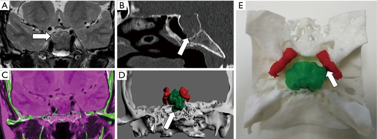
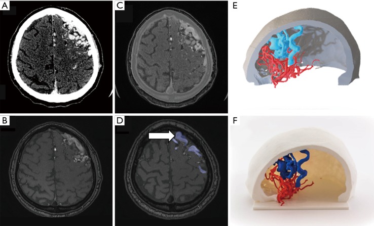
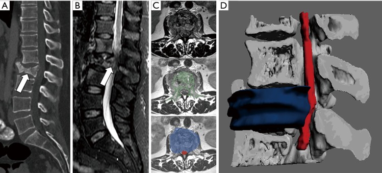
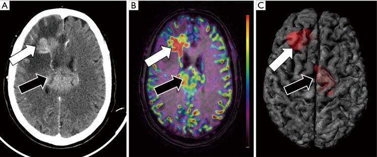
In the last decade, the clinical applications of three-dimensional (3D) printed models, in the neurosurgery field among others, have expanded widely based on several technical improvements in 3D printers, an increased variety of materials, but especially in postprocessing software. More commonly, physical models are obtained from a unique imaging technique with potential utilization in presurgical planning, generation/creation of patient-specific surgical material and personalized prosthesis or implants. Using specific software solutions, it is possible to obtain a more accurate segmentation of different anatomical and pathological structures and a more precise registration between different medical image sources allowing generating hybrid computed tomography (CT) and magnetic resonance imaging (MRI) 3D printed models. The need of neurosurgeons for a better understanding of the complex anatomy of central nervous system (CNS) and spine is pushing the use of these hybrid models, which are able to combine morphological information from CT and MRI, and also able to add physiological data from advanced MRI sequences, such as diffusion-weighted imaging (DWI), diffusion tensor imaging (DTI), perfusion weighted imaging (PWI) and functional MRI (fMRI). The inclusion of physiopathological data from advanced MRI sequences enables neurosurgeons to identify those areas with increased biological aggressiveness within a certain lesion prior to surgery or biopsy procedures. Preliminary data support the use of this more accurate presurgical perspective, to select the better surgical approach, reduce the global length of surgery and minimize the rate of intraoperative complications, morbidities or patient recovery times after surgery. The use of 3D printed models in neurosurgery has also demonstrated to be a valid tool for surgeons training and to improve communication between specialists and patients. Further studies are needed to test the feasibility of this novel approach in common clinical practice and determine the degree of improvement the neurosurgeons receive and the potential impact on patient outcome.

Keywords: Three-dimensional printing (3D printing); brain; computed tomography (CT); hybrid imaging; magnetic resonance imaging (MRI); neurosurgery; spine.


Conflict of interest statement

Conflicts of Interest: The authors have no conflicts of interest to declare.

Figures

Figure 1
Figure 1
Clival chordoma evaluation. A 45-year-old female that undergone MRI for migraine. (A) Axial T2-TSE sequence shows hyperintense lesion within clivus within punctate hypointensities (arrow). (B) Sagittal reconstruction of CT confirms the presence of a lytic lesion within clivus s with probably chondroid matrix inside (arrow). (C) Registration of both MRI and MCDT information allows delimitating brain tissue (magenta) using information from MRI and skull base structures (green) from CT. (D) The 3D virtual model and (E) the 3D printed model enable neurosurgeon to properly identify the location of chordoma (white arrow) concerning the brainstem (black arrow) and the rest of the skull base. This approach allows reduction of the risk of neurological complications before surgery or biopsy.
Figure 2
Figure 2
Meningioma presurgical evaluation. An 81-year-old female with right frontal lobe meningioma. (A) Axial gadolinium-enhanced T1w image shows a large right parasagittal frontal extra-axial lesion consistent with meningioma (arrow). (B) Axial b 1,000 s/mm2 DWI identifies severe restriction of water diffusion within the meningioma which suggests hypercellular lesion (arrow). (C) The fusion of functional information from DWI and morphological information form T1W images demonstrates proper correlation identifying areas of higher hypercellularity within the posterior aspect of the meningioma (arrow). (D) 3D model merging anatomical information from morphological MRI and functional information from DWI allows the evaluation of those areas with a higher restriction of water diffusion within meningioma inside the whole brain anatomy (arrow).
Figure 3
Figure 3
Flowchart for hybrid MRI and CT 3D printed model for epilepsy pre-surgical planning. A 25-year-old male with a drug-resistant epilepsy candidate for focal thermocoagulative therapy. Neurosurgeons ask for the possibility of obtaining a 3D printed model of the whole patient head including soft tissues, skull, and brain for proper planning of electrodes and guides positioning. Information from both (A) axial 3D-T1W and (B) CT images were used for (C) registration and segmentation of (D) gray matter and (E) bone. (F) Hybrid 3D model in STL format was generated and sent to 3D print obtaining a (G) 3D printed model which included information of both the brain, soft tissues, and skull. (H) The printed brain model has undergone a CT scan which perfectly fitted with its source image form MRI (I). 3D printed model enables neurosurgeons to plan electrodes positioning and surgical entry points before surgery.
Figure 4
Figure 4
Presurgical planning of focal epilepsy. A 37-year-old male, with a history of previous surgical left parieto-occipital resection 20 years ago, refers to occipital lobe seizures. Neurologists asked for a presurgical study to minimize the damage of both optical and corticospinal pathways. (A) Axial T1-weighted image shows a large left parieto-occipital area of encephalomalacia (arrow). (B) Color-coded Fraction Anisotropy map, generated from DTI acquisition identifies the involvement of posterior tracts of left corona radiata (white arrow) with apparent preservation of corticospinal tract (black arrow). (C) The fusion of morphological MRI and DTI images allows evaluating the relationships between white matter tracts (black arrow) and post-surgical cavity (white arrow). (D) 3D printed model enables neurosurgeons to address physically the location of white matter tracts for a more secure surgical planning.
Figure 5
Figure 5
Presurgical planning of hypophyseal macroadenoma. A 54-year-old female that undergone MRI for headache and vision loss. (A) Coronal T2 TSE shows a large sellar mass that seems to invade sphenoidal sinus (arrow). (B) Sagittal reconstruction of CT confirms the presence of an enlarged sella turcica with disruption of its floor (arrow). (C) Registration of information of skull base structures from CT (green) and data from MRI (magenta) enables an adequate tissue contrast for adenoma and even internal carotids segmentation. (D) Coronal projection of the 3D combined model allows neurosurgeons to properly evaluate the relationship of the hypophyseal mass (codded in green) with sphenoidal sinus, cavernous sinus and even both internal carotids (codded in red), demonstrating disruption of sella turcica floor (arrow). (E) Zenithal view of 3D printed model shows proper correlation with radiological images and 3D virtual model demonstrating contact between macroadenoma and left internal carotid (white arrow).
Figure 6
Figure 6
Brain arteriovenous malformation assessment. A 35-year-old male with left frontal AVM that undergone both CT-angiography and MRI-angiography studies. Information obtained from (A) CT was used for the skull and both arteries and veins segmentation (B). (C) MR-angiography using time of flight (TOF) technique with arterial velocity codification allows accurately segment high flow veins (arrows at Figure 6D) that compose the AVM as well as differentiate them from the brain parenchyma and skull. Combined 3D modeling (E) and 3D printed models (F) provide neurosurgeons and interventional radiologists a comprehensive assessment of AVM and its relationship with the skull.
Figure 7
Figure 7
Spine and spinal cord evaluation. A 42-year-old female with a history of breast cancer is evaluated in an emergency room for lumbar pain and lower limb paresis. (A) Sagittal reconstruction of CT shows L2 vertebral body collapse which seems to invade the spinal canal (arrow). (B) Sagittal STIR sequence confirms the existence of a metastatic fracture with soft tissue component that invades spinal canal conditioning severe stenosis. (C) Registration of information form axial T2-TSE with CT allows to segment vertebral bone (codded in green) and also soft tissue mass (codded in blue) and spinal cord (codded in red). (D) The 3D model enables neurosurgeons to visualize in a single view all the information form both CT and MRI and identify how the soft tissue component (codded in blue) invades the spinal canal and displaces the spinal cord.
Figure 8
Figure 8
Multimodality assessment of a CNS tumor. A 73-year-old female with changes in behavior and cognitive impairment undergone (A) contrast-enhanced CT that shows two large hyperenancing masses at right frontal lobe (white arrow) and within the corpus callosum (black arrow). (B) MRI was performed using ASL technique for assessment of tumor blood flow before guide biopsy to avoid the administration of gadolinium. The fusion of morphological MRI sequence (FLAIR) and functional MRI sequence (ASL) allows neuroradiologists to characterize the right frontal lesion as a potentially higher grade compared with the corpus callosum lesion as the former shows higher blood flow values (white arrow) compared with the latter (black arrow). (C) The register of morphological MRI and functional (ASL) MRI data allows the generation of a 3D model in a single file with all the information needed for a proper planification of brain biopsy.

References

    1. Liaw CY, Guvendiren M. Current and emerging applications of 3D printing in medicine. Biofabrication 2017;9:024102. 10.1088/1758-5090/aa7279 - DOI - PubMed
    1. Rengier F, Mehndiratta A, Von Tengg-Kobligk H, et al. 3D printing based on imaging data: Review of medical applications. Int J Comput Assist Radiol Surg 2010;5:335-41. 10.1007/s11548-010-0476-x - DOI - PubMed
    1. Wadhwa A, Bhardwaj A, Singh Verma V. A review on brain tumor segmentation of MRI images. Magn Reson Imaging 2019;61:247-59. 10.1016/j.mri.2019.05.043 - DOI - PubMed
    1. Hoang D, Perrault D, Stevanovic M, et al. Surgical applications of three-dimensional printing: a review of the current literature & how to get started. Ann Transl Med 2016;4:456. 10.21037/atm.2016.12.18 - DOI - PMC - PubMed
    1. Waran V, Pancharatnam D, Thambinayagam HC, et al. The utilization of cranial models created using rapid prototyping techniques in the development of models for navigation training. J Neurol Surg A Cent Eur Neurosurg 2014;75:12-5. - PubMed