Review
. 2019 Dec 20:2019:2525984.
doi: 10.1155/2019/2525984. eCollection 2019.

Innate Lymphoid Cells: Regulators of Gut Barrier Function and Immune Homeostasis

Affiliations
Review

Innate Lymphoid Cells: Regulators of Gut Barrier Function and Immune Homeostasis

Hui Fan et al. J Immunol Res. .

Abstract

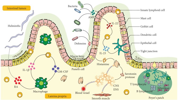
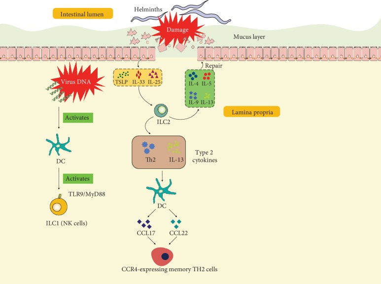
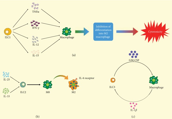
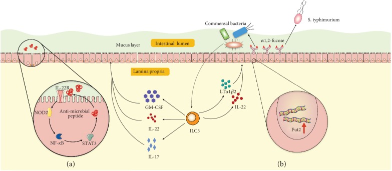
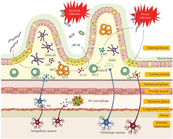
Innate lymphoid cells (ILCs), identified in the early years of this century as a new class of leukocyte family unlike the B or T lymphocytes, play a unique role bridging the innate and adaptive immune responses in mucosal immunity. Their origin, differentiation, and activation process and functions have caught global interest. Recently, accumulating evidence supports that ILCs are vital regulators for gastrointestinal mucosal homeostasis through interactions with other structural and stromal cells in gut epithelial barriers. This review will explore the functions of ILCs and other cells in maintaining gut homeostasis and feature the crosstalk between ILCs with other cells and potential pharmacotherapy targeting ILCs applicable in intestinal innate immunity.


Conflict of interest statement

The authors have declared that no competing interests exist.

Figures

Figure 1
Figure 1
Illustration of intestinal barrier structure and functions. The intestine barrier contains the chemical barrier and the physical barrier. The chemical barrier is composed of antimicrobial peptides (AMPs) such as amphiregulin. It provides chemical agents attacking invading microorganisms including bacteria and helminths. The physical barrier includes the mucus layer and cell junctions between the epithelium. It serves as the wall spatially separating the invading microorganisms and host. There are many types of cells in the gut epithelium regulating the epithelium function. Disruption of the intestinal barrier allows the leak of gut bacteria from the lumen into the lamina propria, inducing excessive immune responses of the host immune cells. Retinoic acid (RA) released by macrophages or dendritic cells assists in host resist helminthic infection. IL-22 released by ILCs promotes epithelial cells secreting AMP in response to bacterial infection, which is regulated by IL-23 from dendritic cells. Moreover, macrophage-derived IL-1β promotes ILCs' production of GM-CSF, which further stimulates more macrophage differentiation from monocytes. The enteric nervous system including neuron and glial cells interacts closely with mast cells and regulates blood vessels. IL: interleukin; AMP: antimicrobial peptide; GM-CSF: granulocyte-macrophage colony stimulating factor; RA: retinoic acid; ENS: enteric nervous system; CNS: central nervous system.
Figure 2
Figure 2
Illustration of interactions of ILCs and DCs in the intestinal tract. At the initial site of viral infection, virus DNA activates DCs, further activating ILC1 (mainly NK cells) by the TLR9/MyD88 pathway. After damage caused by helminths, epithelial cells produce TSLP, IL-33, and IL-25. These cytokines stimulate ILC2s producing type 2 cytokines including IL-4, IL-5, IL-9, and IL-13 which participate in repair of epithelial cells and mucus layer. In addition, ILC2-derived Th2 and IL-13 stimulate DCs, inducing the release of CCL17 and CCL22 and recruitment of CCR4-expressing memory TH2 cells. IL: interleukin; TSLP: thymic stromal lymphopoietin; DC: dendritic cell; ILC: innate lymphoid cell; CCL: chemokine (C-C motif) ligand; Th: helper T cell.
Figure 3
Figure 3
Illustration of interactions of macrophages and different ILCs. (a) TNF-α, IFN-γ, IL-12, and IL-15 released from ILC1 stimulate macrophage differentiating into cytotoxic macrophage and inhibit differentiation into M2 macrophage differentiation. (b) After being stimulated by IL-25 and IL-33, ILC2 promotes the transformation of M2 macrophage from M0 macrophage. (c) The crosstalk of ILC3 and macrophage is mainly induced by GM-CSF and IL-1β. ILC: innate lymphoid cell; TNF: tumor necrosis factor; IL: interleukin; M0: M0 macrophage; M2: M2 macrophage; GM-CSF: granulocyte-macrophage colony stimulating factor.
Figure 4
Figure 4
Illustration of epithelial cells and ILC3 interactions. (a) After bacterial infection, ILC3-derived GM-CSF, IL-22, and IL-17 stimulate epithelial cells by activating the IL-22R-NOD2-NF-κB signaling pathway and result in epithelial cells secreting antimicrobial peptides into the mucus layer. (b) ILC3-derived IL-22 and lymphotoxin (LTα1β2) induce fucosyltransferase 2 (Fut2) gene expression in epithelial cells and result in fucose production in the intestinal tract. Fucose can be utilized by commensal bacteria but not by pathological bacteria such as Salmonella typhimurium. ILC: innate lymphoid cell; GM-CSF: granulocyte-macrophage colony stimulating factor; NOD2: nucleotide oligomerization domain-containing protein 2; NF-κB: nuclear factor- (NF-) κB; STAT3: signal transducers and activators of transcription 3.
Figure 5
Figure 5
Illustration of crosstalk of ILC2 and ILC3 with neurons in helminth infection and gut inflammation, respectively. This image was modified from Reference [54]. Enteric cholinergic neuron-derived NMU activates ILC2 responses and protects against helminth infection. Lamina propria ILC2 function is also regulated by VIP and NE. Glial cell-derived neurotrophic factors stimulate IL-22 production by lamina propria ILC3s, promoting barrier integrity. SNS-derived NE induces a tissue-protective phenotype (M2) in muscularis macrophages. Abbreviations: AMP: antimicrobial peptide; NE: norepinephrine; NMU: neuromedin U; NMUR: neuromedin U receptor; SNS: sympathetic nervous system; GFL: glia cell-derived neurotrophic factor ligand; VIP: vasoactive intestinal peptide; VPAC2: vasoactive intestinal peptide receptor 2.

References

    1. Viggiano D., Ianiro G., Vanella G., et al. Gut barrier in health and disease: focus on childhood. European Review for Medical and Pharmacological Sciences. 2015;19(6):1077–1085. - PubMed
    1. Martin A., Devkota S. Hold the door: role of the gut barrier in diabetes. Cell Metabolism. 2018;27(5):949–951. doi: 10.1016/j.cmet.2018.04.017. - DOI - PubMed
    1. Winer D. A., Luck H., Tsai S., Winer S. The intestinal immune system in obesity and insulin resistance. Cell Metabolism. 2016;23(3):413–426. doi: 10.1016/j.cmet.2016.01.003. - DOI - PubMed
    1. Randolph G. J., Bala S., Rahier J.-F., et al. Lymphoid aggregates remodel lymphatic collecting vessels that serve mesenteric lymph nodes in Crohn disease. The American Journal of Pathology. 2016;186(12):3066–3073. doi: 10.1016/j.ajpath.2016.07.026. - DOI - PMC - PubMed
    1. Bischoff S. C., Barbara G., Buurman W., et al. Intestinal permeability – a new target for disease prevention and therapy. BMC Gastroenterology. 2014;14(1) doi: 10.1186/s12876-014-0189-7. - DOI - PMC - PubMed

MeSH terms

LinkOut - more resources