. 2019 Oct 21;2(10):4667-4674.
doi: 10.1021/acsabm.9b00747. Epub 2019 Sep 26.

Synthesis of Ultrasmall Synthetic Melanin Nanoparticles by UV Irradiation in Acidic and Neutral Conditions

Affiliations

Synthesis of Ultrasmall Synthetic Melanin Nanoparticles by UV Irradiation in Acidic and Neutral Conditions

Jeanne E Lemaster et al. ACS Appl Bio Mater. .

Abstract

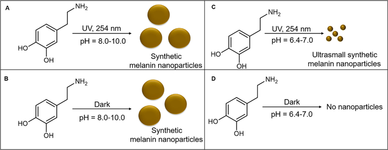
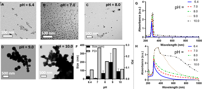
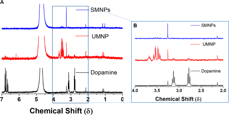
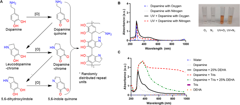
Synthetic melanin nanoparticles have value in metal chelation, photoprotection, and biocompatibility. Applications of these materials have been reported in optics, biomedicine, and electronics. However, precise size control has remained relatively difficult-especially for materials below 1000 nm. In this paper we describe the synthesis of ultrasmall synthetic nanoparticles with size of 9.4-31.4 nm in weakly acidic and neutral conditions via UV-irradiation. Size control of these particles was possible by varying the pH from 6.4-10.0. We then used UV-vis, FTIR, and nuclear magnetic resonance to investigate the mechanism of UV-induced polymerization. The data show that reactive oxygen species from UV irradiation oxidizes intermediates of the reaction and accelerates the formation of these synthetic melanin structures.

Keywords: UV irradiation; facile preparation; mechanism; melanin; polydopamine; synthesis; synthetic melanin nanoparticles.


Conflict of interest statement

The authors declare no competing financial interest.

Figures

Figure 1.
Figure 1.
Synthesis and characterization of ultrasmall synthetic melanin nanoparticles. Ultrasmall synthetic melanin nanoparticles were made by the polymerization of dopamine in aqueous solution with UV irradiation at a wavelength of 254 nm. (A) Dopamine polymerizes under UV in basic conditions at pH = 8.0–10.0 to form synthetic melanin nanoparticles (SMNPs). (B) Dopamine polymerizes in the dark in basic conditions at pH 8.0–10.0 to form synthetic melanin nanoparticles. (C) Dopamine polymerizes under UV in acidic conditions at pH 6.4–7.0 to form ultrasmall synthetic melanin nanoparticles (UMNPs). (D) Dopamine does not polymerize in the dark at pH 6.4–7.0 conditions.
Figure 2.
Figure 2.
TEM and absorption spectrum of SMNP and UMNP at initial concentration of 1 mg/mL dopamine. (A) TEM image of UMNP formed at pH 6.4 with 24 h UV irradiation; the average size is 31.4 nm. The size was confirmed with multispectral advanced nanoparticle tracking analysis (MANTA) analysis, indicating a mode size of 34 nm of all particles measured. (B) TEM image of UMNP formed at pH 7.0 with 24 h UV irradiation indicated an average size of 9.40 nm. (C) TEM image of SMNP formed at pH 8.0 with 24 h UV irradiation indicated average size of 69.2 nm. (D) TEM image of SMNP formed at pH 9.0 with 24 h UV irradiation indicated average size of 200 nm. (E) TEM image of SMNP formed at pH 10.0 with 24 h UV irradiation indicated average size of 92.8 nm. (F) Size dependence on pH. The sample with pH 7.0 yielded the smallest spherical UMNP under UV irradiation. (G) Absorbance spectrum based on pH from 6.4 to 10.0 prior to UV irradiation shows the characteristic dopamine peak at 280 nm. (H) Absorbance spectrum based on pH from 6.4 to 10.0 shows a broadening of the characteristic dopamine peak from 280 to 1000 nm, indicating polymerization of UMNP and SMNP.
Figure 3.
Figure 3.
Characterization of SMNP and UMNP formed in water with UV irradiation at varying concentrations. (A) Basic dopamine solutions with pH 10.0 displayed polymerization with UV irradiation (254 nm) for 24 h and showed a broad peak shift from 290–400 nm at initial concentrations of dopamine of 1–15 mg/mL. (B) Dopamine solutions with pH 10 showed polymerization in the dark after 24 h. (C) Acidic dopamine solutions with pH 6.4 with initial dopamine concentrations from 1 to 15 mg/mL were irradiated with UV light (254 nm) for 24 h to form UMNP. Absorbance increased as concentration of dopamine increased with a red shift in the absorbance spectrum. (D) Acidic dopamine solutions with pH 6.4 in dark conditions for 24 h showed a characteristic dopamine peak at 280 nm and did not show polymerization. (E) UV wavelength of 254 nm irradiated for 24 h causes higher polymerization in dopamine than samples irradiated at 302 and 364 nm for 24 h. Polymerization does not occur when samples are irradiated at 364 nm for 24 h. (F) FTIR spectrum of SMNP (pH 10.0) compared to UMNP (pH 6.4) show similar FTIR spectrum. Characteristic peaks at 3324 and 1636 cm−1 indicate the presence of hydroxyl groups and N–H vibrations, respectively.
Figure 4.
Figure 4.
NMR spectroscopy shows loss of aliphatic side chains in UMNP indicating that polymerization has occurred. (A) Stacked NMR spectrum of SMNPS, UMNP, and dopamine. (B) Dopamine hydrochloride dissolved in D2O yielded two triplets at 2.77 and 3.13 δ corresponding to the aliphatic side chains.
Figure 5.
Figure 5.
Mechanism of UV-initiated polymerization. (A) Under the presence of oxygen, dopamine forms dopamine quinone, dopamine-chrome, and 5,6-indole quinone. Adapted with permission from ref . Copyright 2014 Wiley. (B) Dopamine under a typical atmosphere (inset, O2) and a nitrogen degassed atmosphere (inset, N2) without UV irradiation display a characteristic peak at 280 nm with no polymerization after 24 h. Dopamine irradiated with UV under an oxygenated atmosphere displays polymerization by turning to the characteristic dark brown color (inset, UV + O2). Dopamine irradiated with UV under a nitrous atmosphere displays little polymerization (inset, UV + N2). (C) Dopamine under basic conditions (Tris base) shows increased polymerization as indicated by a broad absorption from 280 to 1000 nm when compared to dopamine with tris base and 25% DEHA (oxygen scavenger), indicating that oxygen plays a critical role in the polymerization mechanism.

References

    1. Lemaster JE; Wang Z; Hariri A; Chen F; Hu Z; Huang Y; Barback CV; Cochran R; Gianneschi NC; Jokerst JV Gadolinium Doping Enhances the Photoacoustic Signal of Synthetic Melanin Nanoparticles: A Dual Modality Contrast Agent for Stem Cell Imaging. Chem. Mater 2019, 31 (1), 251–259. - PMC - PubMed
    1. Li Y; Xie Y; Wang Z; Zang N; Carniato F; Huang Y; Andolina CM; Parent LR; Ditri TB; Walter ED; et al. Structure and Function of Iron-Loaded Synthetic Melanin. ACS Nano 2016, 10 (11), 10186–10194. - PMC - PubMed
    1. Jiang Q; Luo Z; Men Y; Yang P; Peng H; Guo R; Tian Y; Pang Z; Yang W Red Blood Cell Membrane-Camouflaged Melanin Nanoparticles for Enhanced Photothermal Therapy. Biomaterials 2017, 143, 29–45. - PubMed
    1. Tian Y; Zhao Y; Liu W; Liu Y; Tang Y; Teng Z; Zhang C; Wang S; Lu G Photosensitizer-Loaded Biomimetic Platform for Multimodal Imaging-Guided Synergistic Phototherapy. RSC Adv. 2018, 8 (56), 32200–32210. - PMC - PubMed
    1. Zhang R; Fan Q; Yang M; Cheng K; Lu X; Zhang L; Huang W; Cheng Z Engineering Melanin Nanoparticles as an Efficient Drug–Delivery System for Imaging-Guided Chemotherapy. Adv. Mater 2015, 27 (34), 5063–5069. - PubMed

LinkOut - more resources