. 2019 Dec 13;43(1 suppl 1):e20190075.
doi: 10.1590/1678-4685-GMB-2019-0075. eCollection 2019.

ADP-ribosylation: from molecular mechanisms to human disease

Affiliations

ADP-ribosylation: from molecular mechanisms to human disease

Nicolas C Hoch et al. Genet Mol Biol. .

Abstract

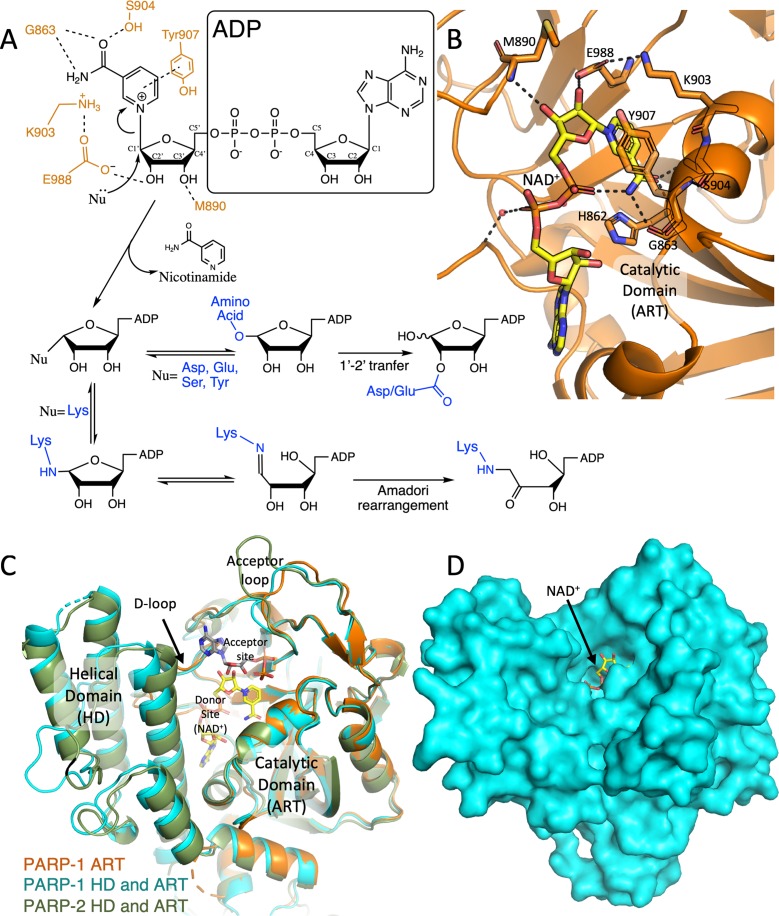
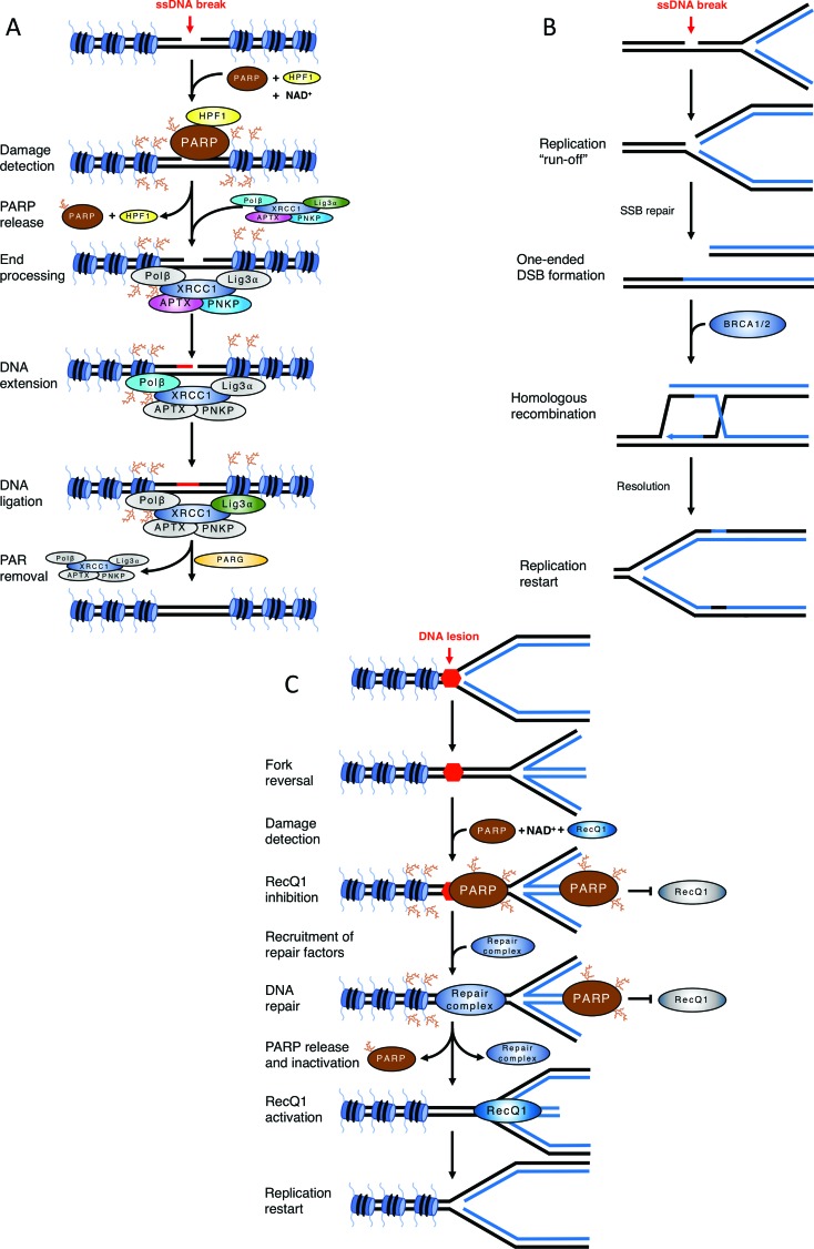
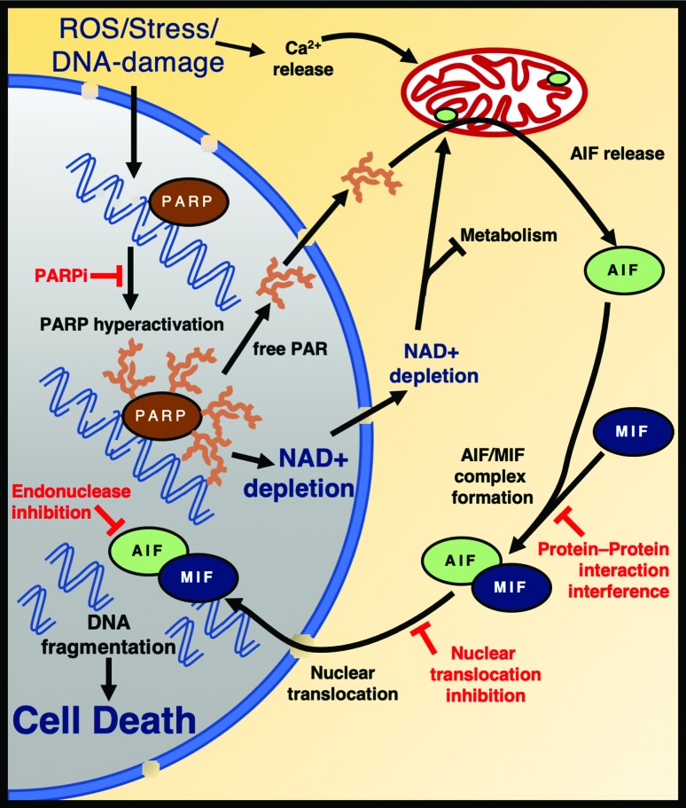
Post-translational modification of proteins by ADP-ribosylation, catalysed by poly (ADP-ribose) polymerases (PARPs) using NAD+ as a substrate, plays central roles in DNA damage signalling and repair, modulates a range of cellular signalling cascades and initiates programmed cell death by parthanatos. Here, we present mechanistic aspects of ADP-ribose modification, PARP activation and the cellular functions of ADP-ribose signalling, and discuss how this knowledge is uncovering therapeutic avenues for the treatment of increasingly prevalent human diseases such as cancer, ischaemic damage and neurodegeneration.


Figures

Figure 1
Figure 1. Schematic mechanism of ADP ribosylation reaction and the catalytic domain of DNA-dependant PARPs. A) A simplified overview of the (ADP)-ribosylation reactions catalysed by PARPs. The final products depend on the acceptor residue acting as a nucleophile (Nu, in blue). PARP1 active-site residues interacting with the ribose-nicotinamide moiety of NAD+ are illustrated in orange. B) The NAD+ (modelled based on the human PARP1 bound to benzamide adenine dinucleotide [PDB: 6BHV], carbon atoms in yellow) in an extended conformation, bound to the catalytic domain of human PARP1 (ART in cartoon, orange, [PDB: 6BHV]). The residues involved in the catalysis are presented as sticks. C) Superposed cartoon view of human PARP-1 ART domain (orange, [PDB: 6BHV]), PARP1 (light blue, [PDB: 5WS1]) and PARP2 (green, [PDB: 3KJD]) showing the structure of the entire catalytic domains (ART and HD). The modelled NAD+ (in yellow) denotes the donor site, while a molecule of ADP (modelled by superimposing the structures of chicken PARP1 [PDB: 1A26] to the human PARP1 [PDB: 3KJD]) indicates the acceptor site. Donor loop (D-loop) and acceptor loop are labelled. D) Surface representation of human PARP1 [PDB: 3KJD] with NAD+ modelled into the active site. The ribose group to be attacked is exposed to the solvent.
Figure 2
Figure 2. Examples of the impact of ADP-ribosylation in DNA damage signalling and repair. A) Mechanism of single-strand break repair. A single-strand break activates PARP1/2, leading to HPF-1 assisted PARylation of chromatin. PARP auto-modification causes its release and PAR chains surrounding the break site recruit XRCC1 complex. APTX and PNKP process break termini, Polβ fills the gap by DNA synthesis and DNA Ligase IIIα seals the remaining nick. PARG removes PAR chains and XRCC1 complex is released, completing the repair. B) Defective single-strand break repair causes a reliance on homologous recombination. An unrepaired single-strand break is encountered by an ongoing replication fork, which converts it into a one-ended double-strand break. This lesion is repaired by BRCA1 and BRCA2-dependent homologous recombination. C) Role of PARP1/2 in fork reversal. A replication fork encounters an obstacle to its progression and reverts. PARP is activated either by the obstacle/lesion itself or by the DNA end at the regressed fork. PAR chains prevent RecQ1 binding/activity. Upon resolution of the block, PARP release (and presumably PAR chain degradation by PARG) allow RecQ1 helicase to access the reversed fork and remodel it back into a canonical replication fork.
Figure 3
Figure 3. PARP1 mediates cell death by Parthanatos. Oxidative damage triggers PARP-hyperactivation, resulting in AIF release from the mitochondria and nuclear translocation of the AIF/MIF complex. Endonuclease activation causes cell death. Some of the potential therapeutic targets are depicted in red. The nucleus is coloured in grey and cytoplasm in orange. AIF: Apoptosis Inducing Factor. MIF: macrophage migration inhibitory factor.

References

    1. Abplanalp J, Leutert M, Frugier E, Nowak K, Feurer R, Kato J, Kistemaker HVA, Filippov DV, Moss J, Caflisch A, et al. Proteomic analyses identify ARH3 as a serine mono-ADP-ribosylhydrolase. Nat Commun. 2017;8:2055. - PMC - PubMed
    1. Ahel D, Horejsi Z, Wiechens N, Polo SE, Garcia-Wilson E, Ahel I, Flynn H, Skehel M, West SC, Jackson SP, et al. Poly(ADP-ribose)-dependent regulation of DNA repair by the chromatin remodeling enzyme ALC1. Science. 2009;325:1240–1243. - PMC - PubMed
    1. Alano CC, Garnier P, Ying W, Higashi Y, Kauppinen TM, Swanson RA. NAD+ depletion is necessary and sufficient for poly(ADP-ribose) polymerase-1-mediated neuronal death. J Neurosci. 2010;30:2967–2978. - PMC - PubMed
    1. Ali AAE, Timinszky G, Arribas-Bosacoma R, Kozlowski M, Hassa PO, Hassler M, Ladurner AG, Pearl LH, Oliver AW. The zinc-finger domains of PARP1 cooperate to recognize DNA strand breaks. Nat Struct Mol Biol. 2012;19:685–692. - PMC - PubMed
    1. Altmeyer M, Messner S, Hassa PO, Fey M, Hottiger MO. Molecular mechanism of poly(ADP-ribosyl)ation by PARP1 and identification of lysine residues as ADP-ribose acceptor sites. Nucleic Acids Res. 2009;37:3723–3738. - PMC - PubMed