. 2020 Apr 1;77(4):500-507.
doi: 10.1001/jamaneurol.2019.4656.

Assessment of the Validity of the 2HELPS2B Score for Inpatient Seizure Risk Prediction

Affiliations

Assessment of the Validity of the 2HELPS2B Score for Inpatient Seizure Risk Prediction

Aaron F Struck et al. JAMA Neurol. .

Erratum in

  • Error in Author's Name.
    [No authors listed] [No authors listed] JAMA Neurol. 2020 Apr 1;77(4):527. doi: 10.1001/jamaneurol.2020.0140. JAMA Neurol. 2020. PMID: 32091539 Free PMC article. No abstract available.

Abstract

Importance: Seizure risk stratification is needed to boost inpatient seizure detection and to improve continuous electroencephalogram (cEEG) cost-effectiveness. 2HELPS2B can address this need but requires validation.

Objective: To use an independent cohort to validate the 2HELPS2B score and develop a practical guide for its use.

Design, setting, and participants: This multicenter retrospective medical record review analyzed clinical and EEG data from patients 18 years or older with a clinical indication for cEEG and an EEG duration of 12 hours or longer who were receiving consecutive cEEG at 6 centers from January 2012 to January 2019. 2HELPS2B was evaluated with the validation cohort using the mean calibration error (CAL), a measure of the difference between prediction and actual results. A Kaplan-Meier survival analysis was used to determine the duration of EEG monitoring to achieve a seizure risk of less than 5% based on the 2HELPS2B score calculated on first- hour (screening) EEG. Participants undergoing elective epilepsy monitoring and those who had experienced cardiac arrest were excluded. No participants who met the inclusion criteria were excluded.

Main outcomes and measures: The main outcome was a CAL error of less than 5% in the validation cohort.

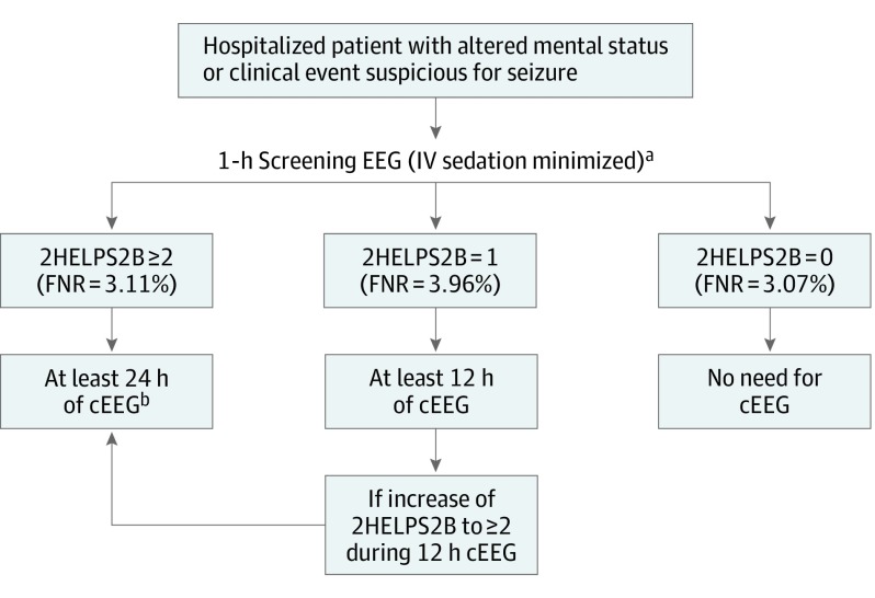
Results: The study included 2111 participants (median age, 51 years; 1113 men [52.7%]; median EEG duration, 48 hours) and the primary outcome was met with a validation cohort CAL error of 4.0% compared with a CAL of 2.7% in the foundational cohort (P = .13). For the 2HELPS2B score calculated on only the first hour of EEG in those without seizures during that hour, the CAL error remained at less than 5.0% at 4.2% and allowed for stratifying patients into low- (2HELPS2B = 0; <5% risk of seizures), medium- (2HELPS2B = 1; 12% risk of seizures), and high-risk (2HELPS2B, ≥2; risk of seizures, >25%) groups. Each of the categories had an associated minimum recommended duration of EEG monitoring to achieve at least a less than 5% risk of seizures, a 2HELPS2B score of 0 at 1-hour screening EEG, a 2HELPS2B score of 1 at 12 hours, and a 2HELPS2B score of 2 or greater at 24 hours.

Conclusions and relevance: In this study, 2HELPS2B was validated as a clinical tool to aid in seizure detection, clinical communication, and cEEG use in hospitalized patients. In patients without prior clinical seizures, a screening 1-hour EEG that showed no epileptiform findings was an adequate screen. In patients with any highly epileptiform EEG patterns during the first hour of EEG (ie, a 2HELPS2B score of ≥2), at least 24 hours of recording is recommended.


Conflict of interest statement

Conflict of Interest Disclosures: Dr Schmitt reported personal fees from American Academy of Neurology and the American Clinical Neurophysiology Society. Dr Rodriguez Ruiz reported educational activity with Neuropace Inc. Dr Haider reported personal fees from Springer Publishing, UpToDate Inc, and Ceribell. Dr Hirsch reported personal fees from Adamas, Aquestive, Ceribell, Eisai, Marinus, Medtronic, and UCB outside the submitted work; royalties for authoring chapters for UpToDate-Neurology and from Wiley for coauthoring the book Atlas of EEG in Critical Care; and spending about 25% of his clinical billable time implementing and interpreting critical care EEG studies. Dr Rosenthal reported grants from Sage Therapeutics and personal fees from Coleman Research Group, and Gerson Lehrman Group, Inc. Dr Westover reported grants from the National Institutes of Health during the conduct of the study. No other disclosures were reported.

Figures

Figure 1.
Figure 1.. Risk-Calibration Graph of the Error for the 2HELPS2B Model in the Initial Study Cohort and the Validation Cohort
The dashed line is an ideal classifier with a calibration error of 0. Data are presented with 95% confidence intervals. The calibration error on the validation cohort was 4.0% (95% CI, 2.8%-6.2%). In the initial study cohort, the t error was 2.7% (95% CI, 2.0%-3.6%); errors between cohorts were not significantly different with a P value of .13.
Figure 2.
Figure 2.. Risk-Calibration Graph of the Error for the 2HELPS2B Model Calculated Only During the First Hour of Electroencephalogram (EEG) in the Validation Cohort, Represented With 3 Risk Levels
One-hour screening EEG, seizure risk calibration. The dashed line is an ideal classifier with a calibration error of 0. Data are presented with 95% confidence intervals. The calibration error was 4.2% (95% CI, 2.5%-7.1%). Comparison of the calibration error between 2HELPS2B calculated during the entire study vs only the first hour of EEG was nonsignificantly different with a P value of .93. The overall seizure risk for each group is as follows: low, 3.1%; medium, 12.0%; and high, 26.6%.
Figure 3.
Figure 3.. Survival Analysis and Time-Dependent Seizure Risk
A, Survival analysis for 72 hours after 1-hour screening electroencephalogram (EEG). Risk is stratified by 2HELPS2B calculated during the preceding 1-hour screening EEG. Dashes in survival lines represent censoring events (low-risk, n = 531; medium-risk, n = 495; high-risk, n = 227). B, Time-dependent risk of seizures stratified by 2HELPS2B score calculated from the first hour of EEG monitoring, The 72-hour risk of seizures decays rapidly as the duration of continuous EEG (cEEG) monitoring increases. The dashed line represents when the false negative rate drops below 5%.
Figure 4.
Figure 4.. 2HELPS2B Clinical Algorithm
If a seizure is detected at any time (including in the 1-hour screening electroencephalogram [EEG]), there is a recommendation for at least 24 hours of EEG monitoring after the last seizure. cEEG indicates continuous EEG monitoring; D/, discontinue; FNR, false negative rate. aFor patients with coma, a screening EEG of up to 90 minutes may be considered. bIn cases of ictal-interictal continuum patterns, which are common in those with a 2HELPS2B score of 2 or greater, a longer duration of monitoring may be required for empirical treatment trials.

References

    1. Ney JP, van der Goes DN, Nuwer MR, Nelson L, Eccher MA. Continuous and routine EEG in intensive care: utilization and outcomes, United States 2005-2009. Neurology. 2013;81(23):2002-2008. doi:10.1212/01.wnl.0000436948.93399.2a - DOI - PMC - PubMed
    1. Kurtz P, Hanafy KA, Claassen J. Continuous EEG monitoring: is it ready for prime time? Curr Opin Crit Care. 2009;15(2):99-109. doi:10.1097/MCC.0b013e3283294947 - DOI - PubMed
    1. Westover MB, Shafi MM, Bianchi MT, et al. . The probability of seizures during EEG monitoring in critically ill adults. Clin Neurophysiol. 2015;126(3):463-471. doi:10.1016/j.clinph.2014.05.037 - DOI - PMC - PubMed
    1. Jordan KG. Continuous EEG and evoked potential monitoring in the neuroscience intensive care unit. J Clin Neurophysiol. 1993;10(4):445-475. doi:10.1097/00004691-199310000-00006 - DOI - PubMed
    1. Young GB, Jordan KG, Doig GS. An assessment of nonconvulsive seizures in the intensive care unit using continuous EEG monitoring: an investigation of variables associated with mortality. Neurology. 1996;47(1):83-89. doi:10.1212/WNL.47.1.83 - DOI - PubMed

Publication types