. 2020 Feb 14;432(4):1109-1125.
doi: 10.1016/j.jmb.2020.01.006. Epub 2020 Jan 10.

Intracellular Neutralization of Ricin Toxin by Single-domain Antibodies Targeting the Active Site

Affiliations

Intracellular Neutralization of Ricin Toxin by Single-domain Antibodies Targeting the Active Site

Michael J Rudolph et al. J Mol Biol. .

Abstract

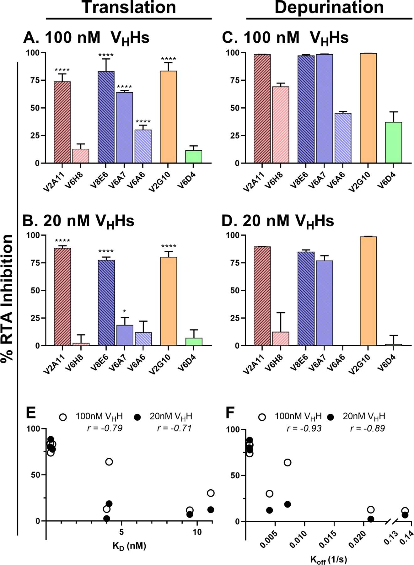
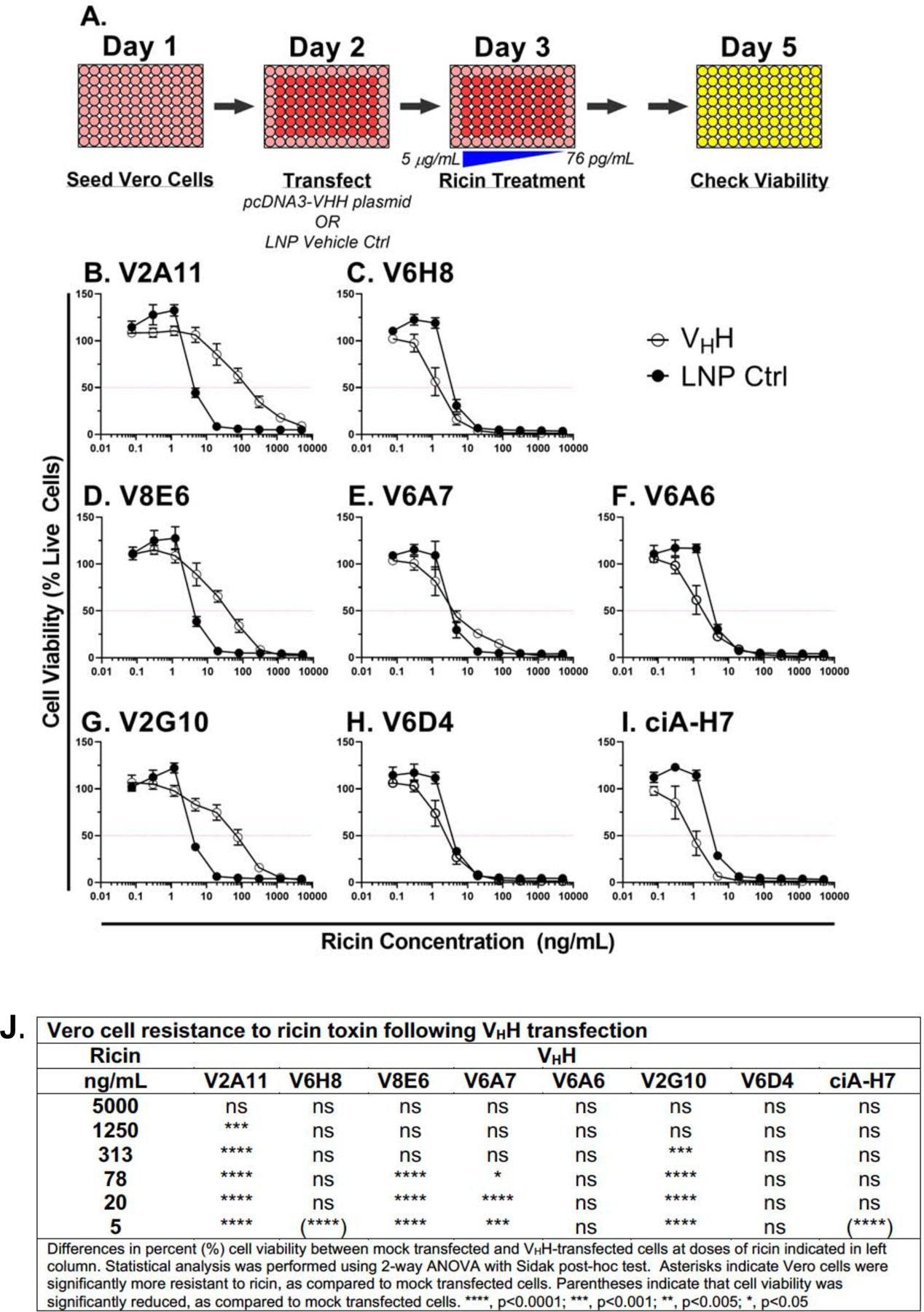
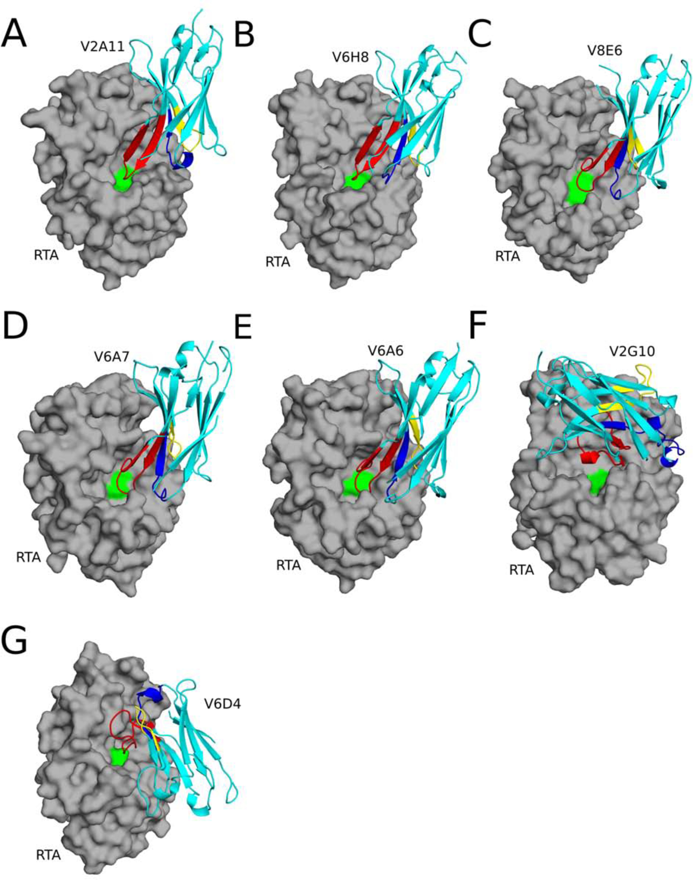
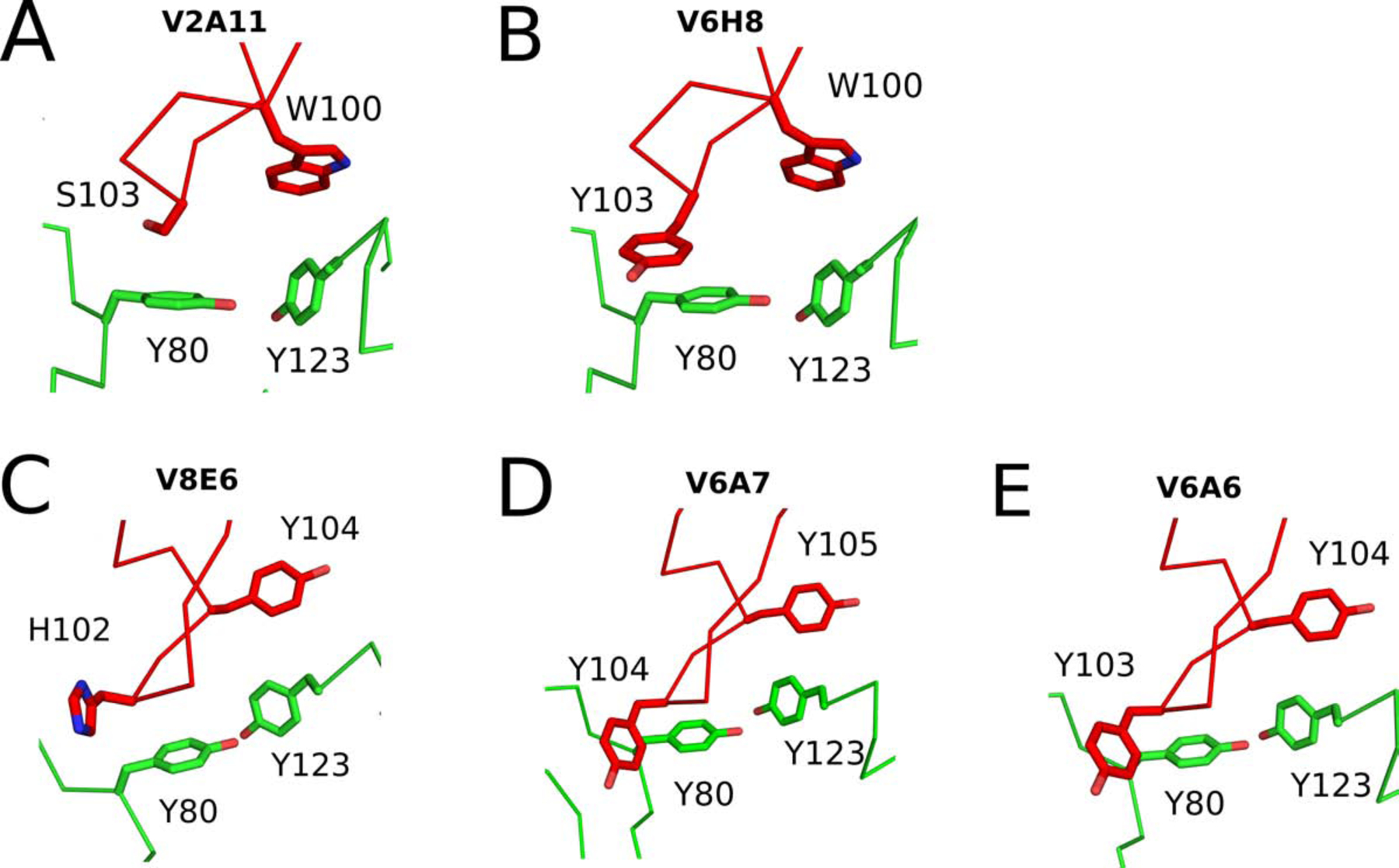
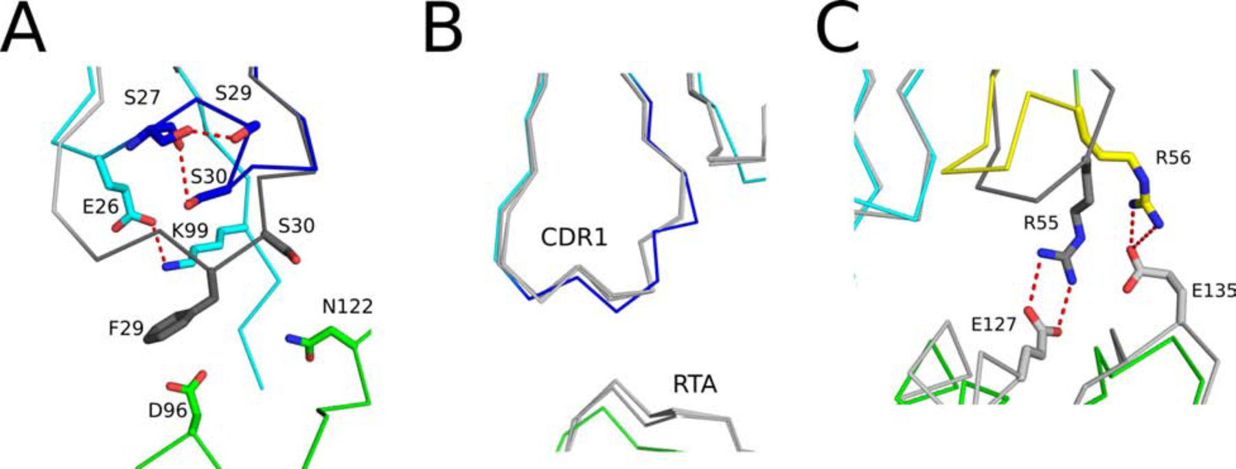
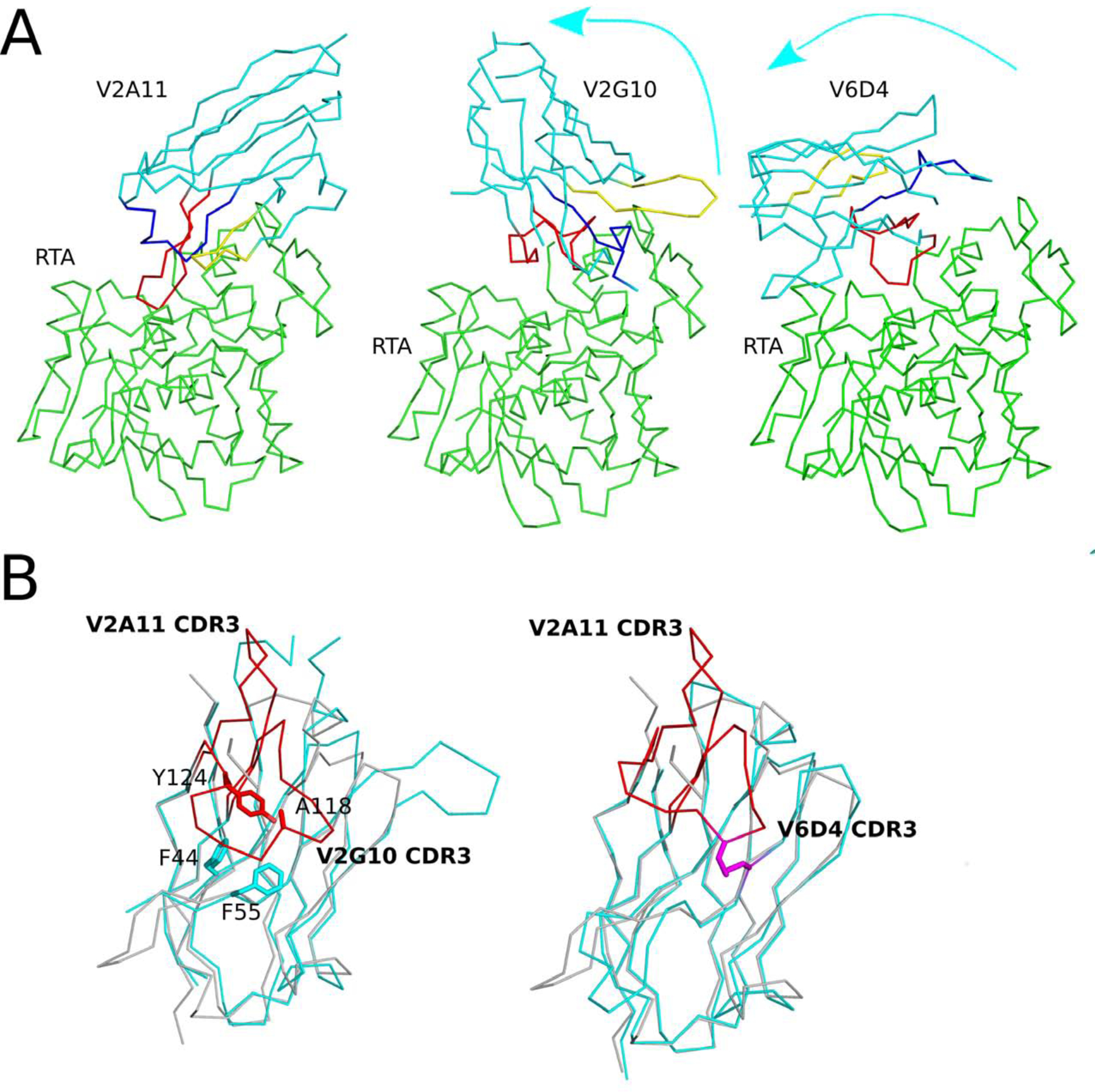
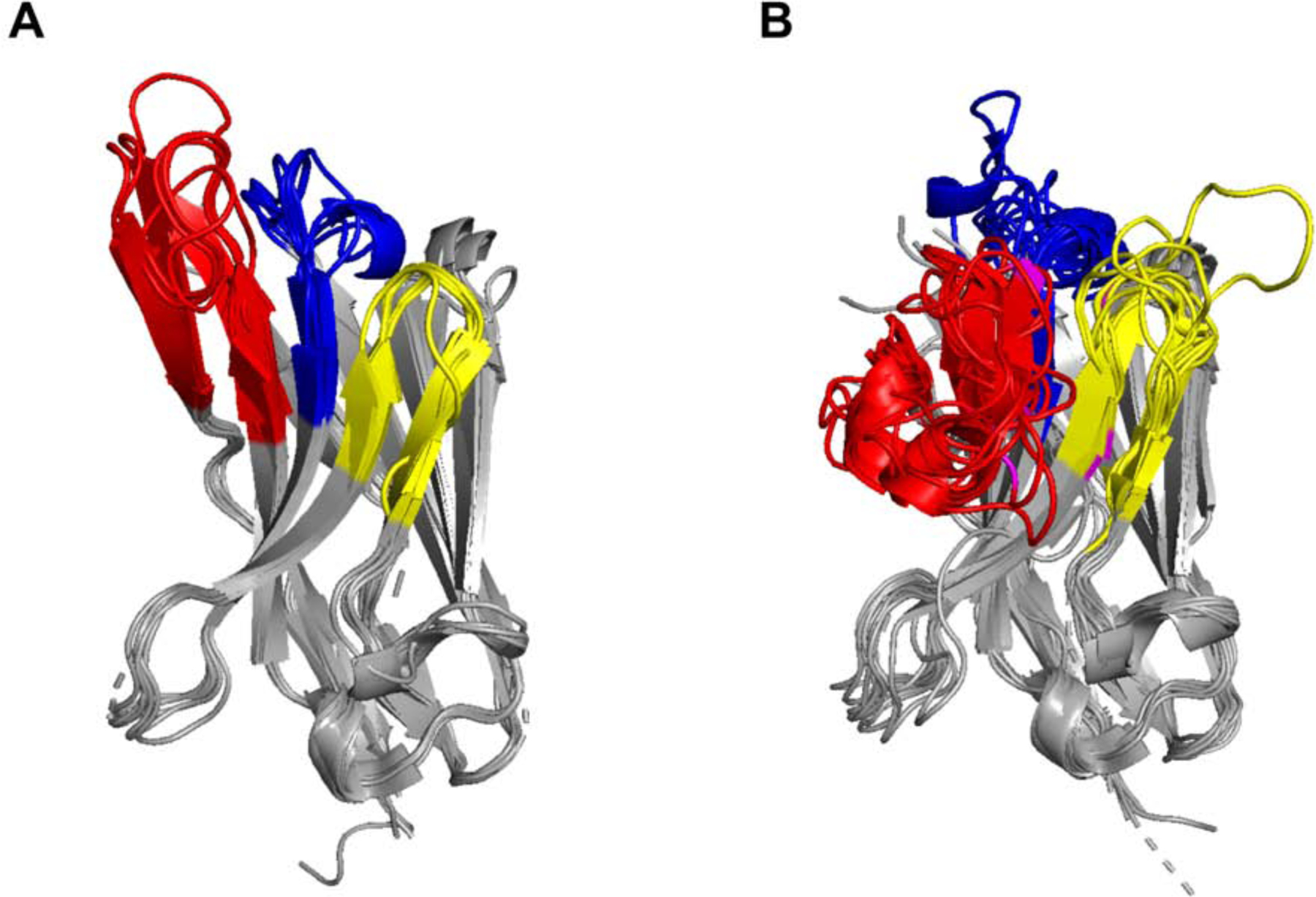
The extreme potency of the plant toxin, ricin, is due to its enzymatic subunit, RTA, which inactivates mammalian ribosomes with near-perfect efficiency. Here we characterized, at the functional and structural levels, seven alpaca single-domain antibodies (VHHs) previously reported to recognize epitopes in proximity to RTA's active site. Three of the VHHs, V2A11, V8E6, and V2G10, were potent inhibitors of RTA in vitro and protected Vero cells from ricin when expressed as intracellular antibodies ("intrabodies"). Crystal structure analysis revealed that the complementarity-determining region 3 (CDR3) elements of V2A11 and V8E6 penetrate RTA's active site and interact with key catalytic residues. V2G10, by contrast, sits atop the enzymatic pocket and occludes substrate accessibility. The other four VHHs also penetrated/occluded RTA's active site, but lacked sufficient binding affinities to outcompete RTA-ribosome interactions. Intracellular delivery of high-affinity, single-domain antibodies may offer a new avenue in the development of countermeasures against ricin toxin.toxin, antibody, structure, intracellular.

Keywords: Antibody; Intracellular; Structure; Toxin.


Conflict of interest statement

Competing interests: The authors have no financial or other competing interests to declare.

Figures

Figure 1.
Figure 1.. Abrogation of RTA’s RIP activity in vitro by single domain antibodies.
(A, B) In vitro translation assays were performed by mixing RTA (0.75 nM) with (A) 100 nM or (B) 20 assays were done with purified yeast ribosomes mixed with RTA (1.0 nM) and (C) 100 nM or without the addition of RTA positive controls (100%). Reactions were performed in triplicate. (E, F) Spearman correlation analysis was performed to determine correlation between IVT **** p<0.0001, *** p<0.001, ** p<0.005, * p<0.05 indicate 50% viability. (J) Significance was calculated using a two-way ANOVA with the Sidak correction test at each ricin concentration to compare viability of cells following intrabody transfection and LNP mock transfection. In two instances (indicated by parentheses in panel J), to their respective main chain color.
Figure 2.
Figure 2.. Protection of Vero cells from ricin toxin by VHH intrabodies.
(A) Cartoon of the experimental set-up. Vero cells were seeded in 96-well plates on day 1 and transiently transfected with VHH-encoding pcDNA3.1 plasmids on day 2. Ricin challenge occurred on day 3 and cell viability was assessed on day 5. (B-I) Cell viabilities of Vero cells transfected with vehicle control (LNP; black circles) or VHHs (open circles) at indicated toxin concentrations. Shown are two biological replicates with three technical replicates each. Horizontal red lines indicate 50% viability. (J) Significance was calculated using a two-way ANOVA with the Sidak correction test at each ricin concentration to compare viability of cells following intrabody transfection and LNP mock transfection. In two instances (indicated by parentheses in panel J), Vero cell viability of VHH transfected cells was less than mock transfected cells, suggesting that VHH expression actually imparts a burden on cells viability. Asterisks: **** p<0.0001; *** p<0.001; ** p<0.005; * p<0.05.
Figure 3.
Figure 3.. Structures of RTA-VHH complexes.
Structures of RTA (gray surface) in complex with (A) V2A11, (B) V6H8, (C) V8E6, (D) V6A7, (E) V6A6 (F) V2G10, and (G) V6D4 depicted as ribbon diagrams. Each VHH is colored cyan, with CDRs 1, 2, and 3 colored blue, yellow, and red, respectively. RTA active site residue Tyr-80 is colored green.
Figure 4.
Figure 4.. Mode 1 VHH interactions within the RTA’s active site residues.
The Cα-traces of RTA (green) in complex with (A) V2A11, (B) V6H8, (C) V8E6 (D) V6A7, and (E) V6A6. The VHH CDR3 elements are colored red. VHH residues forming interactions with RTA active site residues Tyr-80 and Tyr-123 are drawn as sticks. All stick representations are color coordinated to their respective main chain color.
Figure 5.
Figure 5.. Influence of CDR1 and CDR2 configuration on binding affinity.
(Panel A) Superpositioned Cα-traces of RTA-V2A11 complex with RTA-V6H8. The CDR1 element in V2A11 is colored blue while CDR1 region of V6H8 is colored dark gray. (Panel B) Superpositioned Cα-traces of V6A6 with V6A7, V8E6, and V6H8 all bound to RTA showing their similar configurations. In the RTA-V6A6 complex, RTA is colored green and V6A6 is colored cyan with its CDR1 element colored blue. All other RTA-VHH complexes are colored gray. (Panel C) The CDR2 segment in V2A11 is colored yellow while CDR2 element of V6H8 is colored dark gray. RTA is colored green, the framework residues in V2A11 are colored in cyan, and light gray in V6H8 colored light gray in panels A and B. Important residues involving paratope-epitope interactions are drawn as sticks and color coordinated to their respective main chain color. Hydrogen bonds and salt bridges are represented as red dashes. CDR2 Arg-55 and RTA’s Glu-127. V2A11’s CDR2 is also constrained by a salt bridge between CDR2 Arg-56 and RTA’s Glu-135, however, the different position of this salt bridge tethered V2A11’s CDR2 to RTA to a lesser degree (Figure S7C). Thus, the entropic penalty incurred by V2A11 upon RTA binding is likely to be less than that of V6H8, augmenting V2A11’s relative binding affinity for RTA.
Figure 6.
Figure 6.. Additional VHH binding modes with RTA’s active site.
The Cα-traces of RTA (green) in complex with (Panel A) V2A11, V2G10, and V6D4. VHHs are colored in cyan; CDRs 1, 2, and 3 are colored blue, yellow, and red, respectively. Arrows illustrate the direction of the ~99° rotation of V2G10 and the ~165° rotation of V6D4, each relative to V2A11. (Panel B) Superpositioned Cα-traces of V2A11 onto V2G10 and V6D4 demonstrating V2A11’s CDR3 conformation relative to the other two VHHs. CDR3 elements are colored red. 62 V2G10 residues involved in hydrophobic interactions between CDR3 and FR are drawn as sticks and color coordinated to their respective main chain color. The disulfide bond between residues Cys50 and Cys105 of V6D4 is shown in stick representation and colored magenta.
Figure 7.
Figure 7.. Structural analysis of V6D4 and V2G10 with RTA’s active site residues.
Close-up images of the interface between RTA (green) and (A) V2G10 and (B) V6D4. The VHHs CDR3 elements are colored red. VHH residues forming interactions with RTA active site residues Tyr-80 are drawn as sticks. All stick representations are color coordinated to their respective main chain color.
Figure 8.
Figure 8.. Extended and pinned configurations of alpaca VHH CDR3s elements.
Superpositioned Cα-traces of 18 alpaca-derived (Vicuga pacos) VHH crystal structures listed in Table S3. Within this sampling, the CDR3 elements assume two distinct conformations: extended and pinned. (Panel A) In the extended conformations, the CDR3 element (red) projects upwards from β-strand F and away from CDRs 1 (blue) and 2 (yellow), before returning downward to β-strand G. (Panel B) In the pinned or “reefed” conformation, the CDR3 elements (red) fold back onto CDRs 1 (blue) and 2 (yellow), ultimately making contact with vestigial FR2 residues formerly involved in VL interactions, before connecting with β-strand G. Cysteine residues involved in non-canonical disulfides are colored magenta.

Similar articles

Cited by

References

    1. de Virgilio M, Lombardi A, Caliandro R, Fabbrini MS, Ribosome-inactivating proteins: from plant defense to tumor attack. Toxins (Basel) 2, 2699–2737 (2010). - PMC - PubMed
    1. Berger T et al., Toxins as biological weapons for terror-characteristics, challenges and medical countermeasures: a mini-review. Disaster Mil Med 2, 7 (2016). - PMC - PubMed
    1. Grela P, Szajwaj M, Horbowicz-Drozdzal P, Tchorzewski M, How Ricin Damages the Ribosome. Toxins (Basel) 11, (2019). - PMC - PubMed
    1. Sowa-Rogozinska N, Sominka H, Nowakowska-Golacka J, Sandvig K, Slominska-Wojewodzka M, Intracellular Transport and Cytotoxicity of the Protein Toxin Ricin. Toxins (Basel) 11, (2019). - PMC - PubMed
    1. Sapoznikov A et al., Early disruption of the alveolar-capillary barrier in a ricin-induced ARDS mouse model: neutrophil-dependent and -independent impairment of junction proteins. Am J Physiol Lung Cell Mol Physiol, (2018). - PubMed

Publication types